
1. Background
A wound is a localized area of damage to the skin and/or underlying tissue and is recognized as a leading cause of death and health care problems worldwide. Wound healing is a multifaceted biological process characterized by the complex interplay of diverse cellular and molecular events. Critical determinants governing wound-healing progression include inflammation, tissue regeneration, and infection prevention (1). Wound infections are usually caused by several bacteria, including aerobes, facultative anaerobes, and obligate anaerobes (2). Bacterial biofilms play a substantial role in wound development and treatment difficulty (3). One bacterial species that causes wound infections is Klebsiella pneumoniae (4). The alarming global rise in antibiotic resistance among Gram‑negative pathogens has severely limited conventional treatment options and underscored the urgent need for alternative antimicrobial strategies, such as phage therapy (5). The acquisition of plasmids and chromosomal mutations has shifted treatment options for K. pneumoniae infections toward carbapenems as a last-resort therapy (6).
Infections caused by CPK are associated with increased treatment costs, prolonged hospitalization, treatment failure, and increased mortality (7). Conventional strategies to combat microbial infections in wound healing are primarily based on antibiotics. However, the widespread and uncontrolled use of broad-spectrum antimicrobials has detrimental effects on the normal flora. In addition, misuse or overuse of antibiotics in hospitals has led to the development of multidrug-resistant (MDR) strains, which pose a serious threat to human health and safety (8). The World Health Organization (WHO) has prioritized antimicrobial resistance in this group of bacteria as one of the most serious threats to global health. Therefore, research in this field is encouraged to reduce bacterial mutations by identifying the most effective antibiotics (9, 10). Bacteriophages are viruses that infect prokaryotic cells (11). Their mechanism of action is highly specific, as each bacteriophage targets a particular bacterial species, and they can be a suitable alternative to antibiotics for eliminating infectious diseases (12, 13).
Bacteriophages have advantages such as safety, cost-effectiveness, an effective mode of action, ease of extraction, and no effect on eukaryotic cells. Phages have an auto-dosing property because they replicate within the bacterial host (14). However, several significant challenges have limited the widespread clinical adoption of phage therapy. These include: 1) the highly specific nature of phages, requiring precise matching to bacterial strains; 2) the potential for bacterial resistance development through various mechanisms; 3) complex regulatory pathways and a lack of standardized protocols for dosing, administration routes, and treatment duration; 4) potential immune system reactions in human hosts; and 5) limited large-scale clinical trial data supporting efficacy and safety (14). Despite these limitations, phage therapy has shown promising results in specific applications. The effects of phages against K. pneumoniae have also been proven (15, 16). Therefore, phages can be used as a highly effective and efficient tool for treating infections (17).
Hydroxypropyl methylcellulose (HPMC) belongs to the hydrophilic class of polymers and can be used to develop drug delivery systems due to its gelling, thickening, and swelling properties, which can form highly stable, clear, and odorless hydrogels. In this study, HPMC was selected to provide a sustained-release matrix for the phage cocktail and gentamicin, thereby increasing local bioavailability and prolonging the antimicrobial effect at the wound site. In addition, HPMC gels provide a thick but non-tacky feel, disperse easily on the skin, have a cooling effect, and are non-comedogenic (18).
2. Objectives
Given the widespread use of antibiotics and the increasing risk of drug resistance, we aimed to evaluate the antibacterial effects of a topical phage cocktail and gentamicin against K. pneumoniae-caused infectious wounds in Wistar rats.
3. Methods
3.1. Study Timeline
Bacterial isolate collection and bacteriophage isolation from hospital sewage samples were performed between January 2023 and August 2023. All in vivo animal experiments, including wound creation, infection induction, treatment administration, and outcome assessments, were carried out between March 2024 and November 2024. Data analysis and manuscript preparation were completed in December 2024.
3.2. Materials
Sodium Magnesium (SM) solution (5.8 g NaCl, 2.0 g MgSO4.7H2O, 5 mL of 2% gelatin in 1 liter), Polyethylene glycol (PEG) 6000 solution (207 g PEG 6000, 49.9 g NaCl, in 350 mL distilled water), 0.1 M MgSO4.7H2O, 0.1 M CaCl2, Luria Bertani broth medium, and Luria Bertani agar medium. K. pneumoniae ATCC® BAA-1705 was purchased from the Pasteur Center of Iran.
3.3. Bacterial
In this study, we used K. pneumoniae ATCC® BAA-1705 along with 16 clinical bacterial isolates obtained from the Microbiology Department at Al-Muthanna University.
3.4. Isolation and Titration of Phages
Phages active against K. pneumoniae ATCC® BAA-1705 were isolated from hospital sewage samples using the method described by Cerveny et al. (19). Equal volumes of sewage samples and a bacterial culture (LB broth + 5 mM CaCl2) were mixed and incubated overnight at 37°C on an orbital shaker (120 rpm) (Amerex Instruments, Inc., Chest-Type GYROMAX 777). The samples were centrifuged (2,800 RCF, 7 minutes), and the supernatant was first filtered through a 0.45 µm filter (Orange, UK) and then through a 0.2 µm filter (Orange, UK). The resulting lysate was tested for phage activity by spot assay (20). For the spot assay, 3 mL of soft agar (0.7% LB agar) was mixed with 500 µL of fresh bacterial suspension and poured onto plates. After solidification at room temperature for 30 minutes, 10 µL of lysate was spotted in three separate sections of an individual plate. Plates were incubated at 37°C for 24 h and then visually inspected for clear zone (plaque) formation. Phage titration was performed using the double agar overlay method (21). Briefly, 200 µL of each serial dilution was mixed with 500 µL of bacterial culture (mid-log phase, OD600 0.4) and incubated for 20 minutes at 37°C. The mixture was then combined with 3 mL of soft agar (0.7% LB agar + 0.1 M MgSO4, temperature maintained at 48 °C) and poured onto solid LB agar plates (1.5%). Plaques were counted, and the final phage titer was adjusted to 108 PFU/mL in SM buffer; isolates were stored in 50% glycerol at −70°C.
3.5. Amplification of Phage
Phages were amplified using the method of Cerveny et al. (19). A mixture of 10 mL bacterial suspension, 1 mL purified phage solution, and 500 mL of Luria Bertani (LB) broth was incubated for 24 h at 37°C in an orbital shaker incubator. The mixture was then centrifuged at 2,800 × g for 10 min. The supernatant was sequentially filtered through 0.45 and 0.22 µm filters.
3.6. Phage Precipitation
PEG 6000 (Promega Co. USA) was used to enhance bacteriophage precipitation. Cell lysates were centrifuged at 10,000 × g for 30 minutes at 4 °C to pellet bacterial debris. The supernatant was mixed with 10% (w/v) PEG 6000 and 1 M NaCl (final concentrations) and incubated overnight at 4 °C with gentle agitation. The precipitated phages were recovered by centrifugation at 12,000 × g for 45 minutes at 4 °C, and the pellet was resuspended in 1/10th the volume of SM buffer. Residual PEG was removed by low-speed centrifugation (5,000 × g for 10 minutes). The final concentration was rechecked using double agar overlay plaque assays (21).
3.7. Stability of the Bacteriophage at Different Temperatures and pH
To investigate the potential of phages for treating infectious wounds, their biological properties, including stability at different temperatures and pH conditions, host range, adsorption rate, and one-step growth curves, were evaluated. The temperature stability of K. pneumoniae phage A (KPPA), K. pneumoniae phage B (KPPB), K. pneumoniae phage C (KPPC), and K. pneumoniae phage D (KPPD) (108 PFU/mL) was assessed in a water bath at 5, 15, 25, 35, 45, and 550C. To determine phage titers, successive dilutions of KPPA, KPPB, KPPC, and KPPD were spotted in triplicate using the double-layer agar overlay method (22). The stability of phages in SM phage buffer was evaluated by incubation for 10, 20, 30, and 40 min at pH 2, 4, 6, 8, 10, and 12 (22).
3.8. Host Range Determination
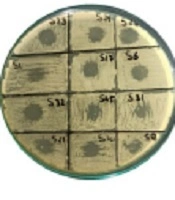
Sixteen bacterial strains, listed in Figure 1, were analyzed for sensitivity to phages KPPA, KPPB, KPPC, and KPPD. The Spot Test (23) was used to determine phage host range. Two microliters of phage lysate (107 PFU/mL) was applied to a 0.5% LB agar plate mixed with bacteria and incubated overnight. Lytic activity was examined after overnight incubation at 37°C and recorded as follows: (N) no plaques, (C) clear plaques.
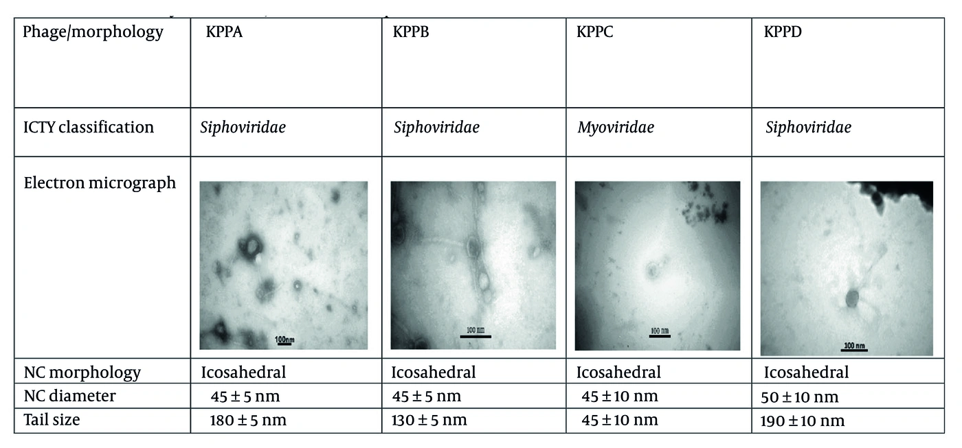
Figure 1.
The morphological features of four phages in this study; ICTV: The International Committee on Taxonomy of Viruses, NC: Nucleocapsid
3.9. Adsorption Rate
The phage adsorption curve was determined by separately mixing phages with log-phase cultures of K. pneumoniae ATCC® BAA-1705 (105 CFU/mL) to obtain a final MOI of 10, followed by incubation at 37°C. Aliquots (100 μL) of the mixture were collected every 4 min for 50 min and filtered through 0.45 µm filters. The titer of unabsorbed phages was calculated from the filtrates. The number of non-adsorbed phages was determined by plating the above mixture using the agar overlay method. Experiments were performed in triplicate.
3.10. One-Step Curve
A one-step growth curve was performed to determine phage burst size and latent time. Burst size represents the ratio of the maximum number of phages obtained after lysis to the initial count of phage particles, and latent time represents the time interval between phage adsorption and the onset of bacterial lysis. For this assay, 5 milliliters of log-phase K. pneumoniae culture was centrifuged and resuspended in 1 mL of LB broth. Each phage was added at an MOI of 10 and incubated at 370C for 15 min. After incubation, the mixture was centrifuged at 13000 rpm for 20 min, and the bacteria–phage pellet was resuspended in 10 mL of LB broth and incubated again at 370C. Every 5 min for 100 min, 100 μL samples were collected, filtered through 0.45 µm filters, and diluted in LB broth. Phage titers were determined using the double-layer technique. Three replicates were performed.
3.11. Preparation of Gel Containing Bacteriophage
Because bacteriophages can maintain antibacterial properties in different pharmaceutical forms, bacteriophages were incorporated into HPMC gel (Dow Chemical International Pvt. Ltd (Mumbai, India) in this study (24). HPMC powder was dispersed in water and stirred. First, a 4% gel was prepared using HPMC and placed in a vacuum oven at 25°C; the hydrogel was then sterilized by autoclaving. Phage stocks were prepared at a concentration of 108 PFU/mL. Finally, a 1:1 solution was mixed with the phage cocktail solution to obtain a final concentration of 2%. Bacterial endotoxins were removed using 0.4 volume of 1-octanol followed by shaking for 1 h. The mixture was centrifuged at 4000 × g for 10 min, and the aqueous phase was collected. Residual organic solvent was removed by centrifuging the phages at 20,000 × g for 1.5 h and then replacing the SM buffer with fresh SM buffer.
3.12. Measurement of Gelation Time
Gelation time was measured according to the tilt method (25). Briefly, 1 mL of each gel formulation (4% HPMC and HPMC-phage gel) was placed into a glass vial, and the vial was equilibrated for 30 seconds in a water bath at 25 ± 0.5°C. After equilibration, the vial was removed from the water bath and tilted to evaluate gelation. Gelation time was defined as the moment when the hydrogel showed no flow for > 5 seconds upon flipping. For reproducibility testing, measurements were performed in triplicate and reported as mean ± standard deviation.
3.13. Bacteriophage Release Pattern Test from the HPMC Gel
Bacteriophage release from HPMC gel was assessed using a modified double-layer agar (DLA) diffusion assay. Briefly, 1.5% LB agar plates were prepared and desiccated in a 37°C incubator for 12 h to minimize excess moisture before use. Then, 100 µL of mid-log phase bacterial culture (OD600 = 0.6) was poured onto the agar plate and incubated at 37°C for 4 h. Subsequently, 100 μL of HPMC gel containing phage was placed in tubes, each containing 900 μL of PBS. Tubes were placed in an incubator at 37°C, and at different time points (1, 12, 24, 36, 48 h), 20 μL of the solution was collected for plaque assay and spread evenly over the bacterial lawn. Following gel application, plates were incubated at 37°C for 72 h, and plaque-forming units (PFUs) were counted to determine phage release kinetics. The cumulative release percentage was calculated using the following equation:
Cumulative Release (%) = (PFUs at time t / Total PFUs in gel) × 100
3.14. Cytotoxicity and Biocompatibility Evaluation
The NIH/3T3 fibroblast cell line was cultured in T75 tissue culture flasks (Corning, USA) using Dulbecco’s Modified Eagle Medium (DMEM) supplemented with 10% fetal bovine serum (FBS) and 1% streptomycin (100 μg/mL) and penicillin (100 IU/mL) for all experiments. Cultures were maintained in a humidified atmosphere at 37°C with 5% CO2. The culture medium was refreshed twice weekly during expansion until cells reached 80% confluency, at which point adherent fibroblasts were detached with 0.25% Trypsin-EDTA (Invitrogen™, USA) and subcultured into 96-well plates for experimental use. MTT assay cytotoxicity testing was performed on sterile gel pieces of uniform weight (50 mg per piece), and gel biocompatibility was evaluated on days 1, 3, and 7.
3.15. Animal Models
Female Wistar rats (4 - 6 weeks old, weighing 120 - 150 grams) were used in this study. The experiments were conducted in accordance with national guidelines for animal studies, and the study was approved by the Ethics Committee of Fasa University of Medical Sciences. Animals were housed in polycarbonate cages in well-ventilated rooms with a 12 h light-dark cycle at 25 ± 2°C and were provided a standard rodent diet and water ad libitum. Rats were acclimatized in the laboratory for approximately 10 days to achieve stable physical conditions before the experiment. Before experiments began, cages and the room were exposed to UV radiation for 24 h and disinfected using Dettol liquid to create a pathogen-free environment.
Rats were randomly allocated into four groups (n = 20 per group) using a computer-generated random sequence (www.randomizer.org) by an independent researcher. Allocation concealment was achieved using sequentially numbered, opaque, sealed envelopes that were opened only immediately before treatment administration. Blinding was not possible for intervention delivery because of the different nature of the treatments; however, blinding was maintained during the measurement of all outcomes. Wound closure was measured from coded images by two blinded assessors. Health scoring was performed independently each day by two blinded researchers. Assessments of bacterial burden and histology were conducted independently by two blinded microbiologists and pathologists, respectively. Sample sizes were calculated assuming a 10% effect size, 80% power, and a 20% attrition rate, consistent with prior studies (26). On the day of the experiment, rats were anesthetized with ketamine (90 mg/kg, IP) and xylazine (10 mg/kg, ip). Hair on the animal's back was shaved, and the area was thoroughly cleaned with depilatory cream. The target areas were disinfected with 10% betadine solution. Using a stencil ruler, a wound with an area of 1.5 cm2 was created using a scalpel blade and a surgical blade, with care taken to remove the full thickness of the skin.
Each group received 200 µL of K. pneumoniae ATCC® BAA-1705 suspension (108 CFU/mL) inoculated onto the wounds. Wounds were monitored for 14 days at 24 h intervals. Wounds were treated with phage cocktail, gentamicin, or their combination. Rats were then placed under a heating lamp, and the animals' eyes were covered with moistened sterile gauze to prevent injury. Gentamicin was administered via intraperitoneal injection at a dosage of 20 mg/kg body weight daily, starting 30 minutes after infection (first dose), followed by additional doses 24 h apart throughout the entire 8 days study period. The phage cocktail treatment consisted of a single application of 0.5 mL HPMC gel suspension containing 108 PFU/mL to the infected area 30 minutes post-infection. For Group 4 (combination therapy), both treatments were initiated 30 minutes post-infection.
The phage titer used (108 PFU/mL) was based on prior in vitro experiments in which optimum killing occurred in bacterial cultures at this dose and was consistent with phage therapy in rodent wound models reported elsewhere (19). The gel volume was set at 0.5 mL to provide adequate coverage of the entire wound area (1.5 cm2, or 0.33 mL/cm2), because lower volumes (0.25 mL) were insufficient and higher volumes (1.0 mL) resulted in spillage. The gentamicin dose used in this study (20 mg/kg/day IP) is the established therapeutic dose reported in the literature for treating Klebsiella infections in rodents (27). Additionally, acetaminophen (1 mg/kg body weight) was administered as an analgesic. A separate room was prepared to house the rats and sterilized using UV emission; similarly, cages, food, and drinking water were sterilized.
Animals were observed twice daily for signs of distress or morbidity. Animals meeting any of the following criteria were euthanized using carbon dioxide (CO2) inhalation followed by cervical dislocation: 1) health scores ≤ 2 over two consecutive observations (very sick animals with exudative swelling around the eyes and partly closed eyes), 2) ≥ 20% body weight loss compared with the starting body weight (at the start of the experiment), 3) inability to eat or drink food (e.g., immobility or extreme weakness), and 4) signs of very severe pain, such as hunched posture, piloerection with lethargy, or a respiratory rate exceeding 100 breaths per minute or breathing difficulty. The health scoring protocol used in this study is outlined in the welfare monitoring section below. None of the animals reached any of the above humane endpoints before the scheduled endpoints, except for mortalities that occurred among several experimental groups, as indicated in Figure 2. Inclusion criteria were good health at arrival, no visible wounds or symptoms of disease before surgery, and successful completion of wound creation and infection without surgical complications. All 80 animals included in this experiment did not meet any exclusion criteria.
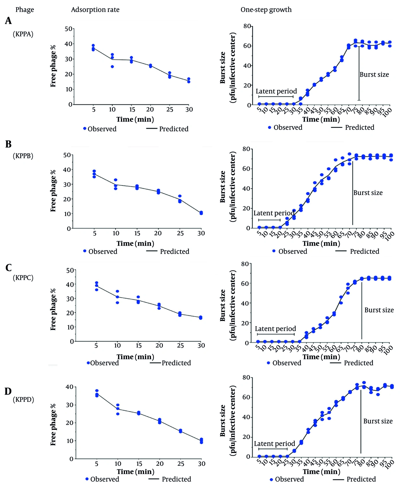
Figure 2.
Adsorption kinetics and one-step growth curves of bacteriophages KPPA, KPPB, KPPC, and KPPD against K. pneumoniae. Data are presented as mean ± SD.
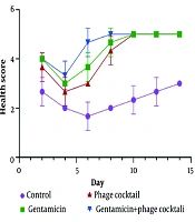
The welfare and clinical status of all animals were assessed twice daily (at 9:00 AM and 5:00 PM) by two trained, blinded observers throughout the 14-day experimental period. Based on each animal’s condition, rats were assigned a score between 0 and 5. The clinical course of skin lesions was monitored daily for blisters, swelling, redness, crusting, bleeding, discharge, granulation tissue, and scar tissue. Animals with a normal condition and no clinical symptoms were assigned a score of five. Animals with mild symptoms, such as tiredness and ruffled fur, were assigned a score of four. A score of 3 was assigned to animals with moderate symptoms, such as severe weakness, ruffled fur, and hunchback. Animals were assigned a score of two if, in addition to the above symptoms, secretions around the eyes led to partial eye closure. A score of one indicated a moribund state, and death was scored as zero. Two independent observers verified the results by scoring the clinical signs (28). Rats were divided into four groups: control (untreated), gentamicin, phage, and phage cocktail + gentamicin (Table 1).
Table 1.
Rat Groups in This Study, Including Control (Untreated), Gentamicin, Phage and Phage Cocktail + Gentamicin
| Group 1 | Control (untreated) |
|---|---|
| Group 2 | Rats infected with 200 µL of 108 CFU/mL K. pneumoniae; single topical application of 0.5 mL HPMC gel containing 108 PFU/mL phage cocktail at 30 min post-infection |
| Group 3 | Rats infected with 200 µL of 108 CFU/mL K. pneumoniae; intraperitoneal gentamicin (20 mg/kg) daily, first dose at 30 min post-infection, then every 24h |
| Group 4 | Rats infected with 200 µL of 108 CFU/mL K. pneumoniae; intraperitoneal gentamicin (20 mg/kg) at 30 min post-infection (then daily) PLUS single topical 0.5 mL phage cocktail gel (108 PFU/mL) at 30 min post-infection |
3.16. Wound Closure in Rats
To evaluate wound closure, photographs of rat wounds were taken using a digital camera on days 0 and 14. Wound size after induction was measured on days 6, 8, 10, 12, and 14 using transparent graph paper. Wound closure was calculated as a percentage (%WC) using the following formula:
Survival rate at day 14 was measured as the primary outcome, whereas secondary outcomes included the percent of wound contraction, changes in bacterial load, improvement in health scores from baseline, and changes in histology. Two blinded investigators independently traced wound outlines on transparent graph paper and calculated wound area using the mean of these three values. Digital photographs were taken for visual records and were not used for quantitative measurements. The formula shown was used to calculate the final percent closure using the baseline area on day 0.
3.17. Phage Toxicity Test
Phage toxicity in rats was investigated according to a previously described method (30). Briefly, 0.25 mL phage (108 PFU/mL) was administered intraperitoneally (IP) to uninfected rats. Health status was monitored for 48 - 72 h. An arbitrary 0 - 2 scale was used: 2 for normal healthy rats, 1 for ailing and weakened rats or those with abnormal health, and 0 for dead rats.
3.18. Examination of Bacterial Load in Wounds
In the present study, we focused on the following points:
1) Cleanliness of the wound before sampling. 2) Obtaining a complete sample of the rat wound for culture. 3) Removal of dense secretions using a sterile needle. 4) Transferring the wound sample using a sterile container and performing culture as quickly as possible to prevent contamination.
Bacterial load was evaluated using the method of Park et al. (31). One rat in each group was randomly euthanized on days 1, 3, 5, 7, 9, and 11 under deep anesthesia. Deep tissue sampling was performed under sterile conditions. Tissue was homogenized in phosphate buffered saline (PBS) (pH 7.2) and then centrifuged (at 3000 rpm) for 10 min. Samples were diluted, plated on LB agar, and incubated overnight at 37°C.
3.19. Pathologic Studies and Wound Healing Process
At the end of the experimental period, animals were euthanized according to institutional guidelines. Tissues from the wound site (approx. 1 x 1 cm) were carefully excised using sterile surgical blades. Tissue samples were fixed in 10% formalin and dehydrated in a graded series of alcohol (70% ethanol (2 h), 80% ethanol (2 h), 90% ethanol (1.5 h), and absolute ethanol (two changes of 1 h each)). Samples were then embedded in paraffin wax, sectioned, and stained with Hematoxylin-eosin. Pathological studies were conducted to evaluate the repair rate of the dermal area. Two blinded pathologists used established semi-quantitative criteria to evaluate biopsy specimens. Epidermal thickness (μm) was measured in three random fields per section using ImageJ image processing software. Fibroblasts were scored by degree of proliferation: 0 - none; 1 - mild; 2 - moderate; 3 - marked. Inflammation was scored as follows: 0 - none; 1 - focal; 2 - multifocal; 3 - diffuse. The final score for each parameter was determined by averaging three individual wounds for each section.
3.20. Statistical Analysis
Data were analyzed using GraphPad Prism 9. Normality was assessed by the Shapiro–Wilk test and equality of variances by Levene's test. For normally distributed continuous data (wound contraction, bacterial load log-transformed), one-way ANOVA with Tukey's post hoc test was used for group comparisons at each time point. For repeated measurements over time (wound contraction, bacterial load, health scores), two-way repeated-measures ANOVA with Bonferroni correction was applied. Survival data (Kaplan–Meier curves, Figure 2) were analyzed using the log-rank (Mantel–Cox) test with pairwise comparisons. Ordinal health scores were analyzed using the Kruskal–Wallis test with Dunn's post hoc test. Histological scores were analyzed using the Mann–Whitney U test. Statistical significance was set at P < 0.05. All tests were two-tailed. Data are presented as mean ± SD unless otherwise stated.
4. Results
4.1. Morphological Characterization of Isolated Bacteriophages
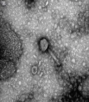
The morphology of the four bacteriophages (KPPA, KPPB, KPPC, and KPPD) isolated in this study was examined by electron microscopy. Their morphological features and dimensions are summarized in Figure 1. Based on their structure, phages KPPA, KPPB, and KPPD were likely classified within the family Siphoviridae, which is characterized by an icosahedral head and a long, non-contractile tail. In contrast, phage KPPC likely belonged to the family Myoviridae, which is identified by an icosahedral head and a characteristic shorter, contractile tail. All four phages exhibited an icosahedral (isometric) nucleocapsid morphology. The average head diameters were relatively consistent across the phages, measuring 45 ± 5 nm for KPPA and KPPB, 45 ± 10 nm for KPPC, and 50 ± 10 nm for KPPD. The primary distinguishing feature was tail length. KPPC, the Myoviridae phage, possessed a short tail of 45 ± 10 nm. Meanwhile, the Siphoviridae phages displayed long tails, with KPPA, KPPB, and KPPD having tail lengths of 180 ± 5 nm, 130 ± 5 nm, and 190 ± 10 nm, respectively.
4.2. Stability of the Bacteriophage at Different Temperatures and pH
Phages KPPA, KPPB, KPPC, and KPPD maintained stability across a broad pH range, with titers of approximately 107 - 108 PFU/mL at pH 6 - 8. Phage viability decreased at certain pH values; titers decreased to about 105 PFU/mL at pH 2, 106 PFU/mL at pH 4 and 12, and about 106.5 PFU/mL at pH 10 (Figure 3). Therefore, phage titers were highest at pH 6 - 8 and decreased at pH ≤ 4 and ≥ 10. Phage KPPA, KPPB, KPPC, and KPPD titers also remained stable at approximately 108 PFU/mL for 40 min at temperatures of 5, 15, 25, 35, 45, and 550C. These results indicate that phage titers remained stable under the tested temperature conditions, as shown in Figure 3.
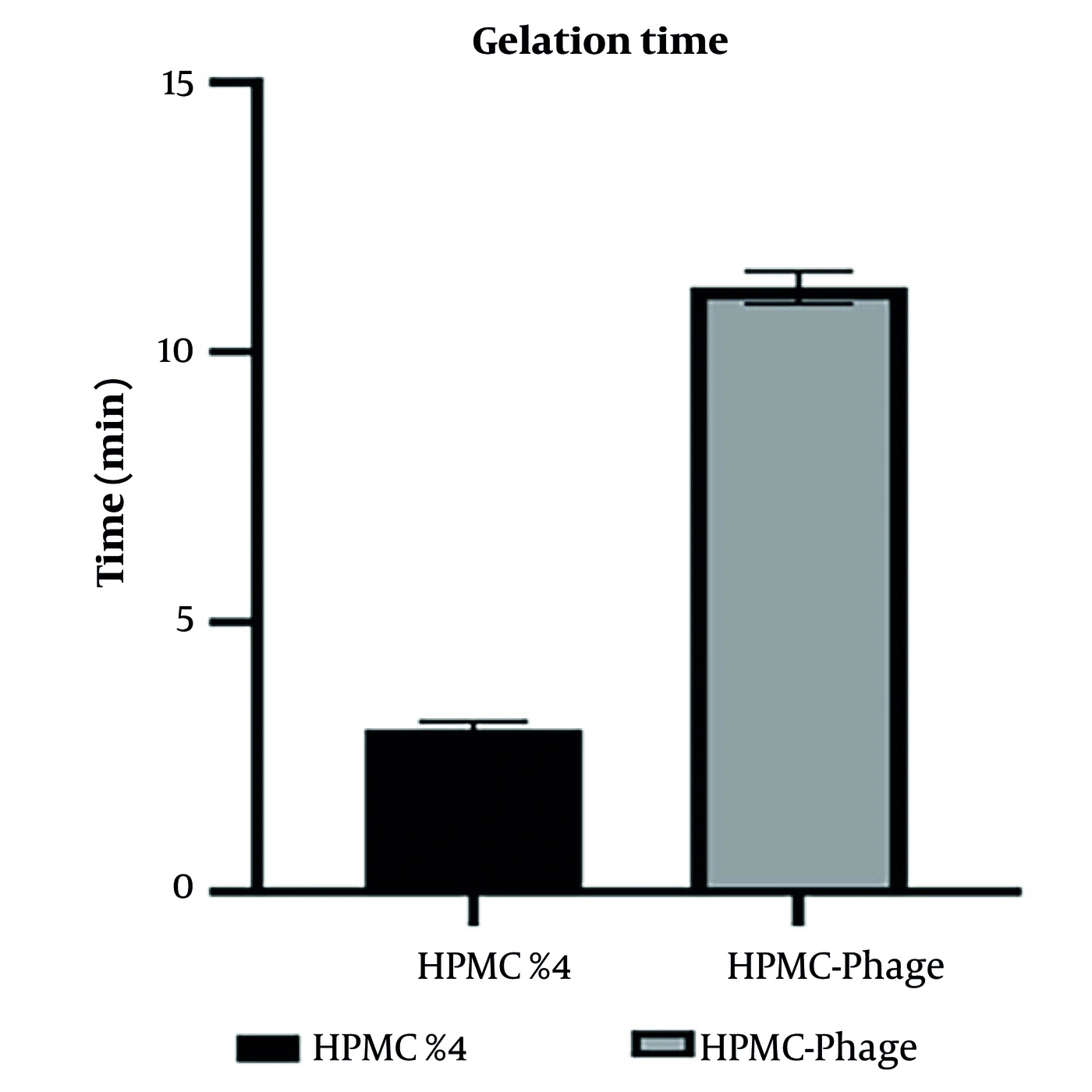
Figure 3.
Gelation times of the 4% HPMC control formulation and the HPMC-phage gel formulation at 25°C. Data are presented as mean ± SD (n = 3).
4.3. Host Range Determination
The host range of phages KPPA, KPPB, KPPC, and KPPD was evaluated against sixteen bacterial strains. Phage KPPB produced clear plaques in 50% of K. pneumoniae strains, indicating that phage KPPB was effective against K. pneumoniae. Phages KPPA, KPPC, and KPPD produced clear plaques on 30, 30, and 40% of K. pneumoniae strains, respectively. A notable result was obtained using the phage cocktail, which produced clear plaques in 70% of K. pneumoniae strains (Table 2).
Table 2.
Host Range Analysis of Bacteriophages Against K. Pneumoniae, S. Aureus, P. Aeruginosa, and E. Coli Strains and Susceptibility Patterns Determined by Spot Test Assay a
| No | Species | Strain | KPPA | KPPB | KPPC | KPPD | Phage cocktail |
|---|---|---|---|---|---|---|---|
| 1 | K. pneumoniae | ATCC 9997 | N | C | N | C | C |
| 2 | K. pneumoniae | ATCC 13883 | N | N | N | N | N |
| 3 | K. pneumoniae | ATCC®BAA-1705 | C | C | C | C | C |
| 4 | K. pneumoniae | ATCC 11296 | N | N | N | N | N |
| 5 | K. pneumoniae | JCM 1662 | C | C | N | N | C |
| 6 | K. pneumoniae | DSM 30104 | N | C | N | N | C |
| 7 | K. pneumoniae | CIP 82.9 | N | N | N | N | N |
| 8 | K. pneumoniae | CCM 7798 | N | N | C | C | C |
| 9 | K. pneumoniae | CCUG 15938 | C | N | C | N | C |
| 10 | K. pneumoniae | CECT 851 | N | C | N | C | C |
| 11 | MRSA | ATCC 43300 | N | N | N | N | N |
| 12 | MRSA | ATCC 33591 | N | N | N | N | N |
| 13 | MSSA | ATCC 12600 | N | N | N | N | N |
| 14 | MSSA | ATCC 29213 | N | N | N | N | N |
| 15 | P. aeruginosa | ATCC 27853 | N | N | N | N | N |
| 16 | E. coli | ATCC 25922 | N | N | N | N | N |
a Abbreviations: C, clear plaque; N, no plaque.
4.4. Adsorption Rate
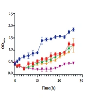
Virus adsorption analysis showed that after 30 min, approximately 85% of KPPA, 90% of KPPB, 80% of KPPC, and 85% of KPPD virions were adsorbed to the host cell K. pneumoniae (Figure 4).
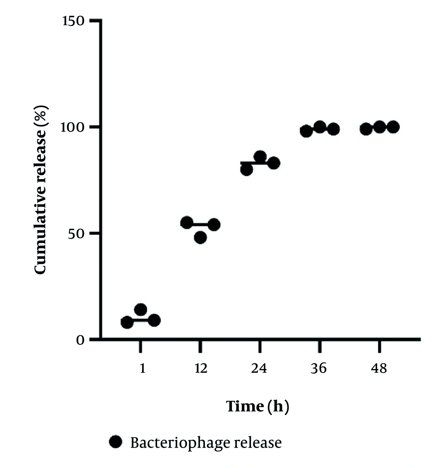
Figure 4.
in vitro release kinetics of bacteriophages from HPMC hydrogel over 36 hour. The graph illustrates the cumulative phage release profile from the HPMC gel. Key time points include an initial burst release (10.33%, 7.01 log PFU/mL at 1 h), progressive diffusion (52.33%, 7.72 log PFU/mL at 12 h), sustained release (83%, 7.92 log PFU/mL at 24 h), and near-complete release (>95%, ~8.00 log PFU/mL) by 36 h. (n = 3).
4.5. One-Step Curve
One-step growth curve analysis (Figure 4) showed that phages KPPA, KPPB, KPPC, and KPPD had latent periods of 30, 20, 35, and 25 min, respectively. The average burst time of the phages was ~70 - 80 min.
4.6. Gelation Time Determination
The gelation properties of the gel formulations were evaluated using the standardized tilt method. Quantitative analysis of each formulation indicated variable gelation. The 4% HPMC control formulation showed rapid thermal gelation, with a mean gelation time of 3.0 ± 0.4 min (n = 3) at 25°C. The HPMC-phage gel demonstrated much slower gelation kinetics and required 11.2 ± 0.7 min (n = 3) to form a complete gel using the same induction methodology (Figure 5).
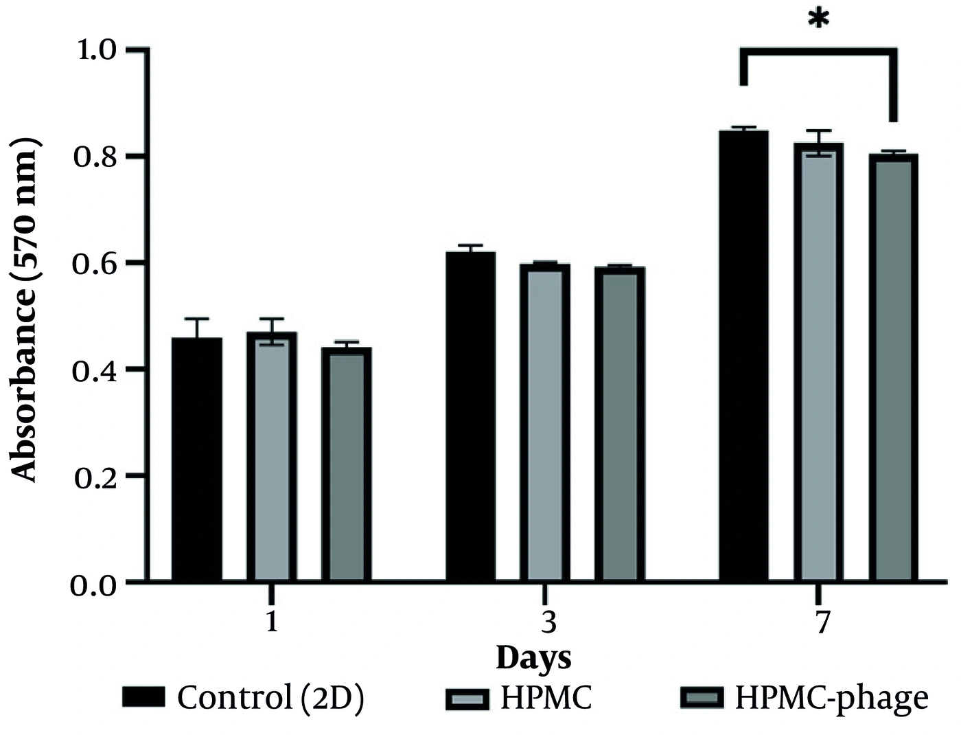
Figure 5.
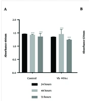
Cytocompatibility evaluation of HPMC and HPMC-phage gels. NIH/3T3 fibroblast viability was assessed via MTT assay after 1, 3, and 7 days of culture. Optical density (OD) measurements at 570 nm demonstrated excellent biocompatibility for both gels, with no significant cytotoxicity (P > 0.05) compared to the 2D control. Data are presented as mean ± SD (n ≥ 3).
4.7. in vitro Release Pattern of Bacteriophages from HPMC Gel
The HPMC gel containing phage exhibited sustained release kinetics, maintained phage viability, and showed potential antibacterial activity over time. The release of phage from the HPMC gel containing phage is depicted in Figure 6. The hydrogel exhibited continued phage release for up to 36 h. At 1 hour, 10.33% release (log10 7.01 PFU/mL) was observed. After 12 hour of exposure in broth, a cumulative release of 52.33% was observed (log10 7.72 PFU/mL), likely reflecting continued diffusion of phage from the gel matrix. At 24 hour, 83% of the phages had diffused out of the gel matrix (log10 7.92 PFU/mL), indicating that phages continued to be released from the gel matrix over 36 hours. By 36 hour, it was loosely observed that almost all phages had been released, with very few phages retained in the gel.
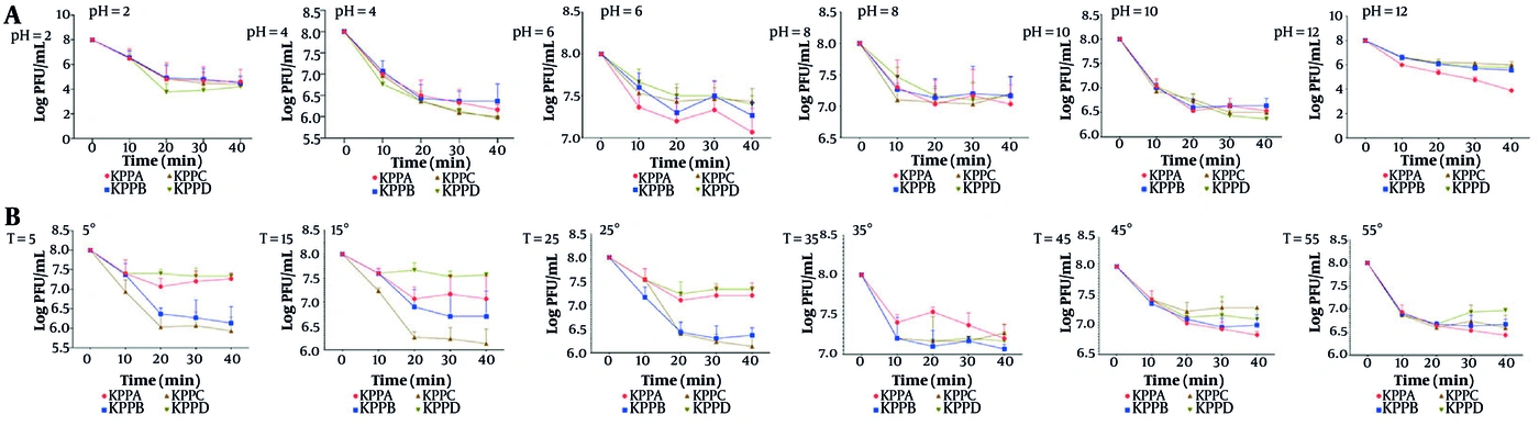
Figure 6.
The stability of phage KPPA, KPPB, KPPC, and KPPD at different pH values (A), and different temperatures (B) and time duration of 40 h.
4.8. Cytotoxicity Study
The cytotoxicity of HPMC gel and HPMC-phage gel was evaluated using the MTT assay (Figure 2), with 2D culture plates serving as the control. Cell proliferation was assessed by measuring optical density (OD) at 570 nm after 1, 3, and 7 days of culture. Both gels showed excellent biocompatibility, and no significant cytotoxicity was observed. The OD for the HPMC gel at 1 day was 0.470, 0.598 at 3 days, and 0.825 after 7 days. The OD of the HPMC-phage gel was 0.441, 0.592, and 0.804, respectively. The 2D control group exhibited the highest proliferation (0.460, 0.622, and 0.849); however, no significant differences were detected (P > 0.05). These results indicate that both HPMC and HPMC-phage gels are cytocompatible and can be evaluated for future biomedical applications.
4.9. Effects of Phage Cocktail on Infected Wounds
To investigate the effect of a phage cocktail on the eradication of K. pneumoniae bacteria in vivo, a phage cocktail was administered as a topical gel; 2% HPMC and a phage concentration of 108 PFU/mL were used. Administration of gel containing the phage cocktail into rat wounds protected 80% (16/20) of infected rats for 14 days. In comparison, control group survival was only 50% (10/20), with 50% mortality by day 14 (Figure 7).
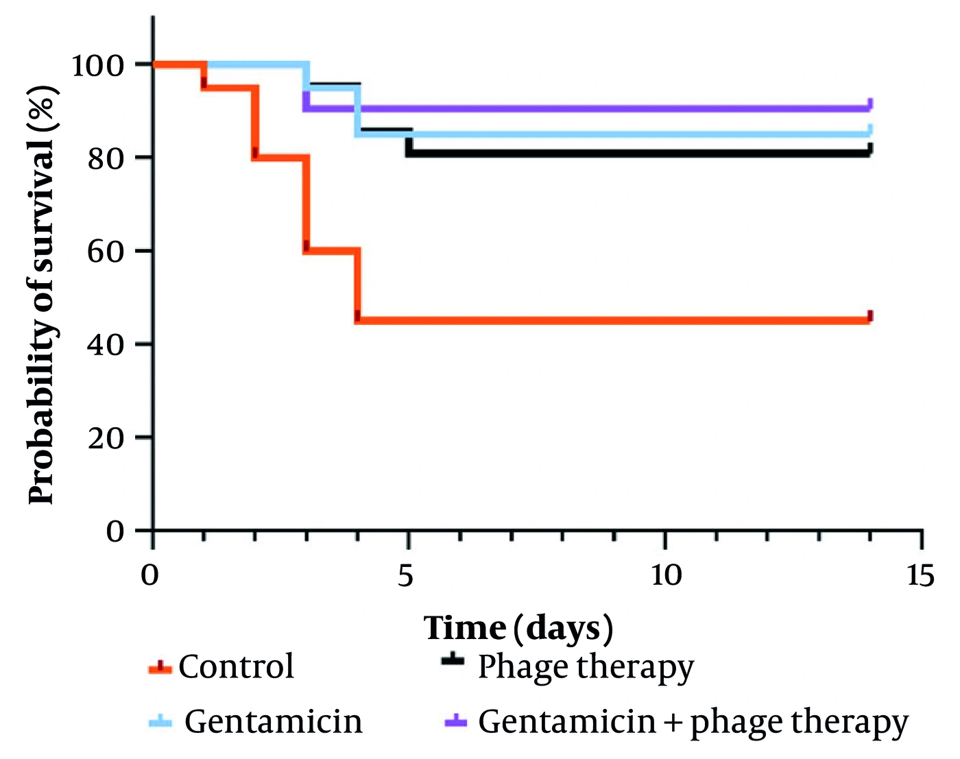
Figure 7.
Diagram of wound contraction in rats. Accordingly, the combination of gentamicin and phage cocktail significantly increased the wound contraction compared to other groups.
4.10. Effects of Gentamicin on Infected Wounds
Gentamicin was used to compare its therapeutic effect with that of bacteriophages in healing infected wounds. The dose used in this study was 20 mg/kg intraperitoneal. Gentamicin protected 85% (17/20) of infected rats (Figure 7).
4.11. Effects of Cocktail Phage + Gentamicin on Infected Wounds
To determine whether phage therapy can be effective as adjunctive therapy, gentamicin and phage therapy were used simultaneously. The survival rate of rats with this treatment was about 95% (19/20). The survival rate of rats is depicted in Figure 7.
4.12. Wound Contraction
Wound contraction results indicated that the simultaneous use of gentamicin and the phage cocktail produced the highest antibacterial activity. Wound contraction in the gentamicin group and the phage cocktail group was lower than that in the combination group (Figure 8).
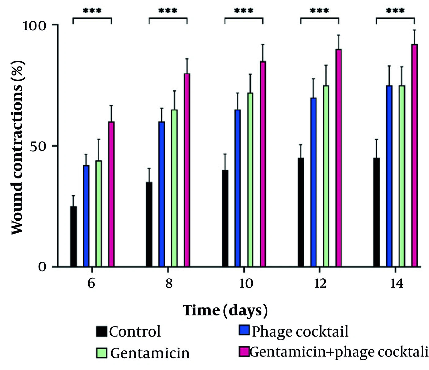
Figure 8.
Diagram of wound contraction in rats. Accordingly, the combination of gentamicin and phage cocktail significantly increased the wound contraction compared to other groups.
4.13. Bacterial Load in Wound
Using the phage cocktail gel with a concentration of 108 PFU/mL for K. pneumoniae, a significant reduction in bacterial load was observed on day 1 (1 log10.7). The bacterial load decreased by 4.2 and 2.5 log CFU/mL on days 5 and 7, respectively (P < 0.01). On the 9th day, the bacterial load reached 1.2 logs CFU/mL, and on the 11th day, it was zero. The bacterial load for gentamicin-exposed wounds infected with K. pneumoniae was 7.2, 6.2, 6, 3.1, 2.1, and 0 log CFU/mL on days 3, 5, 7, 9, and 11, respectively. The greatest decrease in bacterial load was observed in the group of rats treated with phage and antibiotics simultaneously. The decrease in K. pneumoniae bacterial load on the first day was (1 log10.6), and on days 3, 5, and 7, the bacterial load was 5, 3.1, and 1.1 logs CFU/mL, respectively. No isolates were observed in culture on days 9 and 11 (Figure 9).
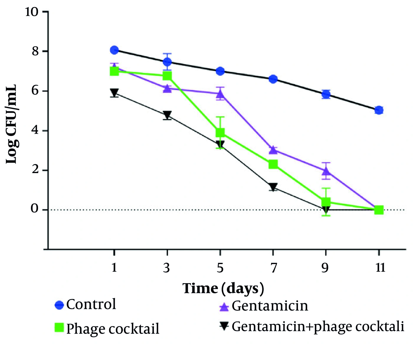
Figure 9.
A: Bacterial load in terms of (Log CFU/mL) in infected rats in various groups after treatment with gentamicin, phage, and a combination of phage and gentamicin. Bacterial load (Log CFU/mL) was measured on specific days after K. pneumoniae injection. Error bars represent the standard deviation (SD) of seven independent values. *** P < 0.001.
4.14. Health Score of Rats
All uninfected rats showed no signs of bacteremia or morbidity, whereas all untreated infected rats had a health score of 0. The group treated with gentamicin had a health score of 5 after 9 days, and the group treated with a phage cocktail had a health score of 5 after 9 days. Meanwhile, the group treated with gentamicin and phage had a health score of 5 after 7 days. The results are depicted in Figure 10.
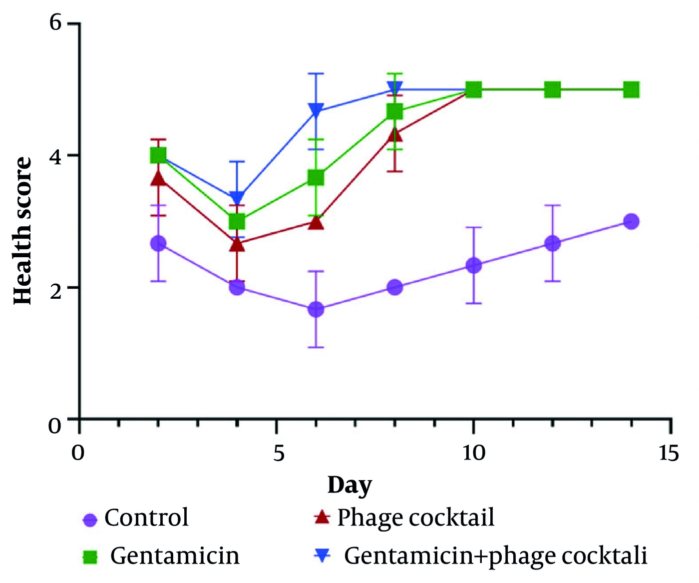
Figure 10.
A rat model of wound infection caused by K. pneumoniae. Comparison of the status of rat at different time intervals in infected rat after treatment with gentamicin, phage, and the combination of phage and gentamicin. Rat was scored on a scale of 0 to 5 in terms of their health status. A score of 5 indicates normal health and unremarkable condition. A mild illness, which is defined as a decrease in physical activity, was given 4 points. Moderate illness, meaning lethargy, was given a score of 3. Severe disease with severe symptoms, such as exudative accumulation around the eyes, was scored as 2. A moribund state or critical state was considered as a score of 1 and at the end of death, 0 points were given. * P < 0.05 and *** P < 0.001 vs. phage therapy group, ## P < 0.01 and ### P < 0.001 vs. gentamicin group.
4.15. Histological Evaluation
Histological results in rats treated concurrently with gentamicin and the phage cocktail showed greater epidermal thickness than in the groups treated with gentamicin or the phage cocktail alone. Light microscopic analyses further showed that inflammatory cell infiltration was reduced in all treated rats, particularly in combination-treated rats compared with the control group. In addition, fibroblast proliferation increased in all treated groups, especially in the combined antibiotic and phage treatment group (Figure 11).
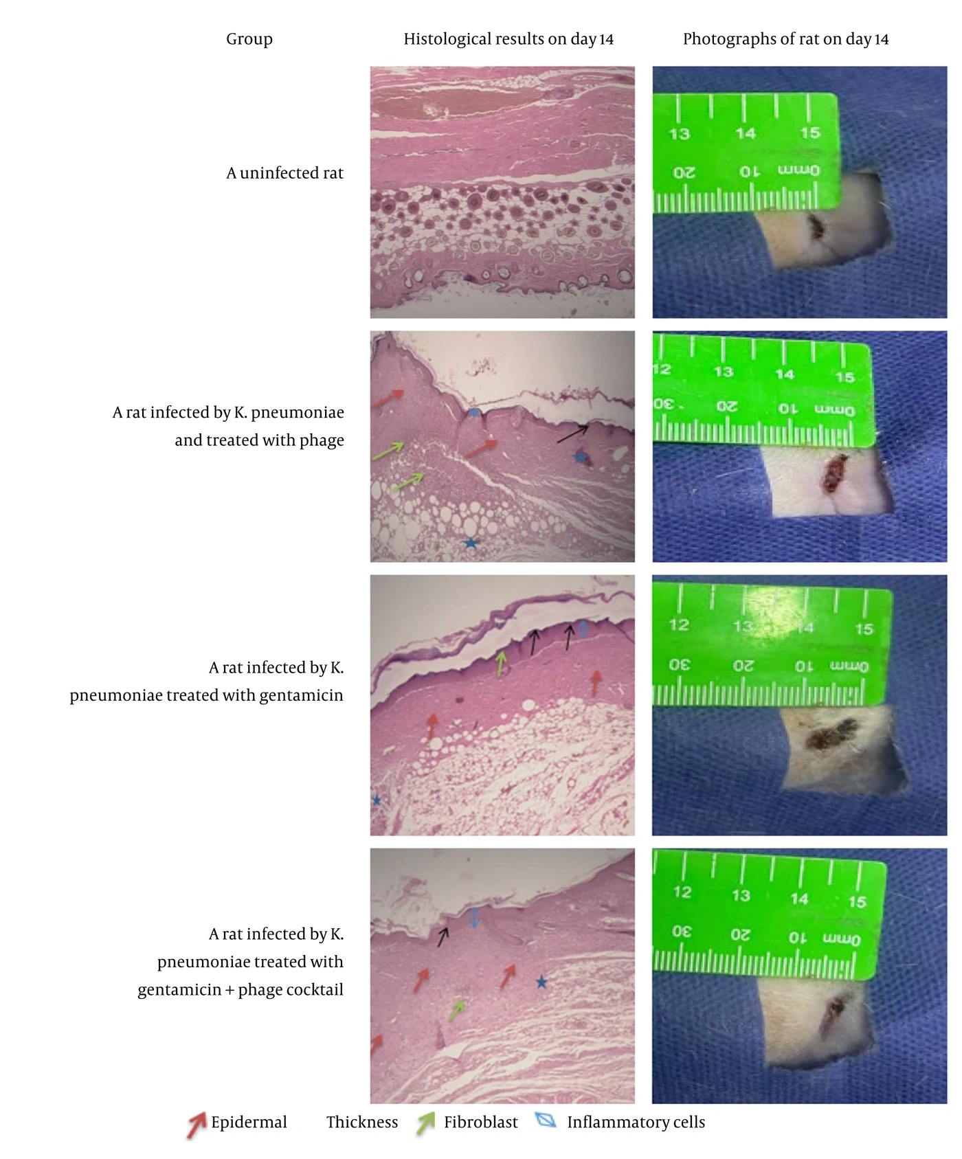
Figure 11.
Wound pathology evaluation and photographs of rat wound after staining with hematoxylin and eosin on day 14
5. Discussion
In the present study, we determined the composition and dose of the phage cocktail (KPPA, KPPB, KPPC, and KPPD). Electron microscopy confirmed that the four isolated phages exhibited diverse morphological features: KPPA, KPPB, and KPPD displayed long non-contractile tails likely characteristic of Siphoviridae, whereas KPPC possessed a short contractile tail typical of likely Myoviridae, suggesting that this phage cocktail comprises functionally distinct viral families with potentially complementary lytic mechanisms (Figure 1). The phage cocktail was designed to (1) increase the spectrum of activity against K. pneumoniae strains and (2) minimize phage resistance. A suitable combination of the obtained phages was prepared as a phage cocktail solution. The use of phage cocktails generally helps to address limitations such as the narrow host spectrum of individual phages and the emergence of phage resistance (32). Therefore, if resistance to a specific phage develops within the cocktail, another phage can compensate by lysing the bacteria, thereby reducing the likelihood of bacterial resistance to the phage cocktail (33).
Host range analysis showed that, whereas the individual phages displayed limited lytic activity against K. pneumoniae strains (30 - 50%), the phage cocktail was effective against 70% of the tested strains, demonstrating that combining phages with different specificities increases the therapeutic spectrum and reduces the likelihood of bacterial resistance (Table 2). HPMC is a biodegradable, cellulose-derived natural polymer that undergoes thermoreversible gelation upon heating. Wound healing was achieved via cost-effective delivery of the phage cocktail in an HPMC-based gel. HPMC’s moisture-retention capacity reduces scab formation and dehiscence, and its semi-occlusive nature can promote autolytic debridement (34). In addition to retaining moisture, the gel’s negative surface charge can further reduce bacterial adhesion (35), complementing the antibacterial effects of phages. Overall, HPMC, through physical barrier protection combined with biological and antimicrobial activity (phages), can create a favorable microenvironment for healing infected wounds. As demonstrated by Chang et al. (36), a 3% (w/v) HPMC formulation showed optimal rheological properties for sustained local delivery of bacteriophages while maintaining phage viability (36).
In this study, the gelation kinetics of the gel, the time-dependent release profile of bacteriophages from the HPMC gel matrix, and the cytotoxic effects on fibroblast cells were investigated (Figure 4). The increased gelation time of the HPMC-phage gel (11.2 ± 0.7 min) compared with the control HPMC gel (3.0 ± 0.4 min) indicates that bacteriophage incorporation altered polymer network formation, possibly due to interactions between phage particles and HPMC chains that delayed cross-linking (Figure 3). Similar changes in gelation kinetics have been reported with other bioactive-loaded systems, in which macromolecules interfere with gelation kinetics (37). Nevertheless, sustained phage release over a 36 h period indicates successful encapsulation and diffusion, which are key factors for sustained antibacterial action in wound-healing applications. The initial burst release (10.33% at 1 h) likely reflected phages associated with the surface, whereas subsequent gradual release (up to 83% at 24 h) was derived from diffusion controlled by the gel matrix and was consistent with previous studies of HPMC-based topical delivery systems (38, 39) (Figure 3). The near-complete release of phage by 36 h suggests that the gel had degraded sufficiently to provide bioaccessible phage. These findings are consistent with other reports of phage delivery using phage-loaded hydrogels, which have described the benefits of sustained release for therapeutic phage efficacy against infections (38, 39).
MTT assay findings indicate that both HPMC and HPMC-phage gels showed excellent cytocompatibility with NIH/3T3 fibroblasts, with viability and proliferation similar to those in the 2D control group over 7 days (P > 0.05) (Figure 5). The relatively constant OD values (0.804 - 0.825 at day 7) suggest that neither the gel matrix nor the phages exerted cytotoxic effects on fibroblasts. This outcome is consistent with previous findings on HPMC-based hydrogels for wound-healing applications (40, 41). A key factor in phage therapy efficacy is phage titer. Phages must have a high titer to reach infected tissues and multiply within the infected tissue. Therefore, phage dosage was also investigated. In this study, we used high-dose bacteriophages at a concentration of 108 PFU/mL. Using a high dose of bacteriophage relative to the target bacteria, without requiring reproduction and completion of the phage life cycle, caused a significant reduction in bacterial load. This observation is consistent with previous studies (42, 43). Previous studies have also shown that the use of low-titer bacteriophages reduces the probability of success (44). In addition, the initial dose must be sufficient because phages undergo phage metabolism (neutralization of the phage through interaction with the immune system) in the animal body or activation through in situ phage propagation; thus, phage levels can be reduced in the animal body. To prevent the development of a resistant bacterial population, a phage cocktail should be used that includes high doses of different phage species; in this way, resistant bacteria can be eliminated (45-47).
In this study, the host range of bacteriophages KPPA, KPPB, KPPC, and KPPD was evaluated (Table 2). Because the therapeutic capacity of phages is often limited by a narrow host range, there is considerable interest in expanding the host range. The use of broad-host-range bacteriophages in phage cocktails has been identified as a strategy to avoid bacterial resistance (48). We also evaluated the effects of phages in a specific medicinal formulation and compared the healing process of infected wounds with that of gentamicin. In addition, we investigated the antibacterial effects of bacteriophages in combination with gentamicin (complementary therapy) and evaluated microbiological and histological parameters in the wounds of infected rats. Another goal of this study was to evaluate the pharmacokinetics of bacteriophages to deliver bacteriophages to bacteria with minimal rejection or inactivation by the immune system. For this purpose, a topical gel containing a phage cocktail was used. Rats infected with K. pneumoniae were treated with a topical gel containing a phage cocktail. The survival rate and wound contraction in rats were then measured for 14 days. Topical treatment is beneficial because the drug can be concentrated on the wound, avoiding the side effects of systemic treatment and achieving a higher drug concentration at the wound site (29).
The decision to administer a one-time injection of phage particles and to apply gentamicin daily was based on differences in the pharmacological characteristics of the two treatments. Phages have “auto-dosage” potential, meaning that they can reproduce until the susceptible bacterial population is depleted, thereby ensuring sustained treatment levels without further interventions (49). In contrast, aminoglycosides such as gentamicin require frequent dosing because of their relatively short half-life and lack of self-amplification. Comparing topical phage application with systemic gentamicin involves route asymmetry. Phage therapy enables delivery of a high dose at the localized infection focus with no significant systemic effects, whereas systemic antibiotics ensure good systemic distribution but low concentrations in the avascular tissue of infected wounds (50). The synergy we observed (95% survival vs. 80 - 85% for monotherapies) can be attributed to complementary actions, with one agent acting at the site of infection and the other acting systemically.
Our data showed that a single dose of the phage cocktail can dramatically reduce mortality in rats infected with K. pneumoniae (Figure 7). Rat viability on day 14 in the topical phage cocktail treatment group was approximately 80%. The survival rate in the gentamicin-treated group was 85%. Combination treatment with gentamicin and the phage cocktail increased the survival rate to 95%. The results of the present study were consistent with those of Sanjay Chhibber et al (51). The synergistic interaction between the phage cocktail and gentamicin likely derived from two phage-related effects on bacterial survival: first, phage-mediated destruction of bacterial cell walls and biofilms, allowing increased antibiotic uptake and penetration; and second, phage-induced bacterial filamentation and physiological stress resulting from lethal phage-mediated interference with cell division, which contributed to increased aminoglycoside susceptibility by lowering the gentamicin MIC (52). The combined strengths of phage-mediated killing and gentamicin killing not only markedly increased bacterial killing rates but also inhibited the emergence of resistant bacteria by perturbing two distinct bacterial survival pathways (53).
In the wound contraction study, the results showed that combination therapy with gentamicin and the phage cocktail had the most potent antibacterial effect, with 92% wound closure observed by day 14, which was substantially higher than that achieved with individual treatments (Figure 8). These findings indicate a synergistic effect, whereby bacterial lysis by the phage cocktail likely increased antibiotic penetration, thereby enhancing antibiotic efficacy in bacterial killing. These results indicate the potential of combination therapy as an effective approach for managing drug-resistant K. pneumoniae wound infections. In addition, bacterial load analysis indicated that combining the phage cocktail with gentamicin resulted in the most rapid clearance of wound infection, with bacteria cleared by day 9 compared with day 11 during monotherapy. The dramatic reduction in wound infection as early as day 1 post-treatment with the phage cocktail at a concentration of 108 PFU/mL indicates the potency of the phage cocktail, while the improved efficacy observed when both agents were used together suggests a synergistic effect. This study therefore suggests the potential of combination treatment as a viable option for treating drug-resistant K. pneumoniae wound infections (Figure 9).
Toxicity test data indicated that all phage-treated rats (without infection) remained normal, with a score of 2 on the conventional scale. Health score results showed that groups treated with single-agent gentamicin or the phage cocktail received a score of 5 (without disease symptoms) after 9 days. Meanwhile, the group treated with complementary therapy (phage cocktail + gentamicin) achieved a score of 5 after 7 days (Figure 10). Histological examination of wound tissues on day 14 indicated that the uninfected group had normal skin structure with standard epidermal thickness, high fibroblast concentration, and low inflammatory cell infiltration. However, wound tissues from groups treated with the phage cocktail or gentamicin alone showed improved wound healing, as indicated by a thickened epidermis, high fibroblast concentration, and low inflammatory cell infiltration, compared with the control group. Moreover, wound tissues from the combination therapy group showed the best histological results, with the greatest epidermal thickness, the highest fibroblast concentration, and the lowest inflammatory cell infiltration, compared with tissues from groups treated with the phage cocktail or gentamicin alone (Figure 11). These results correlate with the wound contraction results shown in Figure 8, in which the combination therapy group exhibited the greatest wound contraction, indicating the potential of combination therapy for managing drug-resistant K. pneumoniae wound infection. Wound healing is a multifaceted biological process characterized by intricate interactions among diverse cellular and molecular events. Critical determinants of wound-healing progression include inflammation, tissue regeneration, and infection prevention.
5.1. Limitations
Some major shortcomings of the study include: 1) the rat wound model does not fully mimic human chronic wounds; 2) there was a difference in the administration route (phages topically vs gentamicin systemically); 3) only 16 clinical strains of K. pneumoniae were screened for susceptibility to phage cocktails; 4) there was no investigation of bacterial resistance development over time; 5) there is a need to optimize the HPMC gel used; and 6) the sample size is small (20 rats/group).
5.2. Conclusions
Based on our findings, the phage cocktail plus gentamicin efficiently eliminated K. pneumoniae infection from a wound site in a rat model. The broad-host-range phage cocktail showed antibacterial activity almost comparable with that of gentamicin. Notably, the combination treatment strategy (topical phage cocktail + systemic gentamicin) was superior to either monotherapy alone in terms of bacterial elimination, wound closure, and health scores. However, interpretation of this synergy must account for the different administration routes employed (topical vs. systemic). Despite the promising results, further investigations are needed to evaluate the feasibility of translating the current regimen (using a single phage dose) to humans. To do so, studies in large animals using clinically meaningful doses are required (e.g. repeating phage therapy treatment and comparing various treatment regimens while matching administration routes). This paper demonstrates that phage-antibiotic combination therapy can be effective in a rat infection model. Further research should focus on investigating the effectiveness of the following two approaches: 1) topical phage and topical gentamicin; 2) systemic phage and systemic gentamicin. As shown in Figure 12, the study workflow illustrates bacteriophage isolation, phage cocktail gel formulation, and its therapeutic effect on Klebsiella pneumoniae-infected wounds in rats.
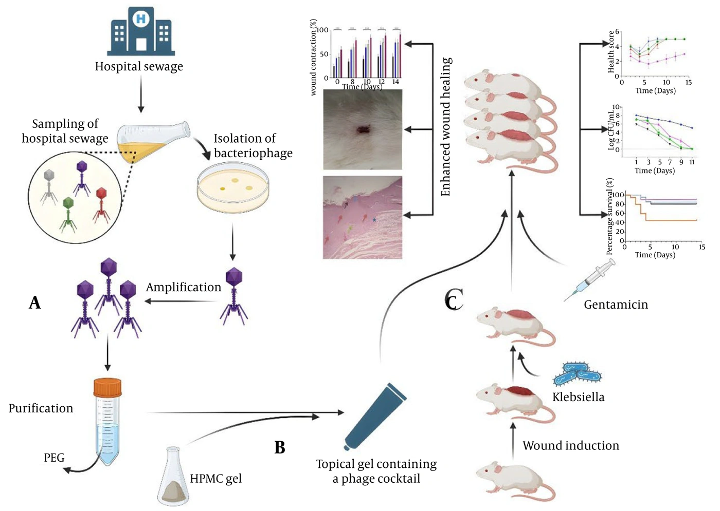
Figure 12.
Schematic Summary of Study Design and Major Findings. (A) Isolation of Klebsiella pneumoniae-specific bacteriophages from hospital wastewater and making the phage cocktail. (B) Formulation of the phage cocktail in hydroxypropyl methylcellulose (HPMC) gel for topical applications. (C) in vivo study with a rat wound infection model. As a complementary therapy, the phage cocktail + gentamicin resulted in better wound healing, reduced bacterial loads, and improved overall survival rates and health scores when compared to monotherapy groups and controls. PEG: Polyethylene glycol, HPMC: Hydroxypropyl methylcellulose.
5.1. Ethical Considerations:
All animal procedures were conducted in accordance with institutional guidelines for laboratory animal care and use. The study was approved by Fasa University of Medical Sciences, Iran.