1. Introduction
Aerococcus viridans is a peroxidase-negative, catalase-negative or weakly positive, facultative anaerobic, Gram-positive coccus that typically appears in tetrads or clusters (1). It can be isolated from clothing, dust, the upper respiratory tract, and the skin of healthy individuals (2). The organism rarely causes disease in humans, except in immunocompromised patients or those with underlying systemic illnesses (3). Previous case reports have described A. viridans causing urinary tract infection (4). However, to date, no cases of chronic osteomyelitis caused by A. viridans have been reported in the English-language literature, based on our search of PubMed (using the terms "Aerococcus viridans" AND "osteomyelitis") and Web of Science databases up to May 2026. We report a case of chronic osteomyelitis caused by A. viridans and provide diagnostic and therapeutic insights to alert clinicians to the possibility of this rare pathogen in chronic bone infections.
2. Case Presentation
A 59-year-old woman presented with a sinus tract on the skin of her left upper arm near the shoulder joint, accompanied by pale yellow purulent discharge and mild swelling for more than 10 years. One week before admission, the skin over the left upper arm became erythematous, with markedly increased swelling that impaired normal movement of the left shoulder joint. A surgical scar approximately 25 cm long was visible at the affected site. The patient had a 7-year history of type 2 diabetes mellitus, managed with oral metformin and glipizide, with recent fasting blood glucose levels ranging from 7.2 - 8.5 mmol/L and an HbA1c level of 7.8%. She reported no history of trauma to the left upper extremity, no prior orthopedic implants or fixation devices, and no previous antibiotic therapy or wound cultures during the 10-year disease course. No immunosuppressive conditions were identified. During the 10-year disease course, the patient had not undergone microbiological culture or received antibiotic therapy for this condition. She reported that she had not sought formal medical attention for the persistent sinus tract because of limited access to healthcare facilities in her residential area, which may have contributed to the chronic, smoldering nature of the infection.
Preoperative photograph showing a 25 cm surgical scar with a sinus tract on the left humerus near the shoulder joint, with surrounding erythema and swelling (Figure 1A).
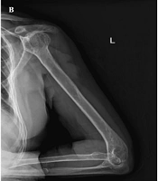
Physical examination revealed marked swelling of the left upper limb, with increased skin temperature compared with that of the contralateral side. Range of motion was limited in the left shoulder and elbow joints, and the left elbow was fixed in extension. Wrist joint mobility was normal, with no significant decline in muscle strength or sensation. Radiographic findings indicated chronic osteomyelitis of the left humerus with cortical thickening, medullary sclerosis, and an intracortical sequestrum (Figure 1B). Initial empirical treatment with ceftriaxone sodium, 2 g intravenously once daily, was started. After stabilization, routine laboratory tests showed a white blood cell count of 8.9 × 10⁹/L, a C-reactive protein (CRP) level of 42 mg/L, and an erythrocyte sedimentation rate (ESR) of 58 mm/h. After surgical contraindications were ruled out, the wound was fully exposed under general anesthesia. Window debridement for chronic osteomyelitis was performed, including resection of devitalized bone and soft tissue, with wide unroofing of the medullary cavity (Figure 2). A vancomycin-impregnated bone cement chain containing 2 g vancomycin per 40 g bone cement was implanted into the medullary cavity. Intraoperatively, five separate specimens, three from the medullary cavity and two from the sinus tract, were collected for bacterial culture and susceptibility testing. A drainage tube was placed to ensure adequate postoperative drainage.
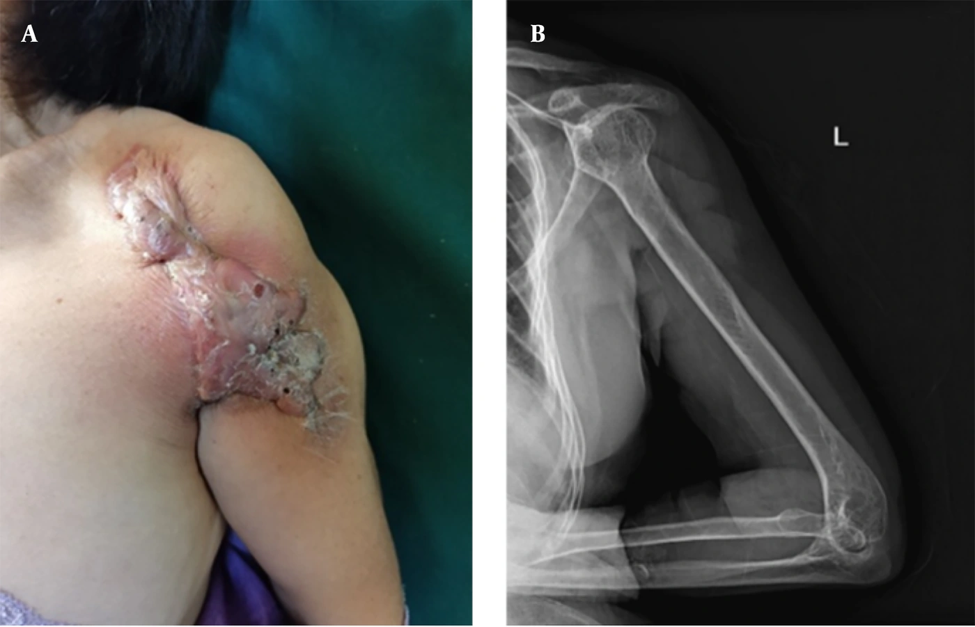
Figure 1.
Before surgery: (A) A scar and a sinus tract draining pus were present on the skin of the left upper arm near the shoulder joint; (B) radiographic findings indicated chronic osteomyelitis of the left humerus.
Intraoperative photograph showing wide window debridement of the osteomyelitic cavity with vancomycin-impregnated bone cement chain implantation.
Postoperative photograph showing closed-suction drainage tube placement to ensure adequate drainage from the affected area (Figure 3A, B).
2.1. Microbiological Identification
All five intraoperative specimens were inoculated onto 5% sheep blood agar and incubated at 35 °C in an atmosphere containing 5% CO2 for 48 hours. Small, α-hemolytic, grayish-white colonies grew in pure culture from all specimens. Gram staining revealed Gram-positive cocci arranged in tetrads and clusters. The isolate was catalase-negative and pyrrolidonyl arylamidase (PYR)-negative. Preliminary identification was performed using the Vitek 2 ID-GPC card (bioMérieux, Marcy-l'Étoile, France), which identified the organism as Aerococcus viridans with a 99.0% confidence level. To confirm species-level identification, the nearly full-length 16S rRNA gene (~1,400 bp) was amplified and sequenced. BLAST analysis revealed 99.8% sequence similarity to the A. viridans type strain ATCC 11563. Phylogenetic analysis using the neighbor-joining method showed that the isolate formed a monophyletic cluster with the A. viridans type strain, distinct from closely related species such as A. urinae (98.2% similarity) and A. sanguinicola (97.9% similarity). Contamination was ruled out based on recovery of the same organism from five independent surgical specimens, the absence of any other pathogenic bacteria, and correlation with the patient's clinical presentation.
Photograph 2 weeks postoperatively showing healed wound and functional recovery with active shoulder abduction to 90° and elbow flexion to 120°.
2.2. Antimicrobial Susceptibility Testing
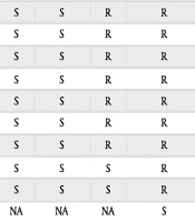
Antimicrobial susceptibility testing was performed using the broth microdilution method. Because the Clinical and Laboratory Standards Institute (CLSI) does not establish interpretive criteria specifically for the genus Aerococcus, minimum inhibitory concentrations (MICs) were interpreted according to the CLSI M100-Ed32 breakpoints for viridans group streptococci. The isolate was susceptible to penicillin (MIC ≤ 0.06 μg/mL), ceftriaxone (MIC ≤ 0.5 μg/mL), vancomycin (MIC ≤ 1 μg/mL), and linezolid, but resistant to erythromycin and clindamycin.
2.3. Follow-Up
At 1 month postoperatively, wound exudate was minimal, with no signs of recurrence. At 1-year follow-up, the patient remained asymptomatic, with no recurrence of infection. Radiographic examination demonstrated healing of the bone defect, with no evidence of sequestrum or new bone destruction. The patient had not required additional surgery or further antibiotic therapy after discharge. Limb function was excellent, with full range of motion in the shoulder and elbow joints, and the patient had returned to all activities of daily living.
Photograph at 1-year follow-up showing excellent functional recovery of the affected limb with full range of motion (Figure 3C).
3. Discussion
Chronic osteomyelitis is a destructive inflammatory bone disease that is often associated with trauma, surgery, or hematogenous spread (5). Although Staphylococcus aureus remains the predominant pathogen (6), infections caused by low-virulence organisms warrant clinical attention. Aerococcus viridans is a Gram-positive, α-hemolytic coccus that commonly appears in tetrads or clusters (7). It is found in the upper respiratory tract and on the skin of healthy individuals, typically exhibits low pathogenicity (1), and is also a common airborne microorganism in hospital environments (8). This report describes chronic osteomyelitis caused by A. viridans, an extremely uncommon presentation in the literature. A literature search of PubMed and Web of Science (search terms: "Aerococcus viridans" and "osteomyelitis"; date range: inception to May 2026) revealed no previously reported cases of chronic osteomyelitis caused by this organism. However, osteomyelitis caused by other Aerococcus species, such as A. urinae vertebral osteomyelitis, has been reported, highlighting the potential of this genus to cause musculoskeletal infection (9). The rarity of A. viridans osteomyelitis may be related to its microbiological characteristics and the host immune status (4). First, this pathogen grows slowly and is easily overgrown on routine blood agar plates by faster-growing, dominant pathogens such as S. aureus (10), often leading to misidentification or dismissal as contamination. Unlike Staphylococcus, A. viridans does not produce catalase (11), a characteristic that can help distinguish these organisms. Furthermore, its low invasiveness and limited virulence factors indicate that it typically requires compromised local defenses, such as orthopedic implants or chronic wounds (12), or mildly impaired host immunity to cause systemic infection (13). In this case, the patient presented with long-standing localized swelling of the left humerus and a history of diabetes, creating an opportunity for A. viridans proliferation.
Regarding treatment, most A. viridans strains remain susceptible to commonly used antimicrobials such as penicillin, cephalosporins, and vancomycin; nevertheless, increasing resistance to aminoglycosides and macrolides has been reported (14). Given its ability to form biofilms and its low metabolic state within osteomyelitic lesions, selecting antibiotics with good bone penetration and efficacy against biofilm-associated bacteria is crucial (15). In general, β-lactam antibiotics such as penicillin and ceftriaxone are recommended as first-line therapy (16), with consideration of prolonged, usually ≥ 2 weeks, intravenous or sequential oral treatment (17). In this case, empirical ceftriaxone was administered for 1 week and then switched to vancomycin for 2 weeks based on susceptibility results. After hospital discharge, the patient continued oral levofloxacin 500 mg daily and rifampin 300 mg twice daily for an additional 4 weeks, as these agents offer good bone penetration and activity against biofilms. At 1 month postoperatively, wound exudate was minimal, indicating a favorable clinical outcome. At 1-year follow-up, the affected limb showed good recovery without significant functional limitations or recurrence.
Aerococcus viridans and other Aerococcus species are often misidentified as staphylococci or streptococci in routine clinical microbiology laboratories, leading to underrecognition of their pathogenic potential. In cases of chronic osteomyelitis with negative or atypical culture results, targeted molecular identification techniques may be used. These strategies include the API 20 STREP system, ID 32 STREP, Vitek 2 ID-GPC cards (bioMérieux), and 16S rRNA amplification and sequencing. Although these techniques are not universally available in clinical microbiology laboratories, they provide high accuracy for detecting A. viridans (1). Clinicians should therefore consider the possibility of rare or low-virulence pathogens, including A. viridans, in chronic bone infections, particularly when routine cultures are negative or show non-dominant growth.
As a single-case report, this study cannot support systematic epidemiological analysis. Future research should accumulate additional cases and investigate the pathogenic mechanisms of A. viridans in musculoskeletal infections to optimize treatment duration and regimens. In summary, this case confirms A. viridans as a rare but definite pathogen in chronic osteomyelitis. Its clinical significance lies in alerting clinicians to the possibility of rare pathogens in chronic bone infections. When faced with chronic bone infections with atypical culture results and a poor response to conventional treatment, clinicians should consider precise microbial identification techniques. Early and accurate pathogen identification is fundamental to establishing effective long-term antimicrobial regimens and performing necessary surgical interventions, ultimately improving the prognosis of such refractory infections.
The ability of A. viridans to establish a chronic bone infection in this patient can be attributed to a combination of pathogen- and host-related factors. From a microbiological perspective, Aerococcus species, including A. viridans, have been demonstrated to form biofilms on abiotic surfaces. Biofilm formation on necrotic bone sequestra is a hallmark of chronic osteomyelitis and confers tolerance to both host immune defenses and antimicrobial agents. The organism's low metabolic activity within the biofilm, coupled with its intrinsically slow growth rate, likely explains the decade-long persistence of infection without systemic dissemination. From the host perspective, the patient's poorly controlled type 2 diabetes mellitus (HbA1c 7.8%) played a permissive role. Chronic hyperglycemia leads to impaired polymorphonuclear leukocyte chemotaxis, phagocytosis, and intracellular killing, creating an immunocompromised niche in which low-virulence pathogens can thrive. This case therefore illustrates how the convergence of a biofilm-forming opportunist and a mildly immunocompromised host can result in a paucisymptomatic yet refractory skeletal infection.
Furthermore, the possibility that A. viridans was initially present as a co-infecting organism alongside a more virulent pathogen, such as Staphylococcus aureus, cannot be excluded. S. aureus may have been eliminated by the host's innate immune response over time, whereas the slower-growing A. viridans persisted asymptomatically within the sclerotic intramedullary cavity. This scenario highlights the importance of not dismissing slow-growing or non-dominant colonies from bone specimens as mere contaminants, particularly when cultures are obtained from a chronic infection site with distorted local anatomy.
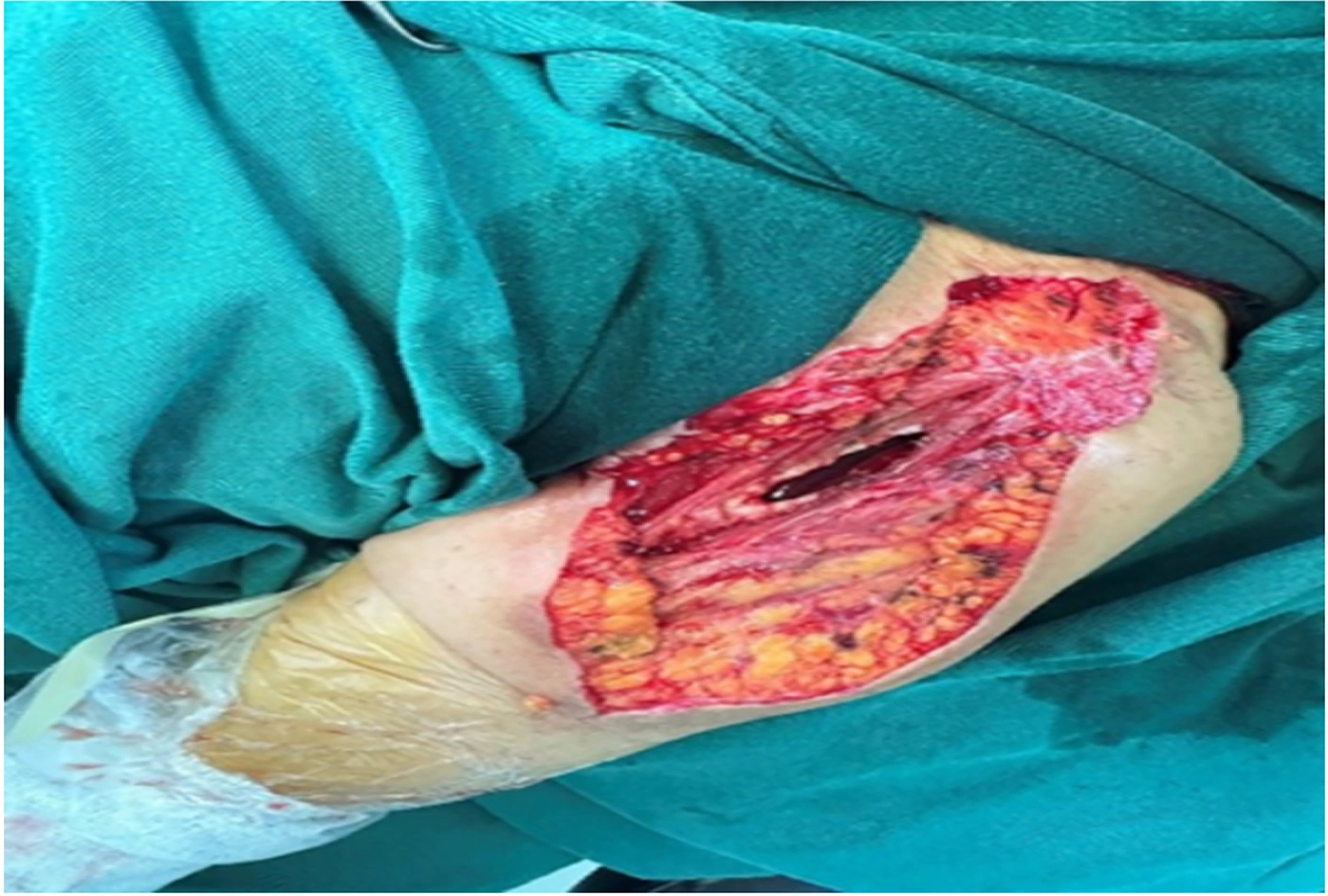
Figure 2.
During surgery, a large amount of necrotic tissue and purulent discharge was observed.
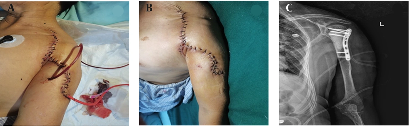
Figure 3.
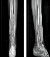
(A, B) The surgical wound was healing well; (C) one year after surgery, radiographs showed that the wound had healed well.
3.1. Conclusions
This report presents the first documented case, to our knowledge, of chronic osteomyelitis of the humerus caused by A. viridans in a patient with type 2 diabetes mellitus. The patient was successfully treated with surgical debridement and targeted antimicrobial therapy. This case underscores the need for clinicians to consider rare, low-virulence organisms in chronic bone infections, particularly when standard cultures are inconclusive. Advanced identification methods, including Vitek 2 and 16S rRNA sequencing, are important for accurate diagnosis and appropriate treatment.