
1. Background
Inflammatory bowel disease (IBD) comprises chronic inflammatory gastrointestinal conditions, including Crohn's disease and ulcerative colitis (UC) (1). Patients with IBD have an increased risk of colorectal cancer, the third most prevalent cancer in humans (2). UC involves inflammation of the gastrointestinal mucosa, primarily in the colon and rectum (3). Although its precise causes remain unknown, several factors have been implicated in its pathophysiology. Inflammatory mediators, including cytokines, reactive oxygen species, and eicosanoids, are thought to play key roles in disease progression (4, 5). Clinical signs include colonic ulceration and edema, which can cause fever, weight loss, weakness, and bloody or mucoid diarrhea (6, 7). Long-term IBD can also lead to numerous psychosocial problems (8). Corticosteroids and salicylates, such as sulfasalazine, are commonly used to treat UC. Biologic agents and immunosuppressants are additional treatment options (9, 10). However, the limited effectiveness and adverse effects of these medications remain major clinical challenges, highlighting the need for new therapeutic strategies (11).
The enterochromaffin (EC) cell population is lower in samples from patients with severe UC than in samples from patients with nonsevere UC or healthy controls; however, no significant difference has been observed between the nonsevere UC and healthy control groups. Moreover, 5-hydroxytryptamine (5-HT) levels and EC cell numbers have been shown to correlate positively in colonic mucosal biopsies from patients with UC (12). These findings suggest that the severity of mucosal injury may influence the 5-HT/EC cell axis, although the effects of inflammation on these parameters across the spectrum of IBD remain incompletely understood. Whether these changes affect intestinal inflammation is uncertain, but the cited studies demonstrate that mucosal 5-HT levels and EC cell numbers change during IBD-related inflammation. Serotonin may play an important role in exacerbating IBD symptoms, including diarrhea and abdominal pain (12). In animals with colitis, intraperitoneal administration of 5-HT markedly increases interleukin (IL)-1β and IL-6 expression and myeloperoxidase activity by activating 5-HT3 and 5-HT4 receptors in the intestinal mucosa; conversely, inhibiting this signaling pathway can reduce pain (11).
Through activation of its receptors, serotonin can act as both a proinflammatory and an anti-inflammatory signaling molecule in the intestinal mucosa (13). Antipsychotic medications may have anti-inflammatory properties; for example, clozapine may reduce inflammatory responses (14). Clinical trials have demonstrated that antipsychotic medications can affect cytokine levels in cerebrospinal fluid and peripheral blood and inhibit proinflammatory cytokine activity.
Olanzapine is a second-generation atypical antipsychotic approved by the US Food and Drug Administration for schizophrenia and bipolar disorder with manic or mixed episodes. In combination with fluoxetine, it is also approved for treatment-resistant depression and bipolar depression. It is used off-label for conditions including anorexia nervosa, psychosis, severe agitation, and chemotherapy-induced nausea and vomiting.
Its primary mechanism involves antagonism of dopamine D2 receptors in the mesolimbic pathway, which helps manage positive symptoms of psychosis. Olanzapine also antagonizes serotonin 5-HT2A receptors in the prefrontal cortex, contributing to the alleviation of negative symptoms.
2. Objectives
Given the potential anti-inflammatory properties of olanzapine, this study evaluated its effects on colonic injury.
3. Methods
3.1. Animals
Thirty-five adult male Wistar rats weighing 200 - 250 g were used. The animals were obtained from Elmbavaran Aftab Company and housed in the institutional animal care facility at 24 ± 2 °C under a 12-hour light/dark cycle, with unrestricted access to standard chow and water. All rats underwent a one-week acclimatization period before the experimental procedures. The experiment lasted eight days. No animals were excluded before randomization, removed from the study, or omitted from the final statistical analyses. All procedures conformed to the ethical standards and guidelines of Kermanshah University of Medical Sciences, Iran (ethical approval code: IR.KUMS.REC.1399.986).
3.2. Experimental Animals and Grouping
The animals were assigned to seven groups by simple randomization, with five rats per group. Treatments were administered for three days before colitis induction and continued for five days afterward. Colitis was induced in groups 2 - 7 after the three-day pretreatment period. Histological assessment using hematoxylin and eosin (H&E) staining and immunohistochemical analyses were performed under blinded conditions; macroscopic assessment was unblinded.
Group 1 (control): Rats received only food and water under standard conditions throughout the eight-day experiment.
Group 2 (acetic acid): Rats received 1 mL of 4% acetic acid rectally after the three-day pretreatment period and otherwise received standard food and water throughout the eight-day experiment.
Group 3 (sulfasalazine): Rats received oral sulfasalazine at 500 mg/kg for eight days: three days before and five days after colitis induction with 1 mL of 4% acetic acid (15).
Group 4 (olanzapine 2.5 mg/kg): Rats received intraperitoneal injections of olanzapine at 2.5 mg/kg for eight days: three days before and five days after colitis induction (16).
Group 5 (olanzapine 5 mg/kg): Rats received intraperitoneal injections of olanzapine at 5 mg/kg for eight days: three days before and five days after colitis induction (17).
Group 6 (serotonin 1 mg/kg): Rats received intraperitoneal injections of serotonin at 1 mg/kg for eight days: three days before and five days after colitis induction (18).
Group 7 (serotonin 1 mg/kg plus olanzapine 5 mg/kg): Rats received combined intraperitoneal administration of serotonin at 1 mg/kg and olanzapine at 5 mg/kg for eight days: three days before and five days after colitis induction.
3.3. Colitis Induction
Before colitis induction, the animals were fasted for 24 hours with free access to water. Acute colitis was induced using acetic acid, as previously described (3). After light anesthesia with a mixture of 10% ketamine and 2% xylazine at a 9:1 ratio, a lubricated 8-cm polyethylene tube was gently inserted into the rectum, and 1 mL of 4% acetic acid was slowly instilled into the colon. The animals were maintained in a head-down position for 10 minutes to prevent leakage. Daily drug administration resumed two hours after induction and continued for five days (19).
3.4. Macroscopic Scoring
On the final experimental day, the rats were euthanized using high-dose ether, and the distal 7 cm of the colon was excised. Investigators performed macroscopic assessments without formal blinding to group allocation. Specimens were graded according to the Gerald scoring method based on the extent of ulceration, hyperemia, and inflammation: 1) localized hyperemia without ulceration; 2) a linear ulcer without significant inflammation; 3) one linear ulcer with one area of inflammation; 4) two or more areas of ulceration or inflammation; and 5) one major ulcerative or inflammatory lesion extending more than 1 cm along the colon, or two or more major ulcerative or inflammatory lesions. The ulcer index was calculated as previously described (20) by adding the ulcerated area in cm2 to the ulcer severity score.
3.5. Histology
The animals were euthanized on the final experimental day. The colons were dissected and washed with normal saline. The distal 7 cm was used for macroscopic assessment, and the specimens were divided into two parts: one for histological assessment using H&E staining and the other fixed in 10% formalin for immunohistochemical assays.
3.6. Immunohistochemical Assay
Nuclear factor κB (NF-κB) p65 and Toll-like receptor 4 (TLR4) expression were evaluated using the avidin-biotin-peroxidase complex immunohistochemical technique. After deparaffinization and rehydration through graded alcohols, 4-µm tissue sections were incubated in 3% hydrogen peroxide prepared in methanol for five minutes to quench endogenous peroxidase activity. Antigen retrieval was performed by heating the slides in citrate buffer in a microwave oven for 20 minutes. The sections were then incubated overnight at 4 °C with NF-κB p65 monoclonal antibody (catalog No. E-AB-22066) and TLR4 mouse monoclonal antibody (sc-293072), each at a dilution of 1:100 (21). Immunohistochemical staining was assessed under blinded conditions. Representative mucosal areas were selected for analysis, and necrotic regions were excluded. NF-κB p65 and TLR4 expression was assessed semiquantitatively based on staining intensity and the proportion of positive cells. Images were processed using color deconvolution to isolate the brown DAB channel. The mean optical density of the brown staining was analyzed in distinct regions of interest within the colon. The average density value was calculated for each animal and used for statistical analysis. Integrated optical density (IOD) was measured using ImageJ software. The mean value per animal was used for statistical analysis. Two observers independently performed all evaluations while blinded to group allocation.
3.7. Hematoxylin and Eosin Staining
Paraffin-embedded tissue sections, approximately 2 - 4 µm thick, were automatically stained with hematoxylin and 0.2% eosin prepared in an ethanolic medium. Periodic acid-Schiff/Alcian blue staining was performed manually. Sections were incubated with Alcian blue solution (0.5% v/v in 3% acetic acid, pH 2.5) for five minutes to visualize acidic mucopolysaccharides and then exposed to Schiff's reagent (0.5% v/v) for 10 minutes to demonstrate neutral mucins. Digital images were acquired at 40× magnification using a high-resolution whole-slide brightfield scanner. Blinded histopathological assessment evaluated immune cell infiltration, epithelial damage, and changes in mucosal architecture (22).
3.8. Data Analysis
The macroscopic ulcer index was predefined as the primary outcome for evaluating colonic injury severity. Secondary outcomes included histopathological injury scores and immunohistochemical NF-κB p65 and TLR4 expression, which characterized treatment-associated inflammatory and molecular changes. Data were analyzed using one-way analysis of variance followed by Tukey's post hoc multiple-comparison test. All analyses were performed using GraphPad version 9 (GraphPad Software, San Diego, CA, USA). Data are presented as mean ± standard error of the mean (SEM), and P < 0.05 indicated statistical significance.
4. Results
4.1. Extent of Colon Tissue Damage and Inflammation
4.1.1. Macroscopic Study
The control group showed normal colonic mucosa on macroscopic examination. Intrarectal administration of acetic acid caused ulcers, adhesions, intestinal wall thickening, and severe inflammation. Both olanzapine doses (2.5 and 5 mg/kg) significantly reduced inflammation and partially improved mucosal lesions compared with the acetic acid group. The greatest anti-inflammatory and mucosal healing effects, reflected by reduced ulcer severity, were observed with olanzapine at 5 mg/kg. Pretreatment with olanzapine at 5 mg/kg before serotonin administration also markedly attenuated colonic inflammation and tissue injury in rats with colitis (Figure 1).
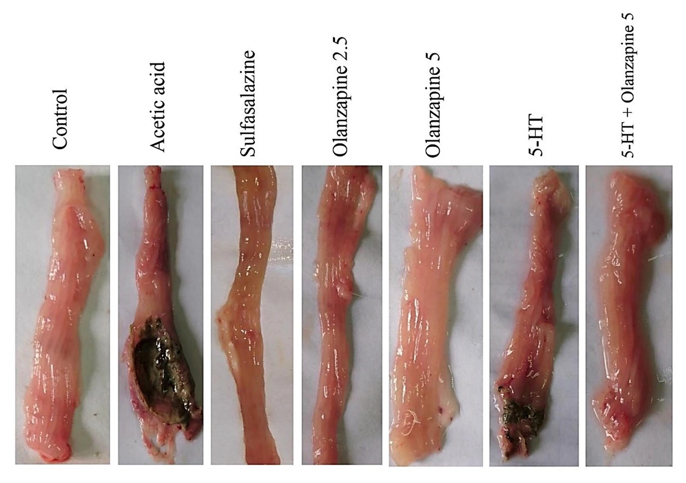
Figure 1.
Macroscopic changes in isolated colon tissue from the control, acetic acid, olanzapine (2.5 and 5 mg/kg), and serotonin (5-HT) groups.
As shown in Figure 2, the acetic acid and serotonin groups had significantly higher macroscopic ulcer indices than the control group (11.8 and 8.5, respectively, vs 0.8; P < 0.05). Sulfasalazine and olanzapine at 2.5 and 5 mg/kg reduced the ulcer index to 2.3, 4, and 2.2, respectively, compared with the acetic acid group (P < 0.05). The greatest decrease was observed with olanzapine at 5 mg/kg. The ulcer index was also significantly lower in the olanzapine 5 mg/kg plus serotonin 1 mg/kg group than in the serotonin-only group (P < 0.05).
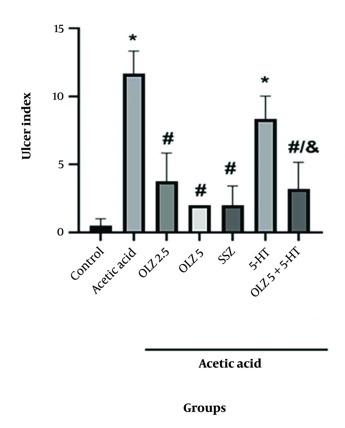
Figure 2.
Changes in the mean ulcer index. Data are expressed as mean ± SEM. Abbreviations: SEM, standard error of the mean; OLZ, olanzapine; SSZ, sulfasalazine; 5-HT, serotonin. *P < 0.05, #P < 0.05, and &P < 0.05 compared with the control, acetic acid, and serotonin groups, respectively.
4.1.2. Histological Examination
Histological damage was assessed semiquantitatively on a 0 - 4 scale according to the severity of mucosal injury: 0, normal mucosa; 1, mild epithelial disruption and minimal inflammatory infiltration; 2, moderate crypt destruction and edema; 3, severe inflammation and loss of goblet cells; and 4, extensive necrosis and complete loss of mucosal architecture.
The control group showed normal mucosal architecture, intact Lieberkühn crypts, and uniformly distributed goblet cells (circle), with an ulcer severity score of 0. The acetic acid group showed severe tissue injury, including epithelial necrosis, crypt destruction, submucosal edema (circle), and extensive infiltration by inflammatory cells and erythrocytes (arrow), with an ulcer severity score of 4. Coadministration of sulfasalazine with acetic acid significantly reduced tissue damage, preserved epithelial continuity and crypt integrity, and markedly decreased mucosal inflammatory cell infiltration (circle), with ulcer severity scores of 1 - 2.
Olanzapine treatment at 2.5 and 5 mg/kg in rats exposed to acetic acid maintained nearly normal mucosal architecture. Goblet cells were preserved, with only mild epithelial disruption and localized inflammatory changes in a few ulcerated regions (circle), corresponding to ulcer severity scores of 1 - 2. Serotonin-treated rats exposed to acetic acid showed a moderate protective effect compared with the acetic acid group (circle), with an ulcer severity score of 3.
Combined olanzapine and serotonin administration in the presence of acetic acid resulted in nearly normal mucosal morphology, with minimal lymphoid hyperplasia and slight leukocyte infiltration (circle), corresponding to an ulcer severity score of 2 (Figure 3).
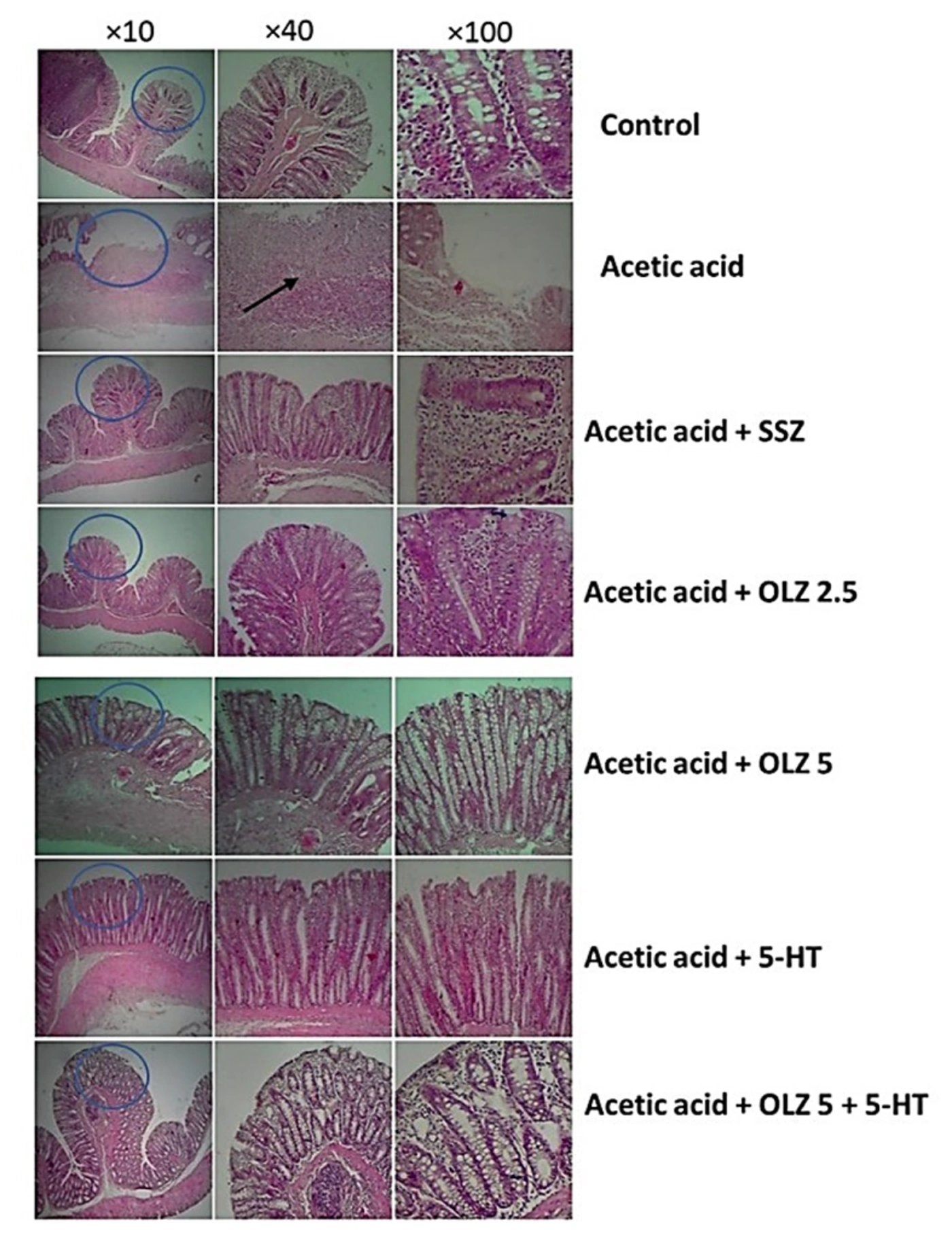
Figure 3.
Photomicrographs of large intestinal tissue from the experimental groups stained with hematoxylin and eosin (H&E) at ×10, ×40, and ×100 magnification. Abbreviations: OLZ, olanzapine; SSZ, sulfasalazine; 5-HT, serotonin.
4.2. NF-κB p65 Expression
NF-κB p65 expression was quantified using IOD measurements and presented as fold changes relative to the control group. The acetic acid and serotonin groups showed expression values of 1.9-fold and 2.15-fold, respectively, compared with the normalized control value of 1.0 (P < 0.01 and P < 0.001, respectively). Sulfasalazine, olanzapine at 2.5 mg/kg, and olanzapine at 5 mg/kg showed expression values of 0.85, 1.15, and 0.95, respectively. These treatments, as well as the combined treatment of olanzapine at 5 mg/kg and serotonin at 1 mg/kg, significantly reduced NF-κB p65 expression compared with the acetic acid group (P < 0.05). Expression was also markedly lower in the combined-treatment group than in the serotonin-only group (P < 0.001) (Figure 4A and B).
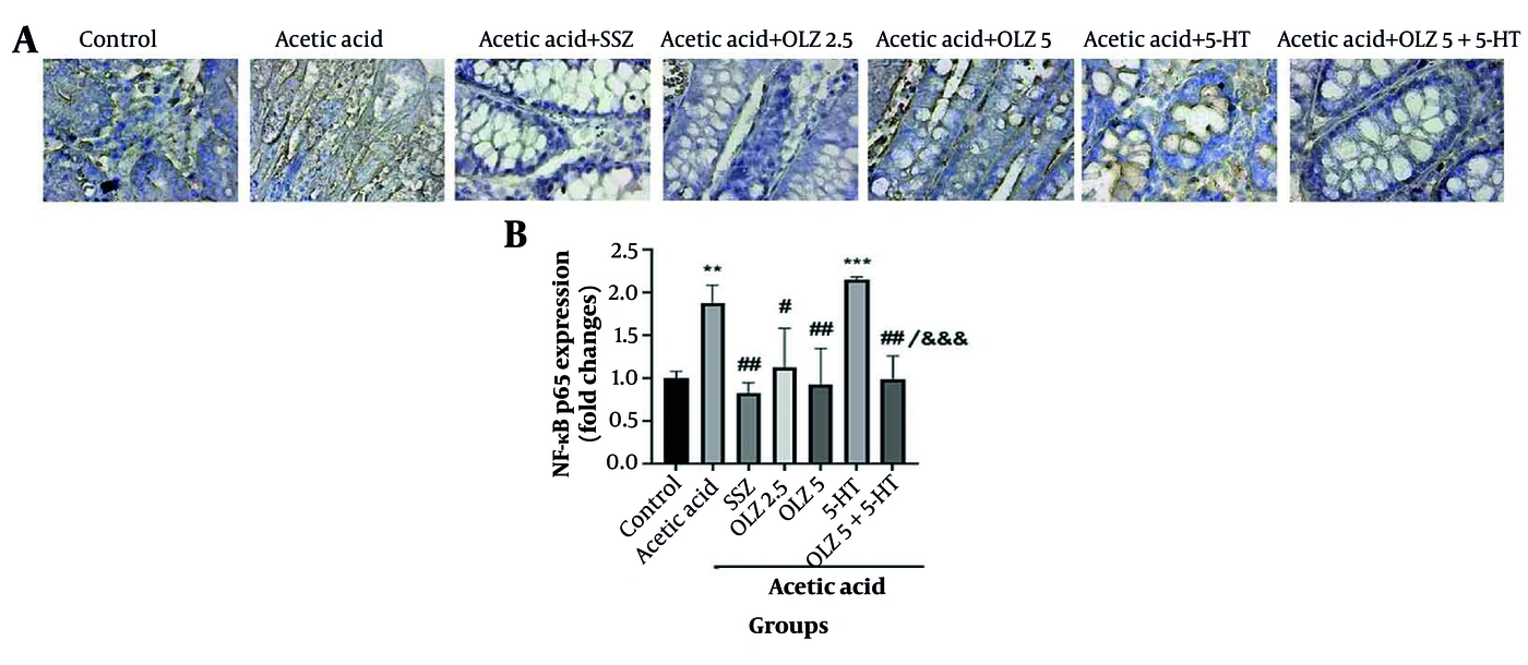
Figure 4.
(A) Immunohistochemical staining of nuclear factor κB (NF-κB) p65 in colon tissue from the experimental groups (×400). (B) NF-κB p65 expression levels. Values are reported as mean ± SEM. Abbreviations: SEM, standard error of the mean; OLZ, olanzapine; SSZ, sulfasalazine; 5-HT, serotonin. **P < 0.01 and ***P < 0.001 compared with the control group; ##P < 0.01 and #P < 0.05 compared with the acetic acid group; &&&P < 0.001 compared with the serotonin group.
4.3. TLR4 Expression
TLR4 staining was quantified as IOD and presented as fold changes relative to the control group. The acetic acid and serotonin groups showed expression values of 1.65 and 1.35, respectively, which were significantly higher than the control value of 0.85 (P < 0.001 and P < 0.01, respectively). Sulfasalazine, olanzapine 2.5 mg/kg (expression value, 0.75), olanzapine 5 mg/kg (expression value, 0.85), and olanzapine 5 mg/kg plus serotonin 1 mg/kg (expression value, 0.85) showed significantly lower TLR4 expression than the acetic acid group (P < 0.01), with values similar to those of the control group. The combined olanzapine 5 mg/kg plus serotonin 1 mg/kg group also showed significantly lower expression than the serotonin-only group (P < 0.05) (Figure 5A and B).
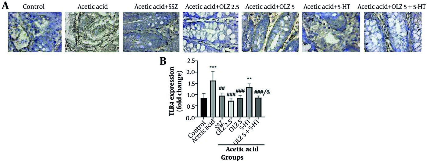
Figure 5.
(A) Immunohistochemical staining of Toll-like receptor 4 (TLR4) in colon tissue from the experimental groups (×400). (B) TLR4 expression levels. All values are reported as mean ± SEM. Abbreviations: SEM, standard error of the mean; OLZ, olanzapine; SSZ, sulfasalazine; 5-HT, serotonin. **P < 0.01 and ***P < 0.001 compared with the control group; ##P < 0.01 and ###P < 0.001 compared with the acetic acid group; &P < 0.05 compared with the serotonin group.
5. Discussion
Colitis, a manifestation of IBD, involves progressive disruption of the mucosal barrier, impairment of epithelial structure and function, and dysregulation of intestinal immune and inflammatory responses. It can arise from chemical injury, oxidative stress, and activation of inflammatory pathways such as NF-κB and TLR4 (23, 24). Colitis is associated with structural changes in intestinal tissue, increased cytokine production, and inflammatory cell infiltration, leading to ulceration, bowel wall thickening, and impaired absorption (3, 25). Identifying agents that reduce inflammation and promote tissue repair therefore remains a priority.
In this study, macroscopic examination showed normal colonic mucosa in the control group, whereas intrarectal acetic acid administration caused severe ulceration, adhesions, and wall thickening. Olanzapine at 2.5 and 5 mg/kg significantly reduced inflammation and improved mucosal lesions, with the 5 mg/kg dose producing the greatest anti-inflammatory effect. These findings are consistent with the effects of sulfasalazine, a reference treatment for colitis (26). Olanzapine pretreatment also attenuated the serotonin-associated increase in the macroscopic ulcer index, indicating a protective effect when serotonin signaling was elevated.
Histopathological analysis supported the macroscopic findings. The control group maintained normal mucosal architecture, healthy crypts, and uniformly distributed goblet cells. The acetic acid group showed extensive injury, including epithelial necrosis, submucosal edema, and dense inflammatory infiltrates. Both olanzapine doses preserved mucosal architecture and reduced inflammatory cell infiltration. Combined treatment with olanzapine and serotonin produced nearly normal mucosal morphology. These findings are consistent with previous reports of the protective effects of anti-inflammatory agents on intestinal tissue integrity (27, 28).
NF-κB p65 expression was markedly elevated in the acetic acid and serotonin groups compared with the control group, reflecting activation of proinflammatory signaling. Olanzapine, alone or in combination with serotonin, significantly reduced NF-κB p65 expression toward control levels. This finding agrees with evidence that attenuation of NF-κB signaling reduces intestinal inflammation and promotes restoration of mucosal integrity (29), suggesting that modulation of NF-κB p65 may contribute to olanzapine's anti-inflammatory effects.
Excessive TLR4 expression may contribute to colitis-associated tumorigenesis by promoting cell proliferation, inhibiting apoptosis in malignant cells, accelerating invasion and metastasis, and supporting a tumor-promoting microenvironment (30). In this study, acetic acid and serotonin significantly increased TLR4 expression, whereas olanzapine reduced expression toward control levels. These findings suggest a potential role for olanzapine in modulating TLR4-dependent inflammatory pathways, consistent with previous studies implicating TLR4 in colitis pathogenesis (31). Downregulation of TLR4 by olanzapine could therefore help limit inflammatory signaling and subsequent tissue damage.
The increases in TLR4 expression following acetic acid-induced injury and serotonin administration suggest that serotonin may exacerbate colitis through TLR4 pathway activation. Olanzapine attenuated this overexpression, bringing expression levels near those of the control group, which suggests that it may modulate TLR4-dependent inflammatory pathways. These findings are consistent with previous literature emphasizing the role of TLR4 in intestinal inflammation. Emerging evidence indicates that serotonin participates in inflammatory processes and modulates immune cell populations through serotonylation (32). Together, these findings support further investigation of TLR4 and serotonin-related pathways as therapeutic targets for reducing inflammation and preventing colitis-associated complications.
5.1. Study Limitations
No formal a priori sample-size calculation was performed. Group sizes were based on commonly used sample sizes in previously published studies of similar experimental colitis models. Although significant differences were observed, the small sample size may limit statistical power and should be considered when interpreting the findings. Further studies with larger sample sizes may help confirm and extend these results.
The acute chemically induced colitis model may not fully represent the complex, chronic pathophysiology of human IBD. The short treatment duration limits assessment of long-term therapeutic outcomes, and the male-only sample prevents assessment of sex-dependent responses. The limited range of systemic inflammatory biomarkers and the lack of a comprehensive safety and tolerability assessment restrict understanding of the drug's clinical potential. The acetic acid model also carries a risk of localized transmural necrosis or focal wall perforation caused by accidental acid pooling or incomplete saline flushing.
5.2. Conclusions
Olanzapine showed anti-inflammatory activity in this rat model of experimentally induced colitis. It reduced neutrophil and erythrocyte infiltration into colonic tissue and improved macroscopic and microscopic outcomes. It also modulated NF-κB p65 and TLR4, supporting further preclinical investigation in IBD. Future studies should examine the molecular mechanisms underlying these effects, including serotonergic and dopaminergic receptor signaling. Given the acute preclinical model and limited duration and scope, these findings are hypothesis-generating. Chronic models, comprehensive safety assessments, and dose-response studies are needed to characterize olanzapine's therapeutic profile in this experimental setting.