1. Context
Nephrolithiasis is among the most common urological disorders worldwide, with steadily increasing incidence and prevalence over recent decades. Epidemiological studies indicate that kidney stone disease affects nearly 10 - 15% of the global population over their lifetime, imposing a substantial burden on healthcare systems and patients’ quality of life (1). The rising prevalence of obesity, diabetes mellitus, metabolic syndrome, dietary changes, and sedentary lifestyles has further contributed to the increasing incidence of urolithiasis (2, 3).
Kidney stones are characterized by a high recurrence rate. Previous studies have demonstrated that recurrence may occur in approximately 50% of patients within 5 years and up to 80 - 90% within 10 years after the initial stone episode. Therefore, surgical removal of stones alone cannot be considered a definitive treatment, as the underlying metabolic abnormalities often persist even after achieving stone-free status (4).
Traditionally, nephrolithiasis was considered primarily a mechanical disorder caused by crystal precipitation within the urinary tract. However, current evidence suggests that stone disease is fundamentally a metabolic disorder involving complex interactions among urinary solute concentration, urine pH, urinary inhibitors of crystallization, dietary habits, genetic predisposition, and environmental factors. Metabolic abnormalities such as hypercalciuria, hyperoxaluria, hypocitraturia, hyperuricosuria, low urine volume, and altered urinary pH play essential roles in stone formation and recurrence (4-6).
Given the metabolic nature of the disease, 24-hour urine analysis has become the cornerstone of metabolic evaluation in stone-forming patients. Comprehensive urinary metabolic assessment enables identification of lithogenic risk factors and facilitates implementation of individualized preventive strategies, including dietary modification and pharmacologic therapy. Current international guidelines recommend extensive metabolic evaluation, particularly in recurrent and high-risk stone formers, often including two separate 24-hour urine collections.
Several minimally invasive surgical techniques are currently used for the management of urinary stones, including percutaneous nephrolithotomy (PCNL), retrograde intrarenal surgery/ureteroscopy (URS/RIRS), and shock wave lithotripsy (SWL). The choice of intervention depends on stone size, location, composition, anatomical factors, and patient characteristics.
Although previous reviews have extensively discussed metabolic evaluation and recurrence prevention in stone formers, most have focused on baseline risk assessment or general preventive strategies rather than the dynamic changes that occur after surgical stone clearance (7).
Technological advances in endourology have significantly improved stone-free rates while reducing perioperative morbidity (8).
Despite successful surgical stone removal, many patients continue to exhibit metabolic abnormalities after intervention. Moreover, postoperative alterations in urinary metabolites may occur secondary to relief of urinary obstruction, resolution of infection, changes in renal tubular function, reduction of inflammation, or lifestyle modifications after treatment. Some studies have suggested that stone removal may improve certain urinary parameters, whereas others have reported persistent or unchanged metabolic derangements after surgery. Consequently, the preoperative metabolic profile may not fully represent the patient’s true baseline metabolic status.
The clinical significance of postoperative urinary alterations remains unclear, particularly regarding whether observed changes represent true metabolic improvement, transient procedural effects, or persistence of the underlying lithogenic phenotype. Therefore, this review aims to evaluate temporal changes in 24-hour urinary metabolic parameters after stone removal and discuss their implications for postoperative risk stratification and prevention strategies.
This narrative review therefore aims to synthesize and critically appraise the available evidence on pre- and postoperative 24-hour urinary metabolic changes in nephrolithiasis and to discuss their clinical implications for long-term stone prevention.
1.1. Pathophysiology of Urinary Stone Formation
Urinary stone disease is a multifactorial metabolic disorder characterized by urine supersaturation with lithogenic substances, crystal nucleation, crystal growth, aggregation, and crystal retention within the renal tubules. Stone formation occurs when the concentration of stone-forming solutes exceeds their solubility threshold, whereas a deficiency of natural inhibitors of crystallization further promotes lithogenesis. Calcium-containing stones, particularly calcium oxalate and calcium phosphate stones, account for nearly 80% of all urinary calculi (4, 9).
Several urinary metabolites play critical roles in nephrolithiasis. Hypercalciuria is among the most common metabolic abnormalities in recurrent stone formers and contributes to calcium-based supersaturation. Hyperoxaluria markedly increases the risk of calcium oxalate crystallization even with small changes in urinary oxalate levels. In contrast, citrate acts as an important inhibitor of stone formation by binding calcium and reducing supersaturation; therefore, hypocitraturia is strongly associated with recurrence (10, 11).
Beyond individual metabolic abnormalities, overall urinary supersaturation is considered the main determinant of stone formation and recurrence, as it integrates the combined effects of urinary solutes and inhibitors. Uric acid metabolism and urinary pH are also important determinants of stone formation. Acidic urine favors uric acid precipitation, whereas alkaline urine predisposes to calcium phosphate and infection-related stones. Dietary factors such as sodium, animal protein, and oxalate intake further influence urinary chemistry, while low urine volume remains a key modifiable risk factor. Importantly, these metabolic abnormalities are not static and may persist or fluctuate after surgical stone removal, supporting the clinical relevance of postoperative metabolic evaluation.
1.2. Importance of 24-Hour Urine Analysis
Twenty-four-hour urine collection is widely considered the clinical standard for metabolic evaluation in patients with nephrolithiasis because it helps identify urinary biochemical abnormalities associated with stone formation and recurrence. International guidelines recommend this assessment, especially in recurrent and high-risk stone formers (4).
Commonly measured parameters include urine volume, calcium, oxalate, citrate, uric acid, sodium, magnesium, phosphate, creatinine, and urinary pH. Urinary supersaturation, which represents the combined lithogenic potential of urine, increases when the concentration of stone-forming solutes rises or when urine volume decreases.
However, 24-hour urine testing is not a perfect gold standard, as results may be influenced by day-to-day variability, patient compliance, dietary intake, and collection errors. Therefore, multiple 24-hour urine collections may improve the reliability of metabolic assessment by reducing the influence of day-to-day biological and dietary variability. The European Association of Urology (EAU) recommends two consecutive 24-hour urine collections for specific metabolic evaluation, whereas the American Urological Association (AUA) considers one or two collections acceptable but prefers two collections when feasible.
The postoperative timing of 24-hour urine analysis remains controversial. Early postoperative samples may be affected by inflammation, hematuria, ureteral stents, infection, and transient changes in hydration status. For this reason, several studies recommend reassessment approximately 4 - 6 weeks after complete stone clearance, although alternative time points (e.g., 2 weeks, 3 months, or 6 months) have also been reported depending on the study design (12).
Importantly, targeted treatment based on 24-hour urine findings—including increased fluid intake, sodium restriction, citrate supplementation, urinary alkalinization, and thiazide therapy—has been shown to reduce stone recurrence (13). Nevertheless, the magnitude of benefit varies across patient populations and depends on baseline metabolic risk and adherence to therapy (Figure 1).
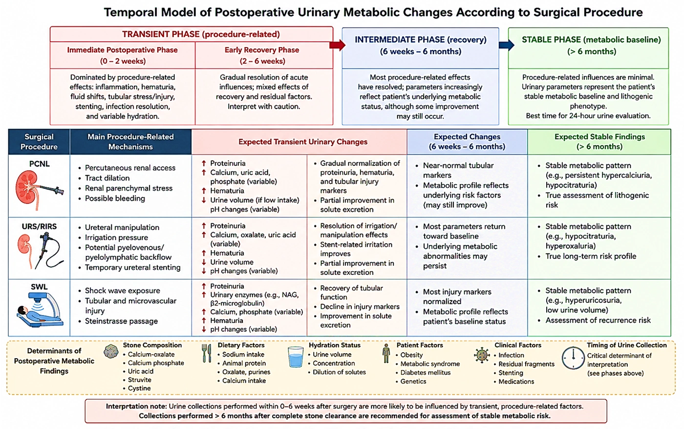
Figure 1.
Temporal model of postoperative urinary metabolic changes according to surgical procedure. The immediate (0 - 2 weeks) and early recovery (2 - 6 weeks) phases represent predominantly transient, procedure-related influences, including renal parenchymal stress following percutaneous nephrolithotomy (PCNL), ureteral manipulation, irrigation pressure, and temporary stenting following ureteroscopy/retrograde intrarenal surgery (URS/RIRS), and shock-wave-related tubular injury following shock wave lithotripsy (SWL). The intermediate phase (6 weeks–6 months) represents progressive resolution of procedure-related effects and recovery toward the underlying metabolic profile. The stable phase (> 6 months) is expected to more closely reflect the patient’s underlying metabolic status and lithogenic risk after complete stone clearance. The indicated time intervals are approximate and are intended as a conceptual framework rather than rigid clinical cutoffs.
1.3. Surgical Management of Kidney Stones
Modern management of nephrolithiasis relies primarily on minimally invasive procedures, including percutaneous nephrolithotomy (PCNL), ureteroscopy/retrograde intrarenal surgery (URS/RIRS), and shock wave lithotripsy (SWL). The choice of intervention depends on stone size, location, composition, anatomical characteristics, and patient-related factors. Advances in endourological techniques have improved stone-free rates while reducing perioperative morbidity (14).
Although the primary objective of these procedures is stone clearance, each surgical modality may produce transient physiological changes that can influence postoperative urinary biochemical measurements. PCNL involves percutaneous renal access and may induce short-term renal parenchymal stress. SWL can cause transient tubular and microvascular injury through shock-wave exposure, potentially resulting in temporary alterations in urinary biochemical markers. For URS/RIRS, ureteral manipulation, irrigation (lavage) pressure, and temporary ureteral stenting may transiently affect renal tubular function and urinary composition and may contribute to short-term changes in urinary citrate excretion through alterations in urinary flow and the local urinary environment (15). These procedure-related effects are expected to be most relevant during the early postoperative period and should therefore be distinguished from persistent metabolic abnormalities that reflect the patient’s underlying lithogenic predisposition (16).
2. Evidence Acquisition
2.1. Review Design and Search Strategy
This narrative review critically synthesized the available evidence regarding changes in urinary metabolic profiles before and after surgical management of nephrolithiasis. Although this study was not designed as a systematic review or meta-analysis, a structured literature search approach was used to enhance transparency and reproducibility.
A comprehensive search was conducted in PubMed, Scopus, and Google Scholar from database inception to March 2026. The search strategy combined controlled vocabulary and free-text terms related to nephrolithiasis, surgical treatment, and urinary metabolic evaluation. The main search terms were: (“nephrolithiasis” OR “kidney stone” OR “urolithiasis”) AND (“24-hour urine” OR “urinary metabolic evaluation” OR “urinary calcium” OR “oxalate” OR “citrate” OR “uric acid” OR “supersaturation”) AND (“percutaneous nephrolithotomy” OR “PCNL” OR “ureteroscopy” OR “URS” OR “retrograde intrarenal surgery” OR “RIRS” OR “shock wave lithotripsy” OR “SWL”). The reference lists of relevant articles were also screened manually to identify additional eligible studies.
2.2. Eligibility Criteria
Studies were considered eligible if they met predefined inclusion and exclusion criteria. Eligible studies included human studies of adult patients (≥18 years) with nephrolithiasis who underwent surgical stone management, including percutaneous nephrolithotomy (PCNL), ureteroscopy/retrograde intrarenal surgery (URS/RIRS), or shock wave lithotripsy (SWL). Studies were included if they reported preoperative and/or postoperative 24-hour urinary metabolic parameters and evaluated one or more urinary risk factors associated with stone formation, including urinary calcium, oxalate, citrate, uric acid, urine volume, urinary pH, or supersaturation indices. Prospective and retrospective observational studies, cohort studies, and relevant clinical guidelines were considered eligible.
Studies were excluded if they involved pediatric populations, animal or experimental models, case reports, conference abstracts, or lacked sufficient clinical data. Studies focusing exclusively on medical dissolution therapy were also excluded. In addition, studies that evaluated only postoperative renal injury biomarkers, such as neutrophil gelatinase-associated lipocalin (NGAL), kidney injury molecule-1 (KIM-1), fatty acid-binding protein (FABP), or cystatin C, without assessment of standard urinary metabolic parameters were not considered eligible.
2.3. Study Selection and Data Extraction
Titles and abstracts of retrieved articles were screened initially for relevance, followed by full-text assessment of potentially eligible studies. Studies were selected based on relevance to the review objectives and the availability of postoperative metabolic data. Because this was a narrative review, formal duplicate screening and assessment of inter-reviewer agreement were not performed.
Data extracted from eligible studies included publication year, study design, sample size, surgical modality, stone characteristics (when available), timing of postoperative urine collection, evaluated urinary parameters, and major findings. Particular attention was given to factors that could influence postoperative metabolic outcomes, including stone composition, baseline metabolic abnormalities, dietary modifications, hydration status, infection resolution, and follow-up duration.
2.4. Evidence Synthesis
Because of substantial heterogeneity among studies in patient populations, surgical techniques, follow-up intervals, and measured outcomes, quantitative meta-analysis was not performed. Findings were synthesized qualitatively by urinary parameter and surgical modality. Studies were interpreted critically according to methodological characteristics, including sample size, study design, timing of urine collection, and potential sources of bias.
During evidence interpretation, we considered the distinction between persistent lithogenic metabolic abnormalities and transient postoperative renal injury responses. Particular emphasis was placed on urinary supersaturation as an integrated marker of stone-forming potential rather than isolated urinary metabolites.
3. Results
3.1. Comparison of Urinary Metabolites Before and After Surgery
Evidence regarding changes in urinary metabolites after stone surgery is inconsistent. Some studies report partial normalization after stone removal, whereas others report persistent metabolic abnormalities, suggesting that nephrolithiasis is largely driven by systemic metabolic factors rather than a purely mechanical process (17).
Postoperatively, urinary calcium may decrease after relief of obstruction, but hypercalciuria often persists. Urinary oxalate may fluctuate in the short term because of diet and hydration; however, long-term levels usually remain unchanged. Citrate, an important inhibitor of stone formation, may improve after resolution of obstruction or infection, but hypocitraturia frequently persists and may require treatment (10).
Urine pH is also clinically relevant: Acidic urine promotes uric acid stones, whereas alkaline urine is associated with calcium phosphate and infection-related stones. After surgery, changes in pH are often related to infection clearance, dietary changes, or improved hydration. Increased fluid intake postoperatively also increases urine volume and reduces supersaturation. Overall, the risk of stone formation is better reflected by urinary supersaturation than by individual metabolites. Supersaturation increases when urinary calcium and oxalate are high and citrate is low. Therefore, postoperative metabolic changes are mainly driven by relief of obstruction, infection control, and behavioral modifications rather than by the surgical procedure itself (11).
3.2. Factors Affecting Postoperative Metabolic Changes
Postoperative urinary metabolic profiles are influenced by multiple patient-related, disease-related, and procedure-related factors. Therefore, differences among published studies should be interpreted in light of variations in baseline metabolic status, stone characteristics, surgical technique, and timing of postoperative assessment.
Common metabolic abnormalities include hypercalciuria, hyperoxaluria, hypocitraturia, and low urine volume. Uric acid stones are frequently linked to persistently acidic urine (low urinary pH), hyperuricosuria, and reduced urinary citrate excretion. Struvite stones are primarily associated with urinary tract infections caused by urease-producing microorganisms, resulting in alkaline urine and altered urinary chemistry. Cystine stones are related to inherited defects in amino acid transport, leading to increased urinary cystine excretion (5, 11). These differences highlight that postoperative metabolic profiles may vary substantially by stone type and underscore the importance of interpreting urinary parameters within the context of stone composition (Table 1).
Table 1.
Summary of Studies Evaluating Postoperative Changes in 24-Hour Urinary Metabolic Parameters After Surgical Management of Nephrolithiasis a
| Studies | Study Design | Sample Size | Surgical Modality | Stone Characteristics | Timing of Urine Collection | Evaluated Urinary Parameters | Main findings | Key Limitations/Confounders |
|---|---|---|---|---|---|---|---|---|
| Zhu et al., 2024 (18) | Prospective observational study | 109 | Stone removal (mixed surgical approaches) | Not fully specified | Before and after stone removal | Calcium, phosphate, oxalate, citrate, uric acid, sodium, magnesium, urinary pH | Urinary calcium and phosphate increased after surgery, whereas oxalate, citrate, uric acid, sodium, magnesium, and urinary pH showed no significant changes | Single-center observational design; limited long-term follow-up; possible effects of diet, hydration, and lifestyle modification |
| Laube et al., 2003 (17) | Observational study | Not reported | Not applicable (metabolic evaluation study) | Not specified | During active stone disease | 24-hour urinary composition | Active urinary stones may influence urinary composition and affect interpretation of metabolic evaluation | Older study; not specifically designed for postoperative assessment; limited clinical follow-up |
| Lycklama et al., 1991 (16) | Clinical observational study | Not reported | SWL | Not specified | Before and after SWL | Metabolic urine profile | Metabolic evaluation after stone treatment may differ from assessment during active stone disease | Older study; limited methodological details; historical treatment protocols |
| Parks and Coe, 2009 (21) | Long-term cohort study | Long-term cohort | Stone removal with medical follow-up | Calcium stone formers | Long-term follow-up | Urinary risk factors and recurrence-related parameters | Metabolic abnormalities frequently persist over time and require long-term preventive management | Not specifically designed to evaluate immediate postoperative metabolic changes |
| Coe et al., 2005 (22) | Review/clinical evidence synthesis | Not applicable | Not applicable | Various stone types | Not applicable | Urinary risk factors and supersaturation | Established nephrolithiasis as a chronic metabolic disorder influenced by urinary supersaturation and persistent metabolic abnormalities | Review article; not a direct pre/post surgical comparison |
| Pearle et al., 2014 (25) | Clinical guideline | Not applicable | All surgical modalities | Various stone types | Not applicable | Metabolic evaluation and recurrence prevention recommendations | Emphasized that stone removal alone is insufficient and postoperative metabolic evaluation is required | Guideline-based evidence; not a primary comparative study |
a Postoperative urine collection intervals varied across studies. Initial postoperative assessments were generally performed within approximately 1 - 14 days after stone removal, whereas follow-up metabolic assessments were typically performed at approximately 4 - 12 weeks after the procedure. These intervals represent reported ranges across the included studies and should not be interpreted as standardized clinical time points. Abbreviation: SWL, shock wave lithotripsy.
In addition to stone composition, several confounding factors may influence postoperative metabolic evaluation, including dietary habits, hydration status, obesity, diabetes mellitus, metabolic syndrome, medication use, residual stone fragments, infection status, and the timing of urine collection. These factors may affect urinary calcium, oxalate, citrate, uric acid, sodium, volume, and pH, thereby contributing to heterogeneity among studies (Table 1).
Baseline metabolic status also plays a critical role in postoperative outcomes. Patients with pre-existing abnormalities in urinary calcium, oxalate, citrate, or uric acid may continue to demonstrate similar metabolic profiles after stone removal, suggesting that surgical intervention does not eliminate the systemic predisposition responsible for stone formation. In contrast, some patients may show partial improvement due to correction of reversible factors such as urinary obstruction, infection, reduced fluid intake, or dietary imbalance (17, 18).
The timing of postoperative 24-hour urine collection represents another major source of heterogeneity among studies. Early postoperative measurements may be affected by transient factors, including hematuria, ureteral stents, inflammation, infection, changes in fluid intake, and temporary alterations in renal tubular function. Consequently, urinary results obtained shortly after surgery may not accurately represent long-term metabolic status. Longer follow-up assessments are more likely to reflect persistent lithogenic risk factors rather than immediate procedural effects (13, 15).
Surgical modality may also influence postoperative biochemical findings, although its impact on long-term metabolic risk remains uncertain. PCNL may induce temporary renal parenchymal stress due to renal access, SWL may cause transient tubular injury through shock-wave exposure, and URS/RIRS may result in short-term changes related to ureteral manipulation, irrigation pressure, or temporary stenting. However, these procedure-related effects should be distinguished from persistent urinary metabolic abnormalities that contribute to stone recurrence (15, 19, 20).
Lifestyle and medical interventions after surgery represent additional confounding factors. Increased fluid intake, dietary modification, reduced sodium consumption, and initiation of preventive medications such as thiazide diuretics or potassium citrate may improve urinary parameters independently of the surgical procedure itself. Therefore, postoperative metabolic changes cannot always be attributed solely to stone removal.
Residual stone fragments may further contribute to persistent metabolic abnormalities and recurrence risk, because incomplete clearance may maintain a lithogenic environment. Moreover, differences in follow-up duration, study design, patient selection, and the absence of control groups contribute substantially to the variability observed across studies.
Overall, postoperative urinary metabolic changes should be interpreted within a multifactorial framework. The persistence or improvement of urinary abnormalities after surgery likely reflects the interaction among underlying metabolic susceptibility, stone composition, surgical effects, and postoperative behavioral modifications rather than the surgical intervention alone.
3.3. Current Evidence and Major Studies
Current evidence regarding postoperative changes in 24-hour urinary metabolic parameters after surgical management of nephrolithiasis remains limited and heterogeneous. Most available studies are observational and differ substantially in patient selection, baseline metabolic status, stone composition, surgical modality, and timing of postoperative urine collection. Therefore, the available evidence should be interpreted cautiously, as direct evidence demonstrating that surgical stone removal modifies the underlying metabolic profile remains insufficient. Studies directly comparing preoperative and postoperative 24-hour urine parameters provide the most relevant evidence for evaluating metabolic changes after intervention, whereas guideline documents and mechanistic studies mainly provide clinical context rather than direct comparative evidence.
A relevant recent study by Zhu et al. in 2024 prospectively evaluated 109 patients undergoing complete stone removal. The authors compared preoperative and postoperative 24-hour urine profiles and reported a significant increase in urinary calcium and phosphate excretion after surgery, whereas urinary oxalate, citrate, uric acid, sodium, magnesium, and urinary pH did not show significant changes. These findings suggest that removal of urinary stones does not necessarily result in normalization of metabolic risk factors. However, interpretation of these findings requires consideration of several limitations, including the observational study design, lack of a control group, and potential effects of postoperative dietary modification, hydration changes, and behavioral interventions (18).
An important methodological issue in postoperative metabolic evaluation was highlighted by Laube et al., who demonstrated that the presence of active urinary stones may influence the composition of 24-hour urine samples. This finding suggests that preoperative urine measurements obtained during active stone disease may not always represent the patient’s stable metabolic status. Consequently, differences between preoperative and postoperative urinary profiles may partly reflect changes in disease activity rather than a direct effect of surgical intervention itself (17).
Long-term observational data have further supported the concept that metabolic abnormalities may persist after stone removal. Parks and Coe demonstrated that urinary risk factors associated with stone formation can remain stable over prolonged periods, emphasizing that nephrolithiasis represents a chronic metabolic disorder requiring continuous preventive strategies rather than a condition corrected solely by stone clearance. These findings highlight the importance of postoperative metabolic assessment and individualized interventions, particularly in patients with recurrent disease or persistent urinary abnormalities (21).
Stone composition represents another important source of heterogeneity among studies. Patients with calcium oxalate, calcium phosphate, uric acid, infection-related, or cystine stones have different underlying metabolic mechanisms and recurrence risks. Therefore, postoperative metabolic changes may vary according to the type of stone removed. For example, calcium stone formers may continue to demonstrate hypercalciuria or hypocitraturia despite successful intervention, whereas uric acid stone formers may remain affected by persistently low urinary pH and systemic metabolic factors. Infection-related stones may also show persistent abnormalities in urinary pH and ammonium metabolism if infection control is incomplete (5, 11).
In addition to long-term metabolic abnormalities, several studies have investigated short-term renal responses following surgical procedures. However, these studies evaluated renal stress or injury biomarkers rather than lithogenic urinary metabolism and should be interpreted separately from standard 24-hour urine parameters. Dzięgała et al. evaluated renal effects after shock wave lithotripsy (SWL) and reported transient changes in urinary biomarkers associated with tubular injury. These findings indicate a temporary renal response to shock-wave exposure rather than persistent alterations in stone-forming metabolic risk (15).
Similarly, Hughes et al. conducted a prospective pilot study evaluating changes in routine laboratory parameters and novel biomarkers after SWL. The authors observed temporary biochemical alterations following treatment, which subsequently improved during follow-up. These findings support the concept that SWL may induce short-term renal stress without necessarily producing long-term metabolic changes associated with recurrence risk (19).
Regarding ureteroscopic procedures, Benli et al. evaluated the early effects of ureterorenoscopy (URS/RIRS) on renal biomarkers and reported increases in urinary NGAL, KIM-1, FABP, and cystatin C shortly after intervention. Although these biomarkers provide valuable information on the acute renal response to instrumentation, they do not directly indicate changes in lithogenic urinary parameters such as calcium, oxalate, citrate, or supersaturation. Therefore, their clinical interpretation should be limited to procedure-related renal stress rather than metabolic correction or deterioration (20).
Another major limitation of current evidence is the variability in postoperative urine collection timing. Studies assessing urine composition shortly after surgery may capture transient effects related to inflammation, hematuria, ureteral stents, infection resolution, changes in hydration status, or temporary alterations in renal tubular function. In contrast, longer-term assessments are more likely to reflect persistent metabolic susceptibility to stone formation. This methodological variability is an important explanation for the inconsistent findings reported across studies (13, 15, 18).
Importantly, isolated urinary metabolites may not fully represent the overall risk of recurrence. Urinary supersaturation integrates multiple factors, including urinary solute concentrations, inhibitors of crystallization, and urine volume, and therefore provides a more comprehensive estimate of lithogenic potential. Previous evidence has emphasized that supersaturation indices may be more clinically relevant predictors of recurrence than individual urinary parameters evaluated separately (21, 22).
International guidelines also emphasize that surgical intervention should primarily be considered a stone-clearing procedure rather than a definitive metabolic treatment. Both European and American guidelines recommend postoperative metabolic evaluation, particularly in recurrent and high-risk stone formers, to identify persistent abnormalities and guide preventive therapy (13, 23).
Taken together, current evidence suggests that postoperative urinary changes occur through two distinct mechanisms. Short-term alterations may result from procedural factors, including renal stress, obstruction relief, infection resolution, or temporary physiological changes. In contrast, long-term metabolic abnormalities generally reflect the underlying systemic predisposition to stone formation and may persist despite successful surgical clearance. Therefore, postoperative management should include continued metabolic evaluation and individualized preventive strategies rather than relying solely on stone removal as a definitive therapeutic approach (Tables 1 and 2).
Table 2.
Postoperative Renal Injury Biomarkers Following Surgical Management of Nephrolithiasis a
| Studies | Study Design | Procedure | Biomarkers Evaluated | Main Findings | Clinical Interpretation |
|---|---|---|---|---|---|
| Dzięgała et al., 2018 (15) | Prospective study | SWL | Urinary renal injury biomarkers | Transient elevation of urinary biomarkers indicating short-term tubular stress after shock-wave exposure | Represents temporary renal response to SWL rather than persistent lithogenic metabolic changes |
| Hughes et al., 2020 (19) | Prospective pilot study | SWL | Serum and urinary biomarkers | Temporary changes in routine laboratory parameters and novel renal biomarkers after SWL | Indicates reversible procedure-related renal effects rather than long-term metabolic alterations |
| Benli et al., 2017 (20) | Prospective clinical study | URS/RIRS | NGAL, KIM-1, FABP, cystatin C | Early postoperative increase in renal injury biomarkers after ureteroscopic intervention | Reflects acute renal stress related to instrumentation rather than persistent stone-forming risk |
a Abbreviations: FABP, fatty acid-binding protein; KIM-1, kidney injury molecule-1; NGAL, neutrophil gelatinase-associated lipocalin; RIRS, retrograde intrarenal surgery; SWL, shock wave lithotripsy; URS, ureteroscopy.
3.4. Limitations of Existing Literature
Despite increasing interest in postoperative metabolic evaluation of nephrolithiasis, the available evidence remains limited by several important methodological challenges. First, most studies investigating postoperative urinary metabolic changes are observational, have relatively small sample sizes, and are frequently conducted at single centers. Consequently, the generalizability of the findings is limited, and the ability to establish causal relationships between surgical intervention and metabolic changes remains uncertain.
A major source of heterogeneity among studies is variability in patient characteristics, including baseline metabolic status, stone composition, recurrence history, dietary patterns, and comorbid conditions. Different stone types, including calcium oxalate, calcium phosphate, uric acid, struvite, and cystine stones, have distinct pathophysiological mechanisms and may demonstrate different postoperative metabolic patterns (24). Therefore, combining heterogeneous stone populations may reduce the ability to identify specific metabolic responses after surgery.
Another important limitation is the lack of standardized protocols for postoperative 24-hour urine assessment. Studies have reported urine collections at different time points, ranging from early postoperative periods to several months after intervention. Early measurements may be influenced by transient factors such as inflammation, hematuria, infection, ureteral stenting, changes in fluid intake, and temporary renal tubular alterations. In contrast, delayed measurements may better reflect persistent metabolic susceptibility but are less frequently available.
The absence of appropriate control groups represents another important limitation. Many studies compare preoperative and postoperative urinary parameters without including nonsurgical stone formers or repeated measurements in untreated patients. Therefore, it remains difficult to distinguish true effects of stone removal from changes related to lifestyle modification, dietary counseling, increased fluid intake, or initiation of preventive medications.
Furthermore, available studies frequently focus on isolated urinary metabolites rather than integrated measures of lithogenic risk. Although parameters such as urinary calcium, oxalate, citrate, and uric acid provide valuable information, urinary supersaturation may better reflect the combined influence of multiple urinary factors on stone formation and recurrence. Limited evaluation of supersaturation indices represents an important knowledge gap.
Publication bias may also influence the current evidence base. Studies reporting significant or clinically interesting postoperative changes may be more likely to be published, whereas studies showing minimal or negative findings may remain unpublished. This possibility should be considered when interpreting the available literature.
Finally, differences in surgical techniques, operator experience, residual stone fragments, and follow-up duration further complicate comparisons among studies. Future investigations should incorporate standardized methodologies, larger multicenter populations, detailed stone composition analysis, and long-term follow-up to better define the relationship between surgical stone clearance and metabolic risk. Because this was a single-author review, a formal independent assessment of risk of bias was not feasible, and potential bias was therefore considered qualitatively.
3.5. Future Perspectives
Future research should focus on establishing standardized approaches for postoperative metabolic evaluation in patients undergoing surgical management of nephrolithiasis. One important priority is defining the optimal timing for postoperative 24-hour urine collection. Standardized assessment intervals would improve comparability among studies and help distinguish transient procedure-related changes from persistent metabolic abnormalities.
Large-scale prospective multicenter studies are needed to clarify whether specific surgical modalities, including PCNL, URS/RIRS, and SWL, produce clinically meaningful long-term effects on urinary metabolic profiles. These studies should incorporate standardized measurements of urinary calcium, oxalate, citrate, uric acid, urine volume, urinary pH, and supersaturation indices before and after intervention.
Future studies should also include detailed stone composition analysis and integrate baseline metabolic characteristics into risk stratification models. Understanding whether different stone types respond differently after surgical clearance may allow more individualized postoperative prevention strategies.
The role of urinary supersaturation deserves further investigation, as it represents an integrated measure of lithogenic risk rather than an isolated biochemical parameter. Future clinical studies should evaluate whether changes in supersaturation after surgery are better predictors of recurrence than individual urinary metabolites.
Emerging technologies, including metabolomics, molecular biomarkers, and artificial intelligence-based predictive models, may provide additional opportunities for personalized risk assessment. These approaches could help identify patients who remain at high risk of recurrence despite successful stone removal and may benefit from intensive preventive interventions.
Randomized controlled trials evaluating standardized postoperative metabolic monitoring and targeted preventive therapies are needed. Such studies may help determine whether systematic metabolic follow-up after surgical stone clearance improves long-term recurrence outcomes and contributes to future guideline development.
Future studies may also benefit from integrating artificial intelligence (AI) and machine learning approaches with urinary metabolomics. Automated metabolite detection, data preprocessing, pattern recognition, and biomarker identification may facilitate the analysis of complex urinary metabolic profiles and help distinguish transient procedure-related alterations from persistent lithogenic abnormalities. Integration of AI-based metabolite analysis with stone composition, surgical modality, dietary factors, and longitudinal clinical data could ultimately support more individualized metabolic risk stratification and recurrence prediction. However, external validation, standardized datasets, and model interpretability will be necessary before such approaches can be reliably translated into clinical practice (25).
4. Conclusions
Current evidence indicates that successful surgical management of nephrolithiasis does not consistently normalize the urinary metabolic abnormalities associated with stone formation. Although short-term biochemical changes may occur after relief of obstruction, resolution of infection, improved hydration, or transient renal stress, these changes should be distinguished from the persistent metabolic abnormalities that underlie long-term recurrence risk. Overall, the available literature supports the concept that nephrolithiasis is primarily a chronic metabolic disorder, whereas surgical intervention should be regarded as a stone-clearing procedure rather than a definitive metabolic treatment.
This review also demonstrates that interpretation of postoperative urinary metabolic findings is complicated by substantial heterogeneity among published studies, including differences in patient populations, stone composition, surgical modality, baseline metabolic status, and the timing of postoperative urine collection. Consequently, direct comparisons across studies remain challenging, and the current evidence should be interpreted with appropriate caution.
From a clinical perspective, postoperative metabolic evaluation using 24-hour urine analysis remains an essential component of recurrence prevention, particularly in recurrent and high-risk stone formers. Assessment should ideally be performed after recovery from the immediate postoperative period, when transient procedure-related physiological changes have largely resolved, allowing a more reliable evaluation of persistent metabolic risk. Whenever possible, urinary supersaturation should be interpreted alongside individual urinary metabolites because it provides a more comprehensive assessment of lithogenic potential.
The principal contribution of this narrative review is the integration of current evidence on temporal postoperative metabolic changes across different surgical approaches while distinguishing persistent lithogenic abnormalities from transient procedure-related biochemical alterations. This perspective may assist clinicians in interpreting postoperative metabolic findings more appropriately and reinforces the importance of individualized long-term preventive strategies rather than relying solely on successful stone clearance.
Future well-designed prospective multicenter studies with standardized postoperative urine collection protocols, comprehensive metabolic assessment, detailed stone composition analysis, and long-term follow-up are required to clarify the true impact of surgical stone removal on urinary metabolic profiles and recurrence risk. Such evidence may contribute to optimizing postoperative surveillance strategies and informing future clinical practice guidelines.