
1. Introduction
Neurological disorders and brain injuries, including ischemic stroke, traumatic brain injury (TBI), and neurodegenerative diseases, represent a substantial global health burden and are frequently associated with long-term functional impairment and reduced quality of life. Recovery from these conditions largely depends on neuroplasticity; however, neuroinflammation, apoptosis, and blood-brain barrier (BBB) disruption often exacerbate neuronal damage and impair neurological recovery (1-3). Despite advances in surgical, pharmacological, and cell-based therapies, clinical outcomes remain limited by challenges such as infection, poor post-transplantation cell survival, and insufficient tissue regeneration. Consequently, extracellular vesicles (EVs), particularly exosomes, have emerged as promising therapeutic vehicles because they can transport diverse molecular cargo, including proteins, lipids, mRNAs, microRNAs (miRNAs), and other nucleic acids involved in intercellular communication and tissue repair (4-6). Exosomes, a specialized class of EVs, retain molecular information from their parental cells and are considered attractive therapeutic vehicles owing to their favorable biocompatibility, low immunogenicity, and capacity to cross physiological barriers such as the BBB. In the nervous system, exosomes released from neural cells have demonstrated therapeutic potential in various central nervous system (CNS) disorders. However, limited accumulation at injury sites, exosome heterogeneity, and the lack of standardized methods for extraction, purification, and large-scale production remain major barriers to clinical translation (3, 4, 6-10). Exercise is a non-pharmacological intervention that may promote neuroprotection through exosome-mediated signaling. Evidence suggests that physical activity increases the release of circulating exosomes and modulates their miRNA cargo, thereby contributing to intercellular communication, tissue adaptation, and repair processes (11-13). Exercise-derived exosomes may mediate the beneficial effects of physical activity on neural function and recovery by transferring regulatory signals to distant tissues (14). Despite growing evidence supporting the role of exercise-derived exosomes in neuroprotection and neural repair, the current literature remains fragmented across different neurological conditions, experimental models, exercise protocols, and exosomal cargo profiles. Previous studies have primarily focused on individual diseases or specific molecular mechanisms, whereas an integrated evaluation of the contribution of exercise-derived exosomes and their associated miRNAs across neurological disorders and brain injuries remains limited. Therefore, this systematic review aimed to synthesize current findings and clarify the mechanisms through which exercise-induced exosomal signaling may contribute to neuroprotection, neuroplasticity, neural repair, and functional recovery.
1.1. Objectives
The present systematic review sought to critically examine existing evidence regarding the role of exercise-derived exosomes and their associated miRNAs in neurological disorders and brain injuries. Specifically, this review investigated how exercise influences exosomal signaling and molecular cargo composition and assessed their potential roles in neuroprotection, neuroplasticity, neural repair, regulation of neuroinflammation and apoptosis, and functional recovery.
2. Methods
2.1. Search Methodology
This systematic review was conducted in accordance with the PRISMA 2020 guidelines. A structured literature search was designed to identify studies investigating the role of exercise-derived exosomes and their miRNA cargo in neurological disorders and brain injuries. The electronic database search was conducted on July 18, 2026, using PubMed, Scopus, and Web of Science. Only studies published in English were considered eligible. In PubMed, the search strategy combined Medical Subject Headings (MeSH) terms with relevant free-text keywords and was limited to articles published between January 1, 2020, and May 31, 2026. An English-language filter and the “Exclude preprints” option were applied. For Scopus and Web of Science, database-specific search strings were developed using comparable concepts and adapted to the indexing characteristics of each database. These searches were restricted to English-language articles published from 2020 to 2026. Boolean operators (AND and OR) were used to combine the predefined search concepts. The complete search strategies for all databases are provided in Table S1 in the Supplementary File.
Because this review was not prospectively registered, no protocol was registered in PROSPERO or any other publicly accessible registry before study initiation.
The literature search was structured around three principal concepts: exosomes and extracellular vesicles, exercise-related interventions, and neurological disorders or brain injuries. These concepts were translated into the following search terms: (Exosomes OR "Extracellular Vesicles" OR exosome OR "extracellular vesicle") AND (Exercise OR exercise OR "physical activity" OR training OR "aerobic exercise") AND ("Nervous System Diseases" OR "Brain Injuries" OR stroke OR "traumatic brain injury" OR "brain injury" OR Alzheimer OR Parkinson OR "multiple sclerosis" OR "spinal cord injury" OR "cerebral palsy").
2.2. Eligibility Criteria
2.2.1. Inclusion Criteria
Eligible studies were selected according to the following predefined criteria:
- Peer-reviewed original research articles involving in vivo experimental models.
- Studies investigating exercise-induced, exercise-derived, or exercise-conditioned EVs/exosomes, including their biological cargo (e.g., miRNAs, proteins, or other bioactive molecules).
- Research studies applying specified exercise protocols to investigate the contribution of exercise-derived EVs/exosomes.
- Studies investigating neurological disorders, CNS injury models, or neurorehabilitation.
- Studies reporting neurological, behavioral, histopathological, cellular, molecular, or mechanistic outcomes related to exercise-derived EVs/exosomes.
- Studies published in English with accessible full-text versions.
2.2.2. Exclusion Criteria
Studies were excluded based on the following predefined criteria:
- Studies involving human participants or in vitro models, as well as secondary publications (reviews, systematic reviews, and meta-analyses) and non-original articles, including editorials, commentaries, conference abstracts, letters, case reports, protocols, corrigenda, and errata.
- Studies investigating exosome- or EV-based interventions without a direct exercise intervention.
- Biomarker-only studies reporting exercise-induced EV/exosome changes without evaluating neurological, functional, histopathological, molecular, or mechanistic outcomes.
- Bioinformatic, computational, prediction-model, or network-analysis studies without experimental validation.
- Studies unrelated to neurological disorders, CNS injury, or neurorehabilitation.
- Studies with insufficient methodological information or unavailable full-text articles.
2.3. Literature Screening and Study Selection
Study selection was performed in two consecutive stages. First, two reviewers independently evaluated retrieved records by screening titles and abstracts to identify potentially eligible studies. Potentially relevant articles were then assessed independently in full text by the two reviewers. Discrepancies were resolved through discussion, and unresolved disagreements were settled by consultation with a third reviewer.
2.4. Risk of Bias Assessment
The quality of the included studies and potential sources of bias were evaluated using the SYRCLE Risk of Bias tool, which was specifically designed to assess methodological limitations in animal-based intervention research. This tool assesses ten items across six bias domains, including selection, performance, detection, attrition, reporting, and other potential sources of bias (15). Each assessment item was classified as having a low, high, or unclear risk of bias based on the methodological details provided in the original articles. Discrepancies in judgments were discussed and resolved, with a third reviewer consulted when consensus could not be reached. Risk-of-bias visualizations were generated using the robvis web application (16). Individual study evaluations and the overall risk-of-bias profile are illustrated in Figure 1.
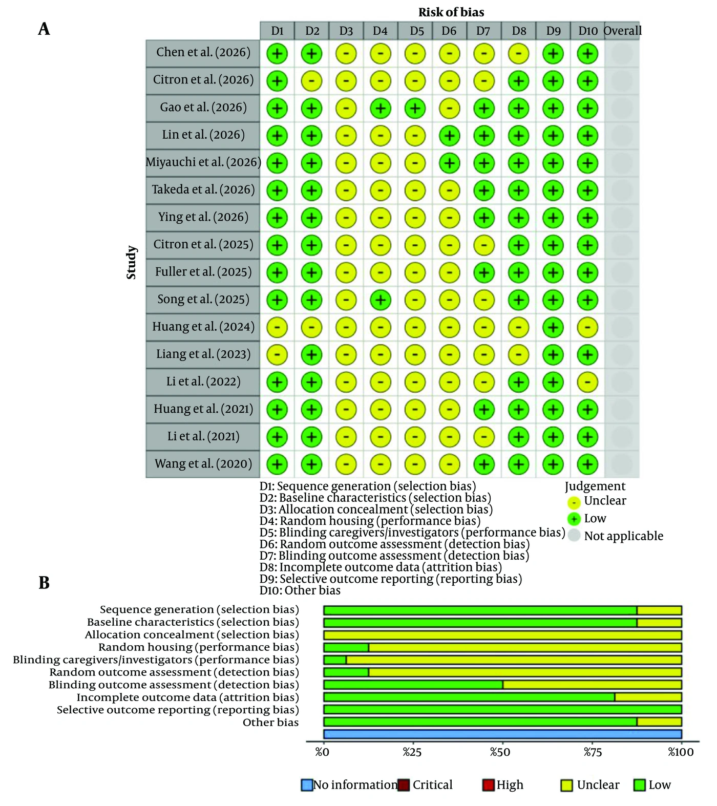
Figure 1.
Methodological quality assessment of the included studies using the SYRCLE Risk of Bias tool; A, Risk-of-bias judgments for each included study across the assessed domains; B, Summary of the proportion of studies classified as having low or unclear risk of bias in each domain. Green indicates low risk of bias, yellow indicates unclear risk of bias, and gray indicates not applicable.
2.5. Data Extraction and Synthesis
Relevant information was collected from each eligible study using a standardized data extraction form. Extracted data included the first author and year of publication, experimental model, neurological disorder or injury model, details of the exercise intervention, characteristics of EVs/exosomes and their associated molecular cargo, major mechanistic and functional outcomes, and the principal findings. Data extraction was performed by two reviewers, and any inconsistencies were resolved through discussion until consensus was reached. Because substantial methodological and clinical heterogeneity was identified across the included studies, particularly regarding neurological disease models, exercise interventions, EV/exosome characteristics, and outcome assessment methods, quantitative pooling of the results was considered inappropriate. Consequently, the evidence was synthesized narratively, with the included studies organized and compared according to the neurological disease model, experimental design, exercise intervention, EV/exosome characteristics, proposed molecular mechanisms, and rehabilitation-related outcomes.
3. Results
3.1. Search Results
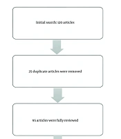
The database search yielded 388 records, including 161 from Scopus, 115 from Web of Science, and 112 from PubMed. After removal of 34 duplicate records, 354 unique publications were retained for title and abstract screening. Of these, 278 records were excluded because they did not meet the predefined eligibility criteria. Full-text evaluation was subsequently performed for the remaining 76 articles. After detailed assessment, 60 articles were excluded because they did not satisfy the eligibility requirements. Consequently, 16 studies met all inclusion criteria and were included in the qualitative synthesis. A flow diagram illustrating the study selection process is presented in Figure 2.
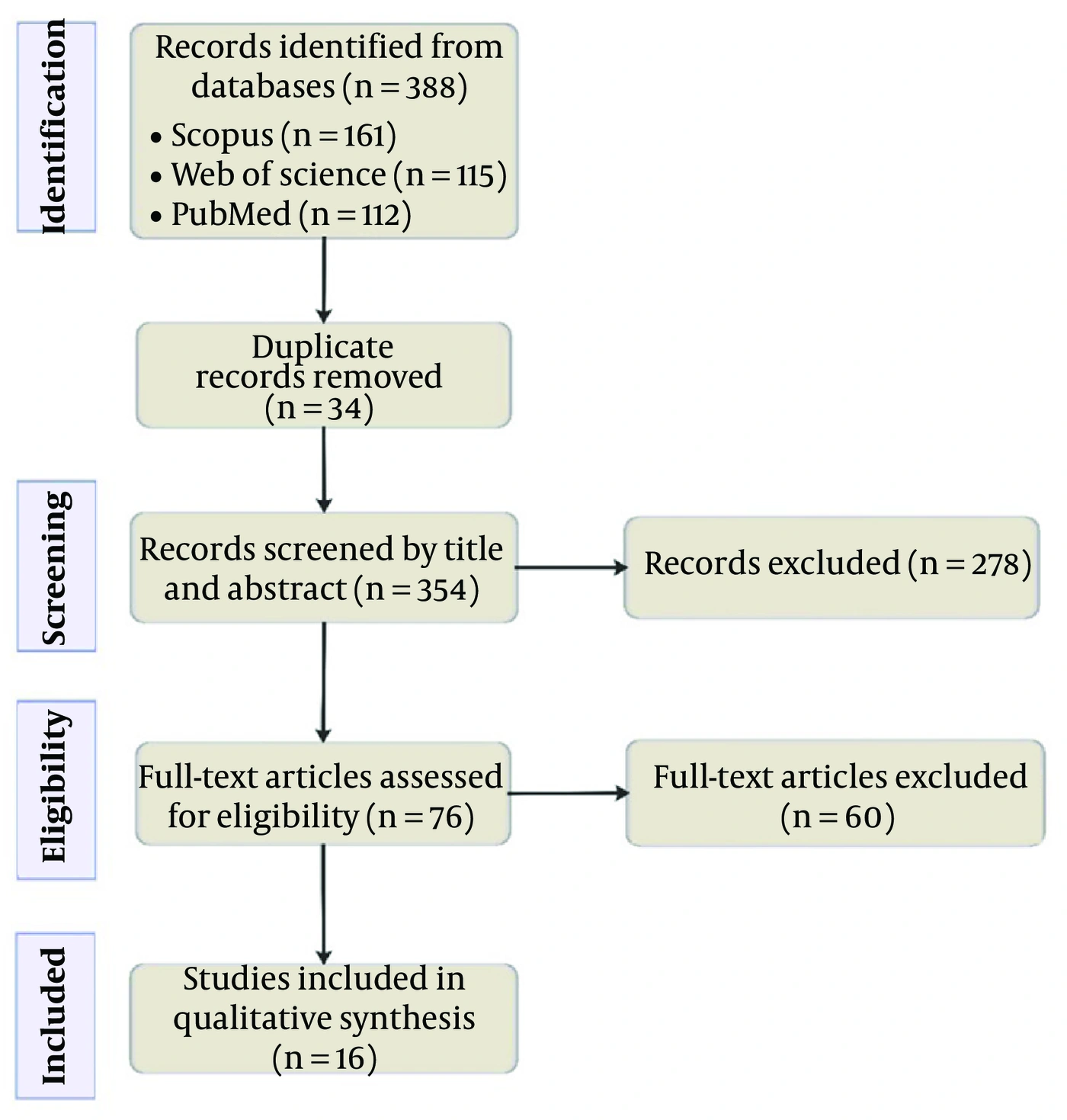
Figure 2.
PRISMA 2020 flow diagram illustrating the screening and selection pathway of the retrieved studies. The figure depicts the progression from database retrieval and removal of duplicate records through eligibility screening and full-text evaluation to the final set of studies included in the qualitative synthesis (11, 17-31).
3.2. Characteristics of the Included Studies
The 16 studies included in this review were published between 2020 and 2026 and were conducted exclusively in experimental animal models, including mice and rats. The investigated disease models included ischemic stroke (n = 9), Alzheimer's disease (AD; n = 4), Parkinson's disease (PD; n = 1), spinal cord injury (SCI; n = 1), and age-related neurodegeneration (n = 1). Exercise interventions varied across studies; however, treadmill training was the most commonly implemented protocol. Most investigations focused on exercise-derived exosomes, whereas fewer studies examined EVs. A comprehensive summary of the characteristics of the included studies is provided in Table 1.
Table 1.
Summary of the Included Studies Describing Experimental Models, Neurological Conditions, Exercise Interventions, Extracellular Vesicle/Exosome Characteristics, Underlying Mechanisms, and Principal Outcomes
| Author (y) | Study Model | Neurological Condition/Model | Exercise Intervention | EV/Exosome Source and Cargo | Main Outcomes and Mechanisms | Key Findings |
|---|---|---|---|---|---|---|
| Chen et al. (2026) (17) | Animal (hypertensive MCAO mice) | Hypertensive ischemic stroke | Treadmill exercise | Exercise-conditioned EPC-derived exosomes (ET-EPC-EXs) | Preserved cerebral blood flow, reduced neuroinflammation, improved vascular integrity via p38 inhibition | ET-EPC-EXs reduced ischemic brain injury and improved early sensorimotor recovery. |
| Citron et al. (2026) (18) | Animal (aged rats) | Aging-related neurodegeneration (PD-related pathways) | Moderate aerobic exercise | Plasma-derived exercise exosomes | Reduced microglial activation, increased BDNF and irisin, modulation of mTOR, MAPK, and cAMP signaling | Exercise-derived plasma exosomes induced exercise-like neuroprotective molecular adaptations in the brains of aged rats. |
| Gao et al. (2026) (19) | Animal (rat, MCAO) | Ischemic stroke | Modified constraint-induced movement therapy (mCIMT) | Brain-derived exosomes and exosomal miRNAs | Increased exosome production; altered miRNA profile; increased MAP2 and VEGF expression; improved neurobehavioral recovery | mCIMT enhanced exosome-mediated neurorepair and promoted functional recovery after ischemic stroke. |
| Lin et al. (2026) (20) | Animal (AD mice) | Alzheimer's disease | Swimming exercise | Skeletal muscle-derived EVs (SKM-EVs), miR-378a-3p | Enhanced amyloid-β plaque clearance; induced disease-associated microglia polarization; regulated lipid metabolism via miR-378a-3p/p110α | Exercise-induced SKM-EVs alleviated cognitive impairment and promoted amyloid-β clearance in AD mice. |
| Miyauchi et al. (2026) (21) | Animal (MCAO rats) | Ischemic stroke | Treadmill exercise | Platelet-rich plasma-derived EVs (aPRP-EVs) | Increased TGF-β1 expression; activated TGF-β/SMAD signaling; reduced neuronal apoptosis; enhanced axonal growth and motor recovery | Exercise-induced PRP-derived EVs enhanced neuroregeneration and improved functional recovery after ischemic stroke. |
| Takeda et al. (2026) (22) | Animal (App(NL-G-F) AD mice) | Alzheimer's disease | Treadmill exercise | Exercise-induced blood EVs enriched with heat shock proteins and cochaperones | Reduced Aβ plaque deposition through exercise-induced blood EV secretion | Exercise-induced blood EVs mediated the beneficial effects of exercise by suppressing Aβ pathology in AD mice. |
| Ying et al. (2026) (23) | Animal (SCI rats) | Spinal cord injury | Treadmill exercise | Exercise-derived exosomes enriched with miR-151-3p | Improved motor recovery and reduced secondary injury via anti-inflammatory and antioxidant mechanisms | Exercise-derived exosomes promoted neuroregeneration and functional recovery after SCI through exosomal miR-151-3p. |
| Citron et al. (2025) (24) | Animal (PD rats) | Parkinson's disease | Moderate chronic treadmill exercise | Plasma-derived exercise exosomes enriched with neuroprotective RNAs | Preserved dopaminergic neurons, altered exosomal RNA cargo, enhanced neuroprotective exerkines | Chronic treadmill exercise modified plasma exosomal RNA cargo and attenuated dopaminergic neuron loss in a rat model of Parkinson's disease. |
| Fuller et al. (2025) (25) | Animal (APP/PS1 mice) | Alzheimer's disease | Voluntary exercise training (VET) | Blood-derived exercise EVs (eEVs) | Improved cognition, reduced resting energy expenditure, EV-mediated neuroprotective effects | Exercise-derived EVs contributed to the beneficial effects of exercise and partially mediated metabolic improvements in APP/PS1 mice. |
| Song et al. (2025) (11) | Animal (MCAO/R rats) | Cerebral ischemia-reperfusion injury | Exercise preconditioning (motorized running wheel) | Plasma-derived exercise exosomes enriched with miR-124 | Reduced neuronal apoptosis, decreased infarct size, improved neurological function via miR-124/STAT3/BCL-2/BAX pathway | Exercise preconditioning increased exosomal miR-124 and alleviated cerebral ischemia-reperfusion injury by inhibiting apoptosis. |
| Huang et al. (2024) (26) | Animal (rats) | Ischemic stroke | Exercise preconditioning | Skeletal muscle-derived exosomes enriched with miR-484 | Reduced neuronal ferroptosis via the miR-484/ACSL4 axis; increased GPX4 and SLC7A11, decreased lipid peroxidation | Exercise-derived skeletal muscle exosomes protected against ischemic brain injury and improved neurological recovery by inhibiting ferroptosis. |
| Liang et al. (2023) (27) | Animal (5XFAD mice) | Alzheimer's disease | Long-term exercise | Brain-derived exosomes enriched with miR-532-5p | Improved blood-brain barrier function via the miR-532-5p/EPHA4 axis | Exercise-derived exosomal miR-532-5p enhanced blood-brain barrier integrity and promoted amyloid-β clearance in Alzheimer's disease. |
| Li et al. (2022) (28) | Animal (MCAO rats) | Ischemic stroke | Early exercise intervention (4 weeks) | Exercise-induced exosomes | Inhibited excessive microglial activation and improved synaptic plasticity through increased Syn and PSD-95 expression | Exercise-induced exosomes enhanced neurological recovery by suppressing microglial activation and promoting synaptic plasticity after ischemic stroke. |
| Huang et al. (2021) (29) | Animal (tMCAO rats) | Ischemic stroke | Running exercise (28 days) | Cerebrospinal fluid-derived exosomal miRNAs | Improved neurological recovery associated with NF-κB and MAPK signaling | Exercise altered cerebrospinal fluid exosomal miRNAs associated with neuroprotection after ischemic stroke. |
| Li et al. (2021) (30) | Animal (MCAO rats) | Ischemic stroke | Exercise intervention (4 weeks) | Exercise-induced serum and brain exosomes | Enhanced synaptic plasticity and corticospinal tract integrity | Exercise-induced exosomes promoted neurological recovery by enhancing synaptic growth and reducing infarct volume after ischemic stroke. |
| Wang et al. (2020) (31) | Animal (MCAO mice) | Ischemic stroke | Moderate treadmill exercise (4 weeks) | Exercise-conditioned EPC-derived exosomes enriched with miR-126 | Enhanced angiogenesis and neuroprotection via the miR-126/PI3K-Akt pathway | Exercise-conditioned EPC-derived exosomes promoted neurological recovery and reduced ischemic brain injury after stroke. |
Abbreviations: Aβ, amyloid-beta; ACSL4, acyl-CoA synthetase long-chain family member 4; APP/PS1, amyloid precursor protein/presenilin-1 transgenic mouse model; aPRP-EVs, athlete platelet-rich plasma-derived extracellular vesicles; BAX, BCL-2-associated X protein; BCL-2, B-cell lymphoma 2; BDNF, brain-derived neurotrophic factor; cAMP, cyclic adenosine monophosphate; CI/RI, cerebral ischemia-reperfusion injury; eEVs, exercise-derived extracellular vesicles; EPC, endothelial progenitor cell; EPHA4, ephrin type-A receptor 4; ET-EPC-EXs, exercise-trained endothelial progenitor cell-derived exosomes; GPX4, glutathione peroxidase 4; MAP2, microtubule-associated protein 2; MAPK, mitogen-activated protein kinase; MCAO, middle cerebral artery occlusion; MCAO/R, middle cerebral artery occlusion/reperfusion; miR-378a-3p, microRNA-378a-3p; mTOR, mammalian target of rapamycin; NF-κB, nuclear factor kappa B; PI3K, phosphoinositide 3-kinase; PRP, platelet-rich plasma; PSD-95, postsynaptic density protein 95; sEVs, sedentary-derived extracellular vesicles; SLC7A11, solute carrier family 7 member 11; STAT3, signal transducer and activator of transcription 3; Syn, synaptophysin; TGF-β1, transforming growth factor-beta 1; tMCAO, transient middle cerebral artery occlusion; VEGF, vascular endothelial growth factor.
3.3. Risk of Bias Assessment
The study-level risk-of-bias assessment is presented in Figure 1A, and the distribution of judgments across the assessed domains is summarized in Figure 1B. Overall, the included studies demonstrated a generally low risk of bias, with no study judged to have a high risk of bias in any assessed domain. Low-risk judgments predominated for sequence generation (D1), baseline characteristics (D2), incomplete outcome data (D8), selective outcome reporting (D9), and other bias (D10). In contrast, allocation concealment (D3) was consistently classified as having an unclear risk of bias across all included studies. Random housing (D4) and random outcome assessment (D6) were predominantly judged as having an unclear risk of bias. Blinding of caregivers and investigators (D5) also received predominantly unclear-risk judgments, whereas blinding of outcome assessment (D7) showed an approximately equal distribution of low- and unclear-risk judgments. Unclear-risk judgments were primarily assigned when the methodological information reported in the original studies was insufficient to permit a definitive assessment.
3.4. Summary of Therapeutic Effects
The included studies consistently demonstrated that exercise-derived EVs and exosomes exert beneficial effects across a range of experimental models of neurological disorders and CNS injury. In ischemic stroke models, the reported therapeutic effects included reduced infarct volume, improved neurological and sensorimotor function, enhanced neuroregeneration, and attenuation of neuronal apoptosis, neuroinflammation, and oxidative stress. Several studies also reported improvements in angiogenesis, synaptic plasticity, axonal remodeling, and vascular integrity, contributing to enhanced functional recovery following cerebral ischemic injury (11, 17, 19, 21, 26, 28-31). In AD models, exercise-derived EVs/exosomes were associated with improved cognitive performance, enhanced amyloid-β clearance, preservation of blood-brain barrier integrity, and exercise-related metabolic adaptations (20, 22, 25, 27). Beneficial effects were also reported in PD, SCI, and aging-related neurodegeneration, including preservation of dopaminergic neurons, reduced secondary tissue damage, improved motor recovery, and enhanced neuroprotective adaptations (18, 23, 24).
3.5. Molecular Mechanisms of Exercise-Derived EVs/Exosomes
To facilitate interpretation, the identified molecular mechanisms were synthesized into three complementary categories: exosomal microRNAs, intracellular signaling pathways, and downstream effector proteins. The included studies demonstrated that the beneficial effects of exercise-derived EVs and exosomes were mediated through diverse molecular mechanisms. Several studies reported the involvement of exosomal miRNAs, including miR-126, miR-124, miR-484, miR-532-5p, miR-378a-3p, and miR-151-3p, which were associated with neuroprotection, angiogenesis, synaptic remodeling, BBB preservation, inhibition of ferroptosis, and functional recovery (11, 20, 23, 26, 27, 31). In addition, multiple intracellular signaling pathways were modulated by exercise-derived EVs/exosomes, including PI3K/Akt, STAT3, TGF-β/SMAD, MAPK, mTOR, NF-κB, and p38 signaling. Several studies also reported increased expression of proteins associated with neuronal survival and regeneration, such as BDNF, VEGF, GPX4, SLC7A11, synaptophysin, and PSD-95, together with reduced neuronal apoptosis, neuroinflammation, oxidative stress, and microglial activation (17-19, 21, 22, 24, 28-31).
4. Discussion
The present systematic review synthesizes available preclinical evidence on the therapeutic potential of exercise-derived EVs/exosomes in neurological disorders. The findings indicate that exercise-derived EVs/exosomes may serve as important biological mediators through which exercise promotes neuroprotection and neurological recovery across diverse experimental models. Rather than acting through a single mechanism, these vesicles appear to exert beneficial effects by simultaneously modulating multiple interconnected pathological processes, including the attenuation of neuroinflammation, oxidative stress, and neuronal apoptosis, together with the promotion of angiogenesis, neurogenesis, synaptic plasticity, and vascular integrity. This coordinated biological activity highlights the potential role of exercise-derived EVs/exosomes in mediating systemic, exercise-induced adaptations that support neural repair and functional recovery. Nevertheless, because the current evidence is derived exclusively from experimental animal studies, further well-designed translational and clinical investigations are required to determine the applicability of these findings to human neurological disorders.
One of the most notable observations of this review is the remarkable consistency of the therapeutic effects of exercise-derived EVs/exosomes across neurological disorders with distinct pathological characteristics. Although ischemic stroke, AD, PD, SCI, and aging-related neurodegeneration differ considerably in their underlying etiologies and disease progression, the included studies consistently reported improvements in neurological recovery following treatment with exercise-derived EVs/exosomes. This convergence of therapeutic outcomes suggests that the biological activity of these vesicles is not disease-specific but instead targets fundamental mechanisms that are commonly disrupted across neurological disorders. Consequently, exercise-derived EVs/exosomes may act as systemic biological mediators that enhance the intrinsic regenerative capacity of the nervous system, rather than exerting isolated effects on individual disease processes.
A key mechanistic finding of this review is that the therapeutic effects of exercise-derived EVs/exosomes appear to be largely mediated by their bioactive molecular cargo, particularly regulatory miRNAs. Wang et al. reported that exercise-induced exosomal miRNAs contribute to maintaining nervous system homeostasis by regulating molecular pathways involved in cellular communication and tissue adaptation (32). Consistent with this concept, this review demonstrated that exercise-derived EVs/exosomes carrying diverse regulatory miRNAs were associated with multiple neuroprotective processes across different preclinical models of neurological disorders. Rather than being attributable to a single regulatory miRNA, the available evidence suggests that the therapeutic effects of exercise-derived EVs/exosomes are more likely mediated through the coordinated activity of multiple exercise-responsive miRNAs.
In addition to the regulatory role of exosomal miRNAs, the findings of this review suggest that the neuroprotective effects of exercise-derived exosomes are mediated by the coordinated modulation of multiple intracellular signaling pathways, rather than by the activation of a single molecular cascade. Across the included studies, signaling pathways including PI3K/Akt, STAT3, MAPK, TGF-β/SMAD, mTOR, NF-κB, and p38 were consistently associated with reduced neuroinflammation and oxidative stress, inhibition of neuronal apoptosis, and enhancement of neuroregeneration and functional recovery. The coordinated regulation of these interconnected pathways may explain the consistent therapeutic benefits observed across diverse experimental models, despite differences in neurological disorders, exercise protocols, and experimental conditions. This interpretation is consistent with the review by Kong et al., who suggested that exercise-derived exosomes regulate multiple signaling networks involved in cellular survival, inflammatory control, oxidative stress responses, and tissue repair rather than acting through a single signaling mechanism (14). Similarly, recent reviews of exosome-based therapies have identified the PI3K/Akt, MAPK, NF-κB, and TGF-β pathways as central molecular mediators underlying neuroprotection and neural regeneration (33, 34). Building on these observations, this review further demonstrates that the coordinated modulation of these signaling pathways is a consistent biological feature of exercise-derived exosomes across diverse preclinical models of neurological disorders, suggesting that convergence on shared intracellular signaling networks may constitute a fundamental mechanism underlying their therapeutic efficacy.
Beyond the coordinated modulation of intracellular signaling pathways, the findings of this review suggest that exercise-derived exosomes facilitate neurological recovery through the regulation of downstream effector proteins involved in neuronal survival, angiogenesis, synaptic plasticity, and redox homeostasis. Across the included studies, increased expression of BDNF and VEGF was consistently associated with enhanced neuronal survival, neurovascular remodeling, and functional recovery. These findings are consistent with the review by Putthanbut et al., who highlighted that extracellular vesicle-based therapies promote neural repair by regulating downstream molecular mediators involved in neuroplasticity, angiogenesis, and tissue regeneration (35). Furthermore, the upregulation of GPX4 and SLC7A11 observed in several studies suggests that exercise-derived exosomes may protect against ferroptotic cell death by enhancing endogenous antioxidant defenses and maintaining redox homeostasis. This observation is supported by the recent review by Bao et al., which identified exosome-mediated regulation of the GPX4-GSH axis, iron homeostasis, and antioxidant pathways as key mechanisms underlying neuroprotection in neurological disorders (36). In addition, increased expression of synaptophysin and PSD-95 provides further evidence of enhanced synaptic remodeling and neuronal connectivity, offering a mechanistic explanation for the improvements in learning, memory, and sensorimotor function reported across the included studies. Collectively, these findings indicate that the coordinated regulation of downstream effector proteins represents a critical molecular interface through which exercise-derived exosomes translate upstream molecular signaling into sustained structural and functional recovery following neurological injury.
Overall, the included studies demonstrated a generally low risk of bias according to the SYRCLE Risk of Bias assessment. However, several domains were frequently judged as having an unclear risk because of insufficient methodological reporting. Therefore, although the overall findings were consistent across studies, the certainty of the available evidence should be interpreted with caution. A formal Grading of Recommendations, Assessment, Development, and Evaluation (GRADE) assessment was not performed because this review synthesized heterogeneous preclinical animal studies with substantial variability in experimental models, interventions, and outcome measures. Although adaptations of the GRADE framework for preclinical evidence have been proposed, no universally accepted approach currently exists for its routine application in heterogeneous preclinical systematic reviews (37).
Despite the overall consistency of the reported therapeutic effects, substantial methodological heterogeneity was evident across the included studies. Variations in animal species, neurological disease models, exercise modalities, training intensity, intervention duration, and treatment protocols may have influenced both the biological characteristics of exercise-derived exosomes and the observed therapeutic outcomes. In particular, differences in exercise prescription are likely to affect exosome release and molecular cargo composition. Consistent with this observation, Abbasian et al. emphasized that exercise intensity and duration are critical determinants of neurological recovery following stroke and highlighted the importance of optimizing exercise protocols to maximize therapeutic benefits (38). Furthermore, considerable variability in exosome isolation, characterization, and reporting methods limits direct comparisons across studies and underscores the need for standardized experimental protocols to improve reproducibility and facilitate the translation of preclinical findings.
Although the preclinical findings summarized in this review are promising, several challenges must be addressed before exercise-derived EVs/exosomes can be translated into clinical practice (14). One major obstacle is the lack of standardized protocols for EV/exosome isolation, characterization, storage, and quality control, which limits reproducibility across studies and hinders direct comparison of experimental findings (39). In addition, the molecular composition and biological activity of exercise-derived EVs/exosomes are influenced by multiple factors, including exercise modality, intensity, duration, and the physiological status of the donor, making it challenging to establish consistent therapeutic products (40). Furthermore, uncertainties regarding delivery efficiency, immunogenicity, and standardized preparation procedures continue to hinder clinical translation (14). Therefore, future translational research should prioritize standardized manufacturing and characterization protocols in accordance with current guidelines, together with well-designed clinical studies to establish the safety and therapeutic efficacy of exercise-derived EVs/exosomes in neurological disorders.
4.1. Limitations
This review has several limitations that should be considered when interpreting the findings. First, all included studies were preclinical animal studies, limiting the direct generalizability of the findings to human neurological disorders. Second, substantial heterogeneity existed across studies with respect to animal models, neurological disease models, exercise protocols, EV/exosome isolation and characterization methods, molecular outcome measures, and functional assessments, precluding quantitative meta-analysis. Third, only English-language publications were included, which may have introduced language bias. Fourth, the review protocol was not prospectively registered, which may limit transparency and increase the potential for methodological bias despite adherence to established systematic review guidelines. Finally, although methodological quality was evaluated using the SYRCLE risk-of-bias tool, a formal GRADE assessment was not performed because the evidence consisted exclusively of heterogeneous preclinical animal studies. Although adaptations of the GRADE framework for preclinical evidence have been proposed, no universally accepted approach currently exists for its routine application in heterogeneous preclinical systematic reviews.
4.2. Conclusions
In conclusion, the available preclinical evidence suggests that exercise-derived EVs/exosomes are promising mediators of neuroprotection and functional recovery across diverse experimental models of neurological disorders. Their therapeutic effects appear to be associated with the coordinated modulation of exosomal miRNAs, intracellular signaling pathways, and downstream effector proteins that collectively support neurogenesis, angiogenesis, synaptic plasticity, and redox homeostasis. Nevertheless, methodological heterogeneity and the lack of standardized approaches for EV/exosome isolation, characterization, and reporting limit direct comparisons across studies and pose challenges for clinical translation. Future studies should prioritize methodological standardization, further elucidate disease-specific molecular mechanisms, and validate these findings in well-designed clinical studies to facilitate the safe and effective therapeutic application of exercise-derived EVs/exosomes in neurological disorders.