
1. Background
Acute kidney injury (AKI) is one of the most challenging and complex complications in critically ill children admitted to pediatric intensive care units (PICUs). Regardless of the primary diagnosis, AKI can substantially increase morbidity, mortality, and treatment costs (1). Recent studies have reported an AKI prevalence of 20% - 40% among critically ill children in PICUs, with higher rates among patients with shock or multiorgan failure (2). Predisposing factors for AKI in children include hypovolemia, shock, sepsis, nephrotoxic drugs, major surgery, and underlying kidney disease (3).
Shock is a major risk factor for AKI (4). Several pathophysiological mechanisms in shock, including hypotension leading to reduced kidney perfusion with subsequent ischemia of kidney tissue, tubular damage, and activation of inflammatory cascades, contribute to AKI (5). Even brief episodes of hypotension can cause kidney injury, highlighting the need for rigorous hemodynamic monitoring to prevent AKI (6).
Shock index (SI), defined as the ratio of heart rate to systolic blood pressure, is a simple, noninvasive parameter used to assess hemodynamic status in critically ill patients (7). SI is a sensitive marker for the early detection of hypovolemia and shock (8). An elevated SI indicates reduced effective circulating volume and impaired tissue perfusion. Multiple studies have linked elevated SI to adverse outcomes, including mortality, the need for mechanical ventilation, and multiorgan failure (9).
Despite strong evidence linking shock to AKI and supporting the prognostic value of SI in critically ill patients, few studies have examined the association between elevated SI and AKI in PICU populations (10). Most research has focused on adults, with limited analyses in pediatric populations (11). Important physiological differences between children and adults in their responses to shock and kidney injury justify studies specifically designed for pediatric populations (12).
2. Objectives
This study aimed to investigate the association between SI and AKI in children admitted to the PICU and to determine whether SI is associated with AKI risk. These findings may improve understanding of the pathophysiological link between hemodynamic disturbances and kidney injury, inform pediatric ICU monitoring protocols, and guide future interventional studies.
3. Methods
3.1. Study Design and Setting
This prospective cohort study investigated the association between SI and the prevalence of AKI among pediatric patients admitted to the PICU of Amir Kabir Hospital, Arak, Iran, over a 6-month period from February to July 2023. Amir Kabir Hospital is a tertiary care facility with a primary focus on pediatric care, and its PICU is dedicated to the care of critically ill children. Ethical approval was obtained from the Research Ethics Committees of Arak University of Medical Sciences (IR.ARAKMU.REC.1401.336).
3.2. Study Participants
The study included pediatric patients admitted to the PICU according to the following criteria.
Inclusion criteria:
1) Age 1 month to 18 years;
2) PICU admission ≥ 24 hours;
3) Complete hemodynamic data, including systolic blood pressure and heart rate, during the first 24 hours;
4) Sufficient data in the patient record for AKI diagnosis using KDIGO criteria, including baseline serum creatinine, baseline glomerular filtration rate (GFR), and urine output; and
5) Parental consent.
Exclusion criteria:
1) Chronic kidney disease stage ≥ 3;
2) History of kidney transplantation;
3) Congenital urinary tract anomalies;
4) Insufficient clinical data;
5) Death within 24 hours; and
6) Voluntary discharge before completion of the evaluation.
The sample size was calculated as 80, including 10% attrition, based on a similar study (13), with α = 0.05 and 90% power. Convenience sampling was used until the target sample size was reached.
3.3. Data Collection
A structured checklist was used to extract and categorize data from patient records. Participants were followed prospectively from PICU admission through the 48-hour AKI assessment window. Data were collected at admission (baseline) and at 24 and 48 hours after admission. The following information was collected: age, weight, serum creatinine, GFR, urine output, systolic blood pressure, heart rate, age-adjusted SI, length of hospital stay, and primary diagnosis.
Serum creatinine and GFR were measured on admission and 48 hours after admission. For the purposes of this study, systolic blood pressure and heart rate were measured and recorded once at hospital admission; however, routine monitoring of hemodynamic stability continued according to PICU protocols.
3.4. Definitions
In this study, AKI was diagnosed as present or absent using KDIGO criteria based on 1) serum creatinine changes measured at a minimum of 2 time points, including admission and 48 hours after admission, to confirm and quantify changes; and 2) hourly urine output measured via a urinary catheter.
A significant limitation was the method used to establish baseline creatinine. For most patients, premorbid values were unavailable, and baseline values were necessarily estimated from admission values or assumptions based on normal GFR for age. This approach may have influenced the accuracy of AKI staging and may have led to overestimation or underestimation of the true incidence; however, consistent use of KDIGO criteria supports internal validity for comparisons within the cohort. Serum creatinine was measured at 2 separate time points to capture changes during the first 48 hours after admission.
AKI was confirmed if ≥ 1 of the following criteria was met:
1) Serum creatinine increased by ≥ 0.3 mg/dL from baseline within 48 hours;
2) Serum creatinine was ≥ 1.5 times baseline within the prior 7 days; or
3) Urine output was < 0.5 mL/kg/h for 8 hours.
Baseline creatinine was assigned using a hierarchical approach:
1) Known premorbid values (n = 26): For patients with documented serum creatinine measurements within 3 months before admission, this value was used as baseline.
2) Admission values (n = 42): For patients without premorbid values, admission serum creatinine was used as baseline, assuming normal renal function before the acute illness.
3) Estimated values (n = 12): For patients without premorbid or admission values, baseline creatinine was estimated using age-specific normal GFR assumptions with the Schwartz formula and back-calculation assuming height at the 50th percentile for age.
For analysis, kidney function status was categorized as serum creatinine increase, GFR reduction, or urine output reduction.
Glomerular filtration rate was calculated using the Schwartz formula:
eGFR = 0.413 × (height in cm / SCr)
GFR reduction was defined as eGFR < 90 mL/min/1.73m2. This threshold was selected based on KDIGO guidelines defining GFR categories and is consistent with previous pediatric AKI studies (14).
All measurements were performed in the Amir Kabir Hospital laboratory, and the results were validated by a pediatric nephrologist.
Systolic blood pressure and heart rate were measured on PICU admission as part of routine clinical assessment. For this study, the first documented measurement at PICU admission was recorded. In patients receiving vasopressor therapy, measurements were obtained via arterial cannulation when available (n = 5); in patients not receiving vasopressors, measurements were obtained using noninvasive oscillometric methods. Routine hemodynamic monitoring continued according to PICU protocols and was not interrupted for this study.
Age-adjusted SI was calculated using age-specific critical thresholds (15):
1) 1 - 6 years: > 1.2;
2) 7 - 12 years: > 1.0; and
3) 12 - 18 years: > 0.9.
Age adjustment accounts for age-related physiological characteristics and provides a more precise measure in pediatric populations.
For patients treated with vasopressors, SI was calculated using arterial-line measurements when available. For patients without arterial lines, noninvasive blood pressure measurements were used. A sensitivity analysis excluding patients treated with vasopressors was performed to assess the robustness of the findings.
3.5. Statistical Analysis
Data were analyzed using STATA version 12. Descriptive statistics, including mean ± SD and frequencies, were used to summarize demographic characteristics. Independent t tests or Mann-Whitney U tests were used to compare continuous variables, and chi-square or Fisher exact tests were used to analyze categorical variables.
Normality of continuous variables was assessed using the Shapiro-Wilk test. Normally distributed variables were compared using independent t-tests, whereas nonnormally distributed variables were compared using Mann-Whitney U tests. Results are presented as mean ± SD.
Logistic regression was used to calculate odds ratios (ORs) with 95% CIs. Univariable logistic regression models examined the association between SI, as the primary exposure, and each outcome, including AKI presence and GFR reduction, without covariate adjustment. Multivariable adjustment was not feasible because of the limited number of non-AKI events (n = 8), which precluded the inclusion of multiple covariates without violating the minimum events-per-variable rule. Therefore, the results should be interpreted as unadjusted associations. Confidence intervals for proportions were calculated using the Wilson method to improve accuracy with small sample sizes. Statistical significance was set at P < 0.05.
4. Results
4.1. Sample Characteristics and Primary Diagnoses
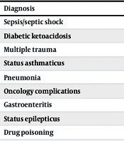
The mean age of the participants was 7.21 ± 4.76 years, the mean weight was 27.32 ± 15.43 kg, and 46.3% were male (n = 37). The prevalence of AKI was 90% (n = 72; 95% CI, 81.0 - 95.0% using the Wilson method; 95% CI, 81.4 - 94.8% using exact binomial). The distribution of primary diagnoses is shown in Table 1.
Table 1.
Primary Diagnoses of the Study Population
| Diagnosis | No. (%) |
|---|---|
| Sepsis/septic shock | 26 (32.50) |
| Diabetic ketoacidosis | 14 (17.50) |
| Multiple trauma | 12 (15.00) |
| Status asthmaticus | 7 (8.75) |
| Pneumonia | 7 (8.75) |
| Oncology complications | 4 (5.00) |
| Gastroenteritis | 4 (5.00) |
| Status epilepticus | 3 (3.75) |
| Drug poisoning | 3 (3.75) |
4.2. Baseline Creatinine Assignment and AKI Classification
Baseline creatinine was derived from known premorbid values in 26 patients (32.5%), admission values in 42 patients (52.5%), and estimated values in 12 patients (15%). Urine output criteria identified 20 AKI cases (27.7%), creatinine criteria identified 20 cases (27.7%), and both criteria identified 32 cases (44.4%).
Variables related to kidney function were compared between patients with normal and high SI. Urine output and 24-hour GFR were nonnormally distributed and were compared using the Mann-Whitney U test. Patients with high SI had significantly lower urine output (P = 0.038) and 24-hour GFR (P = 0.002) than patients with normal SI. Additional results are presented in Table 2.
| Variables | Normal SI | High SI | P-Value |
|---|---|---|---|
| Age (y) | 8.63 ± 4.53 | 7.05 ± 4.79 | 0.382 |
| Weight (kg) | 32.00 ± 13.22 | 26.80 ± 15.64 | 0.325 |
| Urine output (mL/kg/h) | 0.63 ± 0.04 | 0.33 ± 0.11 | 0.038 |
| Creatinine (hour 0, mg/dL) | 0.58 ± 0.18 | 0.62 ± 0.21 | 0.632 |
| Creatinine (hour 24, mg/dL) | 0.64 ± 0.18 | 0.89 ± 0.43 | 0.112 |
| GFR (hour 0, mL/min/1.73m2) | 104.96 ± 34.89 | 85.58 ± 26.35 | 0.061 |
| GFR (hour 24, mL/min/1.73m2) | 88.29 ± 19.90 | 62.86 ± 20.88 | 0.002 c |
| Length of stay (d) | 4.00 ± 1.41 | 4.65 ± 1.59 | 0.270 |
a Values are expressed as mean ± SD. Abbreviations: SI, shock index; GFR, glomerular filtration rate; SD, standard deviation.
b Normally distributed variables (age, weight, baseline creatinine, baseline GFR, and length of stay) were compared using independent t tests. Nonnormally distributed variables (urine output, 24-hour creatinine, and 24-hour GFR) were compared using Mann-Whitney U tests. Normality was assessed using the Shapiro-Wilk test.
c Significance at P < 0.05.
4.3. Association Between Shock Index and Clinical Outcomes
High SI was present in 90.3% of patients with AKI (n = 65; 95% CI, 86.0 - 97.7%) and in 50% of patients without AKI (n = 4; 95% CI, 35.3 - 84.8%). A chi-square test was used to evaluate the association between SI abnormality and the clinical outcome, defined as the presence or absence of AKI. A significant association was observed between SI and AKI. Patients with high SI had 9.28 times higher odds of AKI (95% CI, 1.34 - 59.98; P = 0.001). Additional results are presented in Table 3.
Table 3.
Association Between Shock Index, AKI, and Clinical Outcomes a
| Variables | Normal SI (%) | High SI (%) | P-Value | OR (95% CI) |
|---|---|---|---|---|
| AKI b | 0.001 | 9.28 (1.34 - 59.98) | ||
| Absent | 4 (50.0) | 4 (50.0) | ||
| Present | 7 (9.7) | 65 (90.3) | ||
| Urine output reduction | 0.058 | - | ||
| Present | 8 (14.1) | 49 (85.9) | ||
| Absent | 0 (0.0) | 23 (100.0) | ||
| Serum creatinine increase | 0.604 | 6.26 (0.72 - 290.36) | ||
| Present | 7 (15.6) | 38 (84.4) | ||
| Absent | 1 (2.9) | 34 (97.1) | ||
| GFR reduction b | 0.003 | 8.00 (1.18 - 50.47) | ||
| Absent | 4 (50.0) | 4 (50.0) | ||
| Present | 8 (10.1) | 64 (89.9) |
a Univariable logistic regression models were used. Multivariable adjustment was not performed because of sparse outcome data (only 8 non-AKI cases). Abbreviations: AKI, acute kidney injury; SI, shock index; GFR, glomerular filtration rate; OR, odds ratio.
b Significance: chi-square test (P < 0.05).
Because of the small number of patients without AKI (n = 8), these odds ratios should be interpreted with caution, as reflected by the wide confidence intervals. Sparse data may lead to unstable estimates, and these findings require validation in larger cohorts.
5. Discussion
AKI is a common and challenging complication in children admitted to PICUs and has important clinical consequences. Early diagnosis can substantially affect treatment and prognosis. Diagnostic criteria for AKI in children are generally based on changes in urine output, serum creatinine, and GFR. However, these criteria may manifest differently across patient populations. A better understanding of this variability may improve the diagnosis and clinical management of AKI.
The exceptionally high prevalence of AKI (90%) in our cohort of critically ill children contrasts markedly with the rates reported in most major PICU studies, in which prevalence typically ranged from 10% to 27% (10, 16-18). This discrepancy may reflect several methodological and population-specific factors.
The high prevalence may reflect the study's stringent inclusion criteria, which selected a uniquely severe subpopulation. By including only children who required a PICU stay of ≥ 24 hours and had complete hemodynamic monitoring data, the study may have preferentially captured patients with severe illness requiring intensive surveillance. This interpretation is supported by the finding that 90.3% of patients with AKI had an elevated SI, a marker of substantial hemodynamic compromise. The primary diagnoses in the cohort—septic shock (32.5%), diabetic ketoacidosis (17.5%), and major trauma (15%)—also indicate a high-acuity case mix with an increased risk of kidney injury. Therefore, these findings are most generalizable to this specific high-risk subset of PICU patients rather than to all PICU admissions.
Another factor is that the study was conducted at a single tertiary care center in Iran. Local patient demographics, admission thresholds, underlying disease patterns, and resource availability may influence the case mix and observed AKI prevalence compared with multicenter studies (4, 10) or studies conducted in different health care systems.
Despite the unusually high AKI prevalence, the core finding of a strong, statistically significant association between elevated SI and AKI (OR = 9.28) remained evident within this high-severity cohort of critically ill children. Taken together, the high AKI prevalence and its discrepancy with broader PICU studies highlight the vulnerability of this subgroup of hemodynamically unstable patients to kidney injury and underscore the importance of considering patient acuity, the precise AKI definition (including the role of GFR estimates and fluid overload), and the clinical setting when interpreting AKI epidemiology. Future research should prospectively validate SI in diverse PICU populations using standardized AKI criteria such as KDIGO while accounting for fluid balance, to better define its prognostic utility for AKI risk.
The present study showed that mean urine output and GFR after 24 hours were significantly lower in the high-SI group than in the normal-SI group. In contrast, age, weight, creatinine, baseline GFR, and length of hospitalization did not differ significantly between groups. The odds-ratio results were consistent with these findings: patients with higher SI were more likely to have AKI and reduced GFR.
A comprehensive review of the available literature suggests that the strong association between high SI and AKI can be explained by several interconnected mechanisms. At the macroscopic level, an increase in SI reflects a decrease in effective circulating blood volume, which can impair kidney perfusion (19). This condition is accompanied by activation of the renin-angiotensin-aldosterone system, which causes renal vasoconstriction and further decreases blood flow (20). At the microscopic and cellular level, ischemia-induced hypoperfusion can lead to tubular epithelial cell injury accompanied by cellular debris in the tubular lumen (21). At the same time, the release of proinflammatory cytokines, such as TNF-α and IL-6, and reactive oxygen species during the systemic inflammatory response to shock can exacerbate glomerular endothelial injury (20). These processes can ultimately impair renal blood flow autoregulation and increase renal vascular resistance, thereby reducing glomerular filtration (19).
From a clinical perspective, these mechanisms help explain why patients with high SI in the present study were more likely to develop AKI. The present findings support SI as a simple hemodynamic marker that may help identify patients at increased risk of AKI; however, its role in guiding treatment decisions requires further validation.
5.1. Study Limitations
This study has several limitations. First, the exceptionally high baseline AKI prevalence, although informative for a high-severity subgroup, limits the generalizability of the findings to broader, less critically ill PICU populations. The results are most applicable to children requiring prolonged PICU stays and intensive hemodynamic monitoring.
Second, the use of admission creatinine or estimation formulas to determine baseline creatinine, rather than documented premorbid values, may have influenced the accuracy of AKI classification. Using admission creatinine as baseline may underestimate true baseline values in patients with preexisting renal impairment or overestimate AKI severity in patients with low baseline values. This limitation is inherent in studies of critically ill populations in which premorbid laboratory values are frequently unavailable.
Third, the small number of patients without AKI (n = 8) resulted in insufficient statistical power for robust multivariable adjustment and precise estimation of effect sizes, as reflected by the wide confidence intervals. This limitation reduces the certainty of the reported odds ratios and precludes adjustment for potential confounders such as sepsis, illness severity, or nephrotoxic medication exposure. Future studies with larger sample sizes and adequate numbers of patients without AKI are needed to confirm these findings and enable adjusted analyses.
5.2. Conclusions
Shock index may be a useful marker of acute kidney injury in children admitted to the intensive care unit. An increased SI was associated with the development of AKI and decreased kidney function. These findings support the clinical value of regular SI monitoring as a simple and accessible marker for identifying children at increased risk of kidney injury.