
1. Background
Central venous stenosis or occlusion is an important vascular access-related complication in patients receiving maintenance hemodialysis. The subclavian vein, brachiocephalic vein, and superior vena cava are the central venous segments most often involved (1). Its true incidence remains uncertain because many lesions are asymptomatic and are detected only when symptoms develop or when venography is performed during catheter placement or access intervention. Reported rates in hemodialysis patients range from 3% to 60% (1). In hemodialysis access populations, reported rates of central venous occlusion range from 25% to 40% (2).
The pathogenesis is multifactorial. Previous central venous catheterization contributes through endothelial injury, inflammation, thrombosis, fibrin sheath formation, and neointimal hyperplasia (1-3). In patients with an ipsilateral arteriovenous fistula (AVF) or graft, access-related hemodynamic changes may contribute to venous hypertension and make previously silent lesions clinically apparent (3, 4).
Clinical manifestations are primarily related to venous hypertension and access dysfunction. Patients may present with ipsilateral arm, neck, facial, or chest wall swelling; superficial collateral veins; prolonged bleeding after needle removal; difficult cannulation; increased venous pressure during dialysis; inadequate dialysis blood flow; or access failure (1, 2). In 150 hemodialysis patients with central venous occlusion, Gür et al. reported ipsilateral arm swelling in 145 patients (97.0%), face or neck swelling in 57 patients (38.0%), breast swelling in 5 patients (3.3%), and inability to puncture the access in 19 patients (12.6%) (2). Ultrasound, computed tomography venography, and conventional venography or fistulography help confirm the diagnosis, delineate lesion anatomy, and guide intervention (1, 4).
Endovascular therapy is commonly used for symptomatic central venous stenosis or occlusion. Percutaneous transluminal angioplasty is usually the initial treatment, whereas stenting may be considered for elastic recoil, significant residual stenosis, recurrent stenosis, or angioplasty-resistant lesions (1, 5-7). Technical success is often high, but patency remains variable. Gür et al. reported technical success in 141 of 150 patients (94.0%) (2), whereas Bakken et al. reported 12-month primary patency rates of 29% after angioplasty and 21% after primary stenting (5). More recent studies have continued to highlight variability in patency after angioplasty, stenting, or stent-graft treatment in hemodialysis patients with central venous disease (8, 9).
Local data from Vietnam remain limited. In particular, real-world data describing lesion distribution on angiography, procedural characteristics, immediate anatomical success, and 1-month clinical outcomes after endovascular treatment for symptomatic central venous stenosis or occlusion in patients receiving maintenance hemodialysis are limited.
2. Objectives
This study aimed to describe the clinical and imaging characteristics and to evaluate immediate anatomical success, 1-month clinical outcomes, and early complications following endovascular treatment for central venous stenosis or occlusion in patients receiving maintenance hemodialysis at Cho Ray Hospital.
3. Methods
3.1. Study Design and Setting
This retrospective observational analysis was conducted within a larger longitudinal project evaluating early, mid-term, and long-term outcomes after endovascular treatment for central venous stenosis or occlusion in maintenance hemodialysis patients. The study was conducted at the Department of Vascular Surgery, Cho Ray Hospital, Vietnam. For the present analysis, we reviewed patients treated between January 2018 and December 2025. All patients who met the eligibility criteria during the study period were included.
3.2. Study Population
The study included maintenance hemodialysis patients aged 18 years or older who were diagnosed with symptomatic central venous stenosis or occlusion and underwent endovascular treatment. Eligible patients had clinical manifestations of central venous obstruction, access dysfunction, or reduced arteriovenous fistula flow during hemodialysis. Central venous stenosis or occlusion was confirmed by digital subtraction angiography and involved the subclavian vein, brachiocephalic vein, or superior vena cava. Central venous stenosis was defined as more than a 50% reduction in luminal diameter compared with the adjacent normal venous segment, whereas central venous occlusion was defined as complete interruption of venous flow. Complete electronic medical records and valid contact information were required.
Patients were excluded if they had concomitant peripheral venous stenosis or occlusion, a functioning kidney transplant, extrinsic tumor compression of the central vein, previous surgery involving the central venous system, central venous thrombosis related to hematologic disease, or they refused to participate.
3.3. Endovascular Procedure
All procedures were performed under digital subtraction angiography guidance. The access route was selected according to vascular access anatomy and lesion characteristics. Local anesthesia was administered with 2% lidocaine, and a 7-Fr vascular sheath was inserted via the selected access route. After diagnostic angiography, lesion location, severity, and length were assessed.
Systemic heparin was administered at 50 IU/kg before intervention. A 0.035-inch guidewire was used to cross the lesion. Catheter support was used when needed for complete occlusion. After successful crossing, balloon angioplasty was performed. Balloon size was selected according to the estimated normal venous diameter adjacent to the lesion. Balloons ranged from 9 to 14 mm in diameter and 40 to 80 mm in length. Balloon inflation pressure was selected according to lesion characteristics and procedural response.
Completion venography was used to assess venous flow, residual stenosis, and procedure-related complications. Stent placement was considered when the angioplasty result was inadequate, including significant residual stenosis or recoil. When residual stenosis remained greater than 50% after balloon angioplasty, venous stent placement was performed. Stent diameter ranged from 12 to 14 mm, and stent length ranged from 40 to 60 mm.
3.4. Definitions and Outcomes
Central venous stenosis was defined as more than a 50% reduction in venous luminal diameter on digital subtraction angiography. Central venous occlusion was defined as complete interruption of the venous lumen, with or without distal reconstitution, on digital subtraction angiography. The central venous segments assessed included the subclavian vein, brachiocephalic vein, and superior vena cava. Stenosis severity was categorized as moderate when luminal narrowing was less than 70%, severe when luminal narrowing was 70% to 99%, and complete occlusion when luminal obstruction was 100%.
The main procedural outcome was immediate anatomical success, assessed on completion angiography. Anatomical success was defined as good venous flow or residual stenosis below 30%. Anatomical failure included residual stenosis of 30% or greater, inability to cross the lesion, or procedure-related venous rupture resulting in failure.
Early clinical outcomes and complications were assessed within 1 month after the index endovascular intervention, primarily at the scheduled 1-month follow-up visit. Early clinical outcomes included decreased arm edema or pain, the presence or restoration of an arteriovenous fistula anastomotic bruit, decreased superficial venous collateral circulation, and the ability to undergo hemodialysis through the treated access. Symptom improvement was assessed only among patients who had the corresponding symptom before the intervention. Early complications included hematoma and bleeding recorded within 1 month after the intervention.
3.5. Data Collection
Data were collected from medical records using a standardized data collection form. Baseline data included demographic characteristics, comorbidities, clinical presentation, vascular access characteristics, and catheter or device history. Imaging data included Doppler ultrasound, computed tomography, and angiographic findings. Procedural data included access route, treatment modality, balloon use, procedure time, and hospital stay. Outcome data included immediate angiographic results, the 1-month clinical assessment, and early complications. Data were checked against source records before analysis.
3.6. Statistical Analysis
Data were analyzed using Stata version 17.0. Categorical variables were presented as frequencies and percentages. Continuous variables were presented as mean ± standard deviation or median and interquartile range (IQR), depending on data distribution. Missing observations were not imputed; variable-specific denominators were reported where applicable. Exploratory univariable analyses were performed to assess factors associated with anatomical failure. Fisher exact test was used for categorical variables when appropriate, and univariable logistic regression was used for continuous variables. A 2-sided P value < 0.05 was considered statistically significant. Multivariable regression was not performed because only 10 anatomical failures occurred.
3.7. Ethics Approval and Consent to Participate
The study strictly adhered to the principles outlined in the Declaration of Helsinki. Ethical approval was granted by the Ethics Council in Biomedical Research of the University of Medicine and Pharmacy at Ho Chi Minh City (approval No. 1198/HĐĐĐ-ĐHYD). Written informed consent was obtained from the participants. Personal information was kept confidential and used only for research purposes.
4. Results
A total of 106 patients were assessed for eligibility. Five patients were excluded because follow-up data were unavailable, mainly during the COVID-19 period. Therefore, 101 maintenance hemodialysis patients with symptomatic central venous stenosis or occlusion were included in the final analysis. The mean age was 54.97 ± 14.59 years, and 63 patients (62.4%) were older than 50 years. There were 45 men (44.6%) and 56 women (55.4%). The median body mass index was 20.89 kg/m2 (IQR, 19.48 - 23.31). Hypertension was the most common comorbidity, recorded in 80 patients (79.2%). The most frequent presenting symptoms were arm edema in 78 patients (77.2%) and arm pain in 68 patients (67.3%). A brachial artery-cephalic vein fistula was the most common access type, accounting for 53 patients (52.5%). A history of central venous catheter placement was recorded in 43 patients (42.6%) (Table 1).
Table 1.
Baseline Characteristics and Clinical Presentation (N = 101) a
| Variables | Value |
|---|---|
| Demographic characteristics | |
| Age (y) | 54.97 ± 14.59 |
| Age > 50 (y) | 63/101 (62.4) |
| Male | 45/101 (44.6) |
| Female | 56/101 (55.4) |
| BMI (kg/m2) | 20.89 (19.48 - 23.31) |
| Comorbidities | |
| Hypertension | 80/101 (79.2) |
| Type 2 diabetes mellitus | 14/101 (13.9) |
| Coronary artery disease | 6/101 (6.0) |
| Clinical presentation | |
| Arm edema | 78/101 (77.2) |
| Arm pain | 68/101 (67.3) |
| Superficial venous collateral circulation | 26/101 (25.7) |
| Bruit at the AVF anastomosis | 20/101 (19.8) |
| Hemodialysis access | |
| Radiocephalic fistula | 33/101 (32.7) |
| Brachial artery-cephalic vein fistula | 53/101 (52.5) |
| Brachial artery-superficialized basilic vein fistula | 15/101 (14.8) |
| Left-sided access | 62/101 (61.4) |
| Access and catheter history | |
| Previous failed access creation | 14/101 (13.9) |
| Duration of hemodialysis (mo) | 24 (12 - 48); n = 100 |
| Time from symptom onset to diagnosis (mo) | 1 (1 - 2); n = 100 |
| Previous central venous catheter placement | 43/101 (42.6) |
| Permanent pacemaker | 1/101 (1.0) |
a Values are expressed as mean ± SD, n/N (%) or median (IQR). Abbreviations: AVF, arteriovenous fistula; BMI, body mass index; IQR, interquartile range; SD, standard deviation.
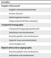
Doppler ultrasound was performed in 86 patients (85.2%). Among 85 patients with available flow measurements, the median preprocedural AVF flow was 1164 mL/min (IQR, 362 - 1673). Computed tomography was performed in 71 patients (70.3%), and subclavian vein involvement was observed in 51 patients (71.8%). Digital subtraction angiography was performed in all patients. The most commonly involved segment on angiography was the brachiocephalic vein, observed in 60 patients (59.4%), followed by the subclavian vein in 50 patients (49.5%). Severe stenosis and complete occlusion were present in 44 patients (43.5%) and 34 patients (33.7%), respectively (Table 2).
Table 2.
Imaging, Lesion, and Procedural Characteristics a
| Variables | Value |
|---|---|
| Doppler ultrasound | |
| Doppler ultrasound performed | 86/101 (85.2) |
| Venous stenosis | 21/86 (24.4) |
| Initial-segment stenosis | 16/86 (18.6) |
| Preprocedural AVF flow (mL/min) | 1164 (362 - 1673); n = 85 |
| Computed tomography | |
| Computed tomography performed | 71/101 (70.3) |
| Subclavian vein involvement | 51/71 (71.8) |
| Brachiocephalic vein involvement | 27/71 (38.0) |
| Superior vena cava involvement | 3/71 (4.2) |
| Lesion length (cm) | 2 (2 - 2); n = 65 |
| Digital subtraction angiography | |
| Brachiocephalic vein involvement | 60/101 (59.4) |
| Subclavian vein involvement | 50/101 (49.5) |
| Superior vena cava involvement | 8/101 (7.9) |
| One involved venous segment | 82/101 (81.2) |
| Two involved venous segments | 18/101 (17.8) |
| Three involved venous segments | 1/101 (1.0) |
| Moderate stenosis | 23/101 (22.8) |
| Severe stenosis | 44/101 (43.5) |
| Complete occlusion | 34/101 (33.7) |
| Degree of stenosis or occlusion (%) | 90 (90 - 100) |
| Lesion length (cm) | 3 (2 - 5) |
| Procedural access route | |
| Cephalic vein access | 82/101 (81.2) |
| Superficialized basilic vein access | 14/101 (13.9) |
| Femoral vein access | 4/101 (4.0) |
| Treatment modality | |
| Balloon angioplasty alone | 81/94 (86.2) |
| Balloon angioplasty plus stent placement | 13/94 (13.8) |
| Procedure and hospitalization | |
| Balloon inflation pressure (atm) | 12 (12 - 14; 10 - 24); n = 94 |
| Procedure time (min) | 115 (90 - 130); n = 81 |
| Postprocedural hospital stays (d) | 3 (2 - 4) |
| Total hospital stays (d) | 11 (7 - 17) |
a Values are expressed as n/N (%) or median (IQR). Lesion sites and procedural access routes were not mutually exclusive. Abbreviations: AVF, arteriovenous fistula; IQR, interquartile range.
The cephalic vein was the most frequently used procedural access route, recorded in 82 patients (81.2%). Among 94 patients who underwent balloon-based intervention, balloon angioplasty alone was performed in 81 patients (86.2%), whereas angioplasty combined with stent placement was performed in 13 patients (13.8%). The median balloon inflation pressure was 12 atm (IQR, 12 - 14; range, 10 - 24; n = 94). The median procedure time was 115 minutes (IQR, 90 - 130; n = 81), and the median postprocedural hospital stay was 3 days (IQR, 2 - 4) (Table 2).
Overall anatomical success was achieved in 91 patients (90.1%). Immediate angiographic results showed good venous flow in 69 patients (68.3%) and residual stenosis of less than 30% in 22 patients (21.8%). Anatomical failure occurred in 10 patients (9.9%), including 7 patients in whom the lesion could not be crossed, 2 patients with residual stenosis of 30% or greater, and 1 patient with venous rupture. Among anatomically successful cases, the median postprocedural AVF flow was 1126 mL/min (IQR, 762 - 1322) (Table 3).
Table 3.
Immediate Anatomical Success, 1-Month Clinical Assessment, and Early Complications a
| Variables | Value |
|---|---|
| Immediate angiographic result | |
| Good venous flow after intervention | 69/101 (68.3) |
| Residual stenosis < 30% | 22/101 (21.8) |
| Residual stenosis ≥ 30% | 2/101 (2.0) |
| Unable to cross the lesion | 7/101 (6.9) |
| Venous rupture | 1/101 (1.0) |
| Overall anatomical outcome | |
| Anatomical success | 91/101 (90.1; 95% CI, 82.7 - 94.5) |
| Anatomical failure | 10/101 (9.9; 95% CI, 5.5 - 17.3) |
| Postprocedural AVF flow | |
| AVF flow (mL/min) | 1126 (762 - 1322); n = 91 |
| AVF flow range (mL/min) | 450 - 3101 |
| 1-month clinical assessment | |
| Decreased arm edema | 68/78 (87.2; 95% CI, 78.0 - 92.9) |
| Decreased arm pain | 54/68 (79.4; 95% CI, 68.4 - 87.3) |
| Bruit at AVF anastomosis present or restored | 71/101 (70.3) |
| Decreased superficial venous collateral circulation | 16/26 (61.5; 95% CI, 42.5 - 77.6) |
| Hemodialysis through the treated access feasible | 74/101 (73.3) |
| Early complications | |
| No early complication | 92/101 (91.1) |
| Hematoma | 8/101 (7.9) |
| Bleeding | 1/101 (1.0) |
a Values are expressed as n/N (%) or median (IQR) unless otherwise indicated. Abbreviations: AVF, arteriovenous fistula; IQR, interquartile range.
At the 1-month follow-up after intervention, arm edema decreased in 68 of 78 patients with baseline edema (87.2%; 95% CI, 78.0 - 92.9), arm pain decreased in 54 of 68 patients with baseline pain (79.4%; 95% CI, 68.4 - 87.3), and superficial venous collateral circulation decreased in 16 of 26 patients with baseline collateral veins (61.5%; 95% CI, 42.5 - 77.6). Bruit at the AVF anastomosis was present or restored in 71 patients (70.3%), and hemodialysis through the treated access was achieved in 74 patients (73.3%). No early complications were recorded in 92 patients (91.1%). Hematoma occurred in 8 patients (7.9%), and bleeding occurred in 1 patient (1.0%) (Table 3). In exploratory univariable analysis, longer lesion length on digital subtraction angiography was associated with anatomical failure (OR = 1.32; 95% CI, 1.09 - 1.59; P = 0.004).
5. Discussion
This study described the clinical profile, imaging characteristics, procedural features, and early outcomes of endovascular treatment for central venous stenosis or occlusion in maintenance hemodialysis patients at a tertiary hospital in Vietnam. The main findings were that most patients presented with symptoms of venous hypertension; the brachiocephalic and subclavian veins were the most frequently involved segments on angiography; balloon angioplasty was the predominant treatment modality; and anatomical success was achieved in 90.1% of patients. At 1 month after intervention, symptom improvement was recorded in most patients with baseline arm edema or pain, whereas early complications mainly consisted of access-site hematoma and bleeding.
The patients in this cohort were middle-aged to older maintenance hemodialysis patients, with a mean age of 54.97 years. This profile is comparable to large hemodialysis cohorts in which the mean age was approximately 54 years (10, 11). Hypertension was the most common comorbidity, followed by diabetes mellitus and coronary artery disease. Patients receiving maintenance hemodialysis often have a substantial chronic disease burden, including impaired health-related quality of life, poor sleep quality, and malnutrition (10-12). In this clinical context, central venous stenosis or occlusion may further compromise dialysis access function and exacerbate symptoms related to venous hypertension.
The dominant clinical manifestations in this study were arm edema and arm pain. These findings are consistent with the mechanism of central venous obstruction, in which impaired central venous outflow increases venous pressure in the ipsilateral limb and dialysis access circuit. Tabriz and Arslan described ipsilateral extremity swelling or pain, venous collateral development in the neck, chest, or arm, difficult cannulation, prolonged bleeding after needle removal, and abnormal thrill or bruit as typical clinical findings of central venous stenosis or occlusion in dialysis patients (1). Gür et al. reported arm swelling in 97.0% of hemodialysis patients with central venous occlusion, with additional face or neck swelling in 38.0% and inability to puncture the access in 12.6% (2). Nasser et al. also described ipsilateral swelling of the face, neck, or arm, increased venous pressure during hemodialysis, and access failure as common manifestations of central venous occlusion (3). The lower proportion of arm edema in our study compared with that reported by Gür et al. may be partly explained by differences in case mix, as our cohort included both stenosis and complete occlusion rather than central venous occlusion alone.
Angiography remained central to defining lesion anatomy in this study. On digital subtraction angiography, the brachiocephalic vein was the most frequently involved segment, followed by the subclavian vein. Severe stenosis and complete occlusion accounted for most lesions. These findings are broadly consistent with previous reports showing that central venous lesions in hemodialysis patients commonly involve the brachiocephalic and subclavian veins (2, 4). Tian et al. analyzed 385 lesions in 309 patients with symptomatic central venous stenosis and emphasized that lesion distribution, catheter-related injury, and access-related factors may influence clinical outcomes (4). In our study, longer lesion length on digital subtraction angiography was associated with anatomical failure in an exploratory univariable analysis. This finding is clinically plausible because longer lesions may be more difficult to cross and dilate; however, it should be interpreted cautiously because the number of anatomical failures was small and no multivariable model was constructed.
Balloon angioplasty alone was the main treatment modality in our cohort, whereas stent placement was used in a smaller proportion of patients. This reflects a conservative, stepwise endovascular strategy in which angioplasty is generally performed first and stenting is reserved for selected cases such as recoil, significant residual stenosis, recurrent stenosis, or angioplasty-resistant lesions (1, 5-7). The anatomical success rate of 90.1% in our study is within the range reported in the literature. Gür et al. achieved technical success in 94.0% of patients with central venous occlusion (2), whereas Tian et al. reported technical success of 91.22% in patients with single-segment lesions treated by endovascular therapy (4). Nasser et al. reported technical and clinical success in 90.0% of patients undergoing sharp recanalization for chronic central venous occlusion (3). Direct comparisons should be interpreted cautiously because studies differ in patient selection, lesion severity, lesion length, treatment technique, stent use, and definitions of technical or anatomical success.
At 1 month after intervention, clinical improvement was recorded in patients with baseline symptoms. Arm edema decreased in 68 of 78 patients with baseline edema, arm pain decreased in 54 of 68 patients with baseline pain, and hemodialysis through the treated access was feasible in 74 patients. These findings suggest short-term symptom relief and access usability in a selected treated cohort. However, because this was an observational study without a control group, these early postintervention findings should not be interpreted as definitive evidence of treatment effectiveness. Bakken et al. reported 12-month primary patency rates of 29% after primary angioplasty and 21% after primary stenting (5). In contrast, selected stent or covered-stent series reported higher patency in specific patient groups (6, 7). Therefore, continued surveillance after early anatomical success remains necessary.
The median AVF flow was 1164 mL/min before intervention and 1126 mL/min after intervention among anatomically successful cases. This apparent lack of increase in median flow should be interpreted cautiously. Preprocedural and postprocedural AVF flow values were summarized using available data with different denominators and were not analyzed as a complete paired dataset in the present report. Therefore, these descriptive values should not be used to infer a statistically significant change in access flow after intervention. Clinical assessment of access usability, symptom improvement, and angiographic outcome may better reflect the early findings of this study than comparison of unpaired flow summaries.
Early complications were limited to hematoma and bleeding in this report. Hematoma occurred in 8 patients, and bleeding occurred in 1 patient. No early complication was recorded in 92 patients. Procedure-related venous rupture was classified as anatomical failure when it resulted in failure of the intervention, whereas early complications within 1 month were reported separately as access-site hematoma and bleeding. Nasser et al., in a cohort requiring sharp recanalization, reported immediate postoperative complications in 17.5% of patients, including major complications such as hemothorax and pneumothorax (3). The lower complication rate in our cohort may reflect differences in lesion complexity, recanalization technique, and the limited use of more aggressive approaches.
5.1. Study Limitations
This study has several limitations. First, this was a single-center retrospective observational analysis conducted within a larger longitudinal project; therefore, selection bias and information bias could not be excluded. Second, only symptomatic patients who underwent endovascular treatment were included; therefore, the findings cannot be generalized to all hemodialysis patients with central venous stenosis or occlusion. Third, treatment selection was based on clinical and procedural judgment rather than randomization; therefore, comparisons between balloon angioplasty and stent placement were not performed. Fourth, some variables had missing observations, and variable-specific denominators were reported. Fifth, preprocedural and postprocedural AVF flow measurements were not analyzed as a complete paired dataset. Sixth, only 10 anatomical failures occurred, limiting the ability to perform multivariable analysis. Finally, this article focused on immediate anatomical success and 1-month clinical outcomes; mid-term and long-term patency, restenosis, reintervention, and access survival require separate follow-up analysis.
5.2. Conclusions
In maintenance hemodialysis patients with symptomatic central venous stenosis or occlusion, lesions mainly involved the brachiocephalic and subclavian veins and commonly presented with arm edema or pain. Endovascular treatment, predominantly balloon angioplasty, achieved immediate anatomical success in most patients and was followed by clinical improvement within 1 month after intervention. Early complications consisted of access-site hematoma and bleeding. Further follow-up is needed to evaluate restenosis, reintervention, and long-term patency.