A female patient was referred to our interventional radiology department for management of inadvertent arterial catheterization after RIJ cannulation. The patient was a 57-year-old white woman with a history of type II diabetes, hypertension, and end stage renal disease (ESRD). She was on hemodialysis three times per week through the formerly placed right jugular vein catheter which was malfunctioned. Earlier that day, the vascular surgeon placed the tunneled RIJ hemodialysis catheter under general anesthesia using ultrasonography guidance and removed the antecedent catheter in the operating room. This procedure was done in a scheduled nonemergency situation. Pulsatile bright red blood backflow through the newly placed catheter was noted in the recovery room. Our team was consulted for removal of the misplaced catheter.
The patient had stable vital signs and was conscious at arrival. Physical examination was unremarkable unless for a bruit over the supraclavicular region, multiple scars and other cutaneous injuries on the right side of the neck due to previous cannulation attempts. Distal pulses were intact; the body mass index (BMI) was 29 and the neck anatomy appeared normal. 12-Fr tunneled double lumen hemodialysis catheter (Arrow; Teleflex, Pennsylvania, United States) was placed through the right internal jugular approach. The patient had one episode of significant bleeding from the puncture site in the recovery unit which dropped her blood pressure and required fluid resuscitation and direct pressure in order to control the hemorrhage. Post catheterization chest x-ray revealed that the catheter tip was not in the venous system; no other significant finding was noted (e.g. pleural and/or pericardial effusion, and pneumothorax).
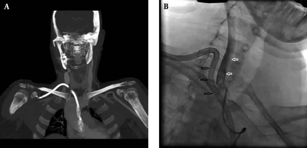
CT angiography was performed after suspected inadvertent catheter placement. The catheter entered the subclavian artery slightly distal to the brachiocephalic bifurcation and proximal to the right vertebral artery origin (
Figure 1A).
CT angiogram (CTA) of a 57-year-old female patient with significant bleeding after hemodialysis catheter placement showing the catheter (black arrows) penetrating in both internal jugular vein and subclavian artery (white arrows). A, The tip of the catheter is in the ascending aorta over the aortic valve. B, Angiogram confirms CTA findings.
After administrating 450 milligrams of clopidogrel orally, the patient underwent conventional angiography through femoral artery access under regional anesthesia in order to remark possible percutaneous/endovascular management of the lesion. CT angiography findings were confirmed and the carotid-vertebral systems were normal (
Figure 1B). We obtained the written informed consent from the patient and his family concerning potential vertebrobasilar circulation stroke and explained other probable complications including myocardial infarction, excessive uncontrollable hemorrhage, and possible open surgery. The vascular surgical team and their operating theatre were prepared for emergency situations.
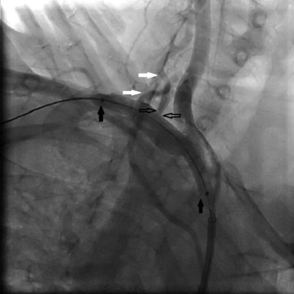
Angiogram after hemodialysis catheter removal showing the balloon occlusion catheter is in place (black arrows), but the arteriovenous communication is still ongoing (open black arrows). Right vertebral artery (white arrows) is also noted.
First, we decided to attempt both temporary balloon occlusion and external compression simultaneously to obtain hemostasis. Removing the catheter and applying pressure without any other intervention was not an option since there was no direct pressure route and the arterial injury was remarkable. We inflated a 6 × 60 mm PTA balloon catheter (FoxCross; Abbott Vascular, Illinois, United States) over the dialysis catheter entry area. At the same time, we withdrew the dialysis catheter and applied external compression on the affected site for 20 minutes under fluoroscopic guidance. This procedure failed to seal the injury and the subsequent angiogram revealed the 4 mm right subclavian artery defect 13mm distal to the brachiocephalic bifurcation and 9 mm proximal to the right vertebral artery origin. This defect was in connection with the jugular vein and consistent with high flow fistula formation (
Figure 2).
Right side subclavian artery branched off the vertebral artery 22 mm from its origin. Since the left vertebral pathway was the dominant route for posterior circulation and it had no obvious arterial disease on antecedent angiographic examination, in this case, it was not necessary to perform the right vertebral artery occlusion tolerance test prior to sacrificing.
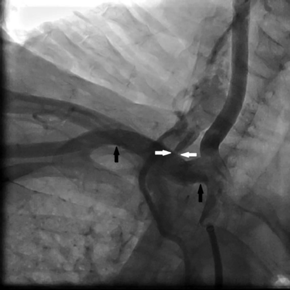
The uppermost arterial diameter at the site of the right subclavian arterial defect was 7 mm at angiography image. Given that, after intravenous administration of 5000 units of heparin, we introduced 7 × 37 mm balloon-expandable covered peripheral stent graft (BeGraft; Bentley, Hechinger, Germany) transfemorally and inflated it over the arterial laceration. Having in mind the need for exact stent placement, controlled deployment, and flexible sizing, we preferred balloon-expandable covered stent over the self-expandable stent. Also, using balloon expansion helps to further expand the stent by using larger balloons. The proximal end of the stent was at the right subclavian artery origin and 37 mm away from the origin was the distal end. The placed stent covered both the arteriovenous fistula and vertebral artery. This intervention also failed. The angiogram revealed continuous extravasation and fistulous communication despite placement of the stent graft, possibly due to both small stent diameter and short stent length selection. The placed stent did not migrate distally (
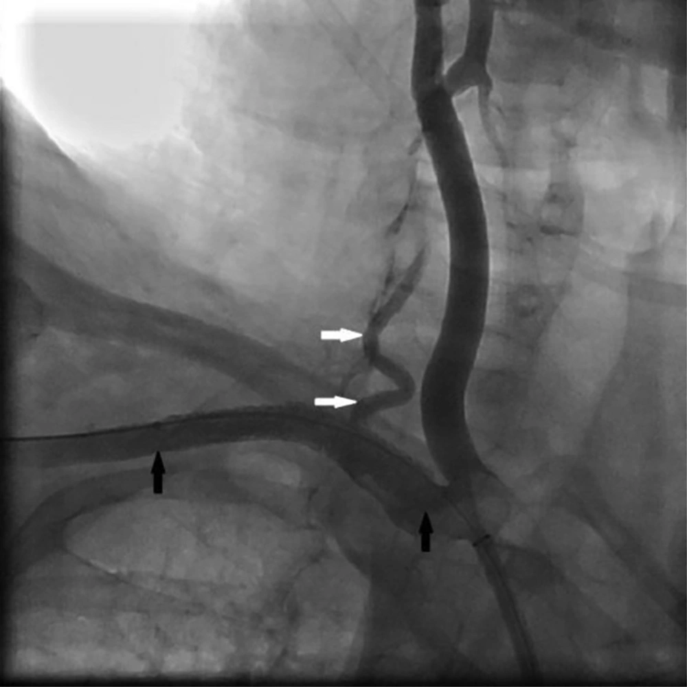
Figure 3). So, we chose a wider and longer stent and the second 8 × 60 mm balloon expandable covered peripheral stent (Atrium Advanta V12; Maquet Getinge Group, Rastatt, Germany) was placed over the same area again. The proximal end of the stent was at the right subclavian artery origin and 60 mm away from the origin was the distal end. Finally, total occlusion of the fistula, as well as patency of the right subclavian artery was achieved (
Figure 4).
First stent has landed in its proper site (black arrows) but the arteriovenous connection is not sealed (white arrows)
Angiography after successful management shows patent subclavian artery and stent in place (black arrows). There is no extravasation. Right vertebral artery (white arrows) is also noted.
After successful treatment, we started clopidogrel for the patient along with continuing her other medication including lifelong aspirin. The patient was discharged from the hospital after 4 days without any endovascular treatment-related complication. The patient was appointed to regular visits to evaluate new arterial insufficiency symptoms and physical examination of his right hand. Clopidogrel was discontinued after two months. Imaging follow-up after 12 months revealed no significant complications (kinking, fracture, displacement, and significant stenosis) on CT angiogram.