Central vein stenosis or occlusion is a common complication of the chronic hemodialysis patient, resulting in considerable edema of the arm with vascular access that is unable to drain normally. Surgical repair of these central veins can be difficult. That is why the percutaneous approach is preferable to the surgical approach (
20). Angioplasty alone may represent a reasonable option for the treatment of central venous lesions. Stents are thought to add a beneficial effect to angioplasty by limiting the elastic recoil present in compliant veins, excluding damaged and dissected intravascular tissues, and acting as an intravascular support to counteract extrinsic compression (
12). This is especially relevant for the central venous lesion, described as having high elastic recoil and poor results with PTA alone (
12). It is natural to expect that reports concerning PTA as the only management option would have lower patency rates.
Previous findings in the patency rates of central vein obstruction in PTS vary widely from ours. We believe that the main reasons for this variation are the different PTS protocols, types of stents, study populations, age of access, access thrombosis at the time of intervention, and veins treated (
8,
10,
12,
21,
22).
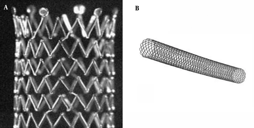
A stent can be classified as a closed-cell stent or an open-cell stent, depending on the density of struts (
15). Closed-cell stents are characterized by small free cell areas between struts, whereas open-cell stents have larger uncovered gaps (
Figure 3) (
23). Closed-cell stents with a small cell size have a dense, metallic mesh and therefore, may provide more effective plaque coverage and reduce the risk of particle embolization (
16). However, closed-cell stents are known to be less flexible and more rigid and are more likely to be used in straight morphologies (
16). In contrast, open-cell stents are flexible; kinked lesions are ideally treated with these stents (
16).
Examples of an open-cell stent (A) and a closed-cell stent (B)
New stents were designed to incorporate visibility, flexibility, and expandability, permitting the treatment of the most complex venous lesion through endovascular means. Potential advantages of this approach include its less invasive nature, safety, ability to be done on an outpatient basis, better preservation of native veins, and, most important, its association with greater patient satisfaction and less discomfort (
12).
Previous studies using closed-cell stents (Wallstent, Boston Scientific, Natick, MA) have failed to demonstrate improved survival or patency compared with angioplasty (
7,
11,
12). Reports concerning the use of the Wallstent in central vein obstruction are common (
13,
24,
25). With Wallstents, technical success was between 96% and 100%. Primary patency ranged from 42% to 84% at 6 months, but was less than 31% at 12 months in four studies (
8,
10-
12), with Haage et al. (
13) demonstrating 56% primary patency at 12 months. Unfortunately, no one has since reported results as successful as those that the Haage group did.
Early experience with open-cell stents (Zilver nitinol stent, Cook, Bloomington, IN, USA) for central venous occlusion appears to indicate that they confer longer mid-term patency than historically observed results for central venous occlusion and central venous stenosis using Wallstents (
22). In a study conducted by Vogel and Parise (
14), use of the SMART stent demonstrated 67% primary patency for central venous stenosis at 12 months. In a study performed by Rajan and Saluja (
22), use of the Zilver nitinol stent (Cook, Bloomington, IN, USA) demonstrated 66.7% primary patency for central venous stenosis at 6 and 12 months.
There have been two reports comparing the closed-cell stent with open-cell stent (
26,
27). The authors of those reports reported no significant difference between the patency of the Wallstents and the nitinol-based Memotherm or Luminexx stent in their small groups (
26,
27).
We have reported our large group study in stent treatment of central vein stenosis or occlusion with open-cell stent or closed-cell stent. Primary patency rates and clinical outcomes were comparable for the two-stent groups.
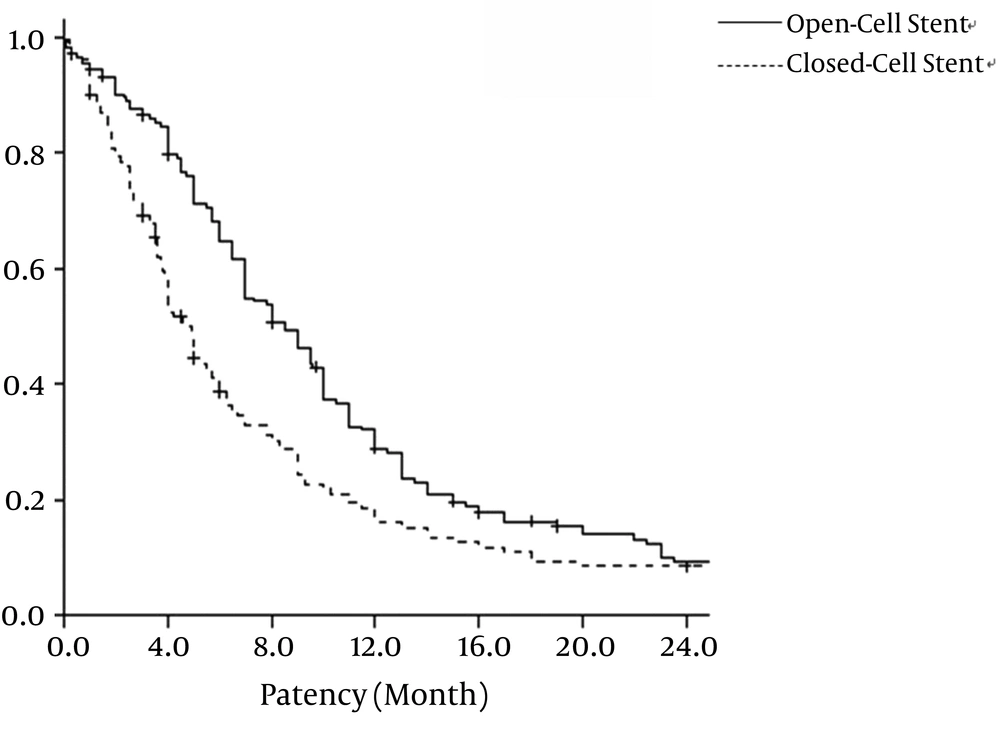
We found that the primary patency rate of open-cell stent was significantly higher than that of the closed-cell stent. The 6-month and 12-month primary patency rates with the open-cell stent were 64.8% and 28.8%, respectively, and primary patency rates with closed-cell stent were 38.7% and 16.0%, respectively. Open-cell stents and closed-cell stents had mean patency rates of 10.9 ± 0.80 months and 8.5 ± 10.87 months, respectively (P =0.002). The reported patency rate of 66.7% at 6 and 12 months following nitinol stent placement by Rajan and Saluja (22) might be the result of a small patient population (n = 6).
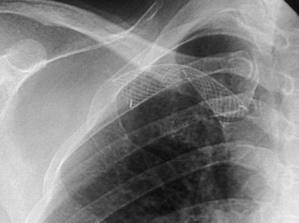
In this study, the mean patency rate of the subclavian vein with open-cell stent was 11.1 ± 1.66 months and the mean patency rate with closed-cell stent was 4.9 ± 0.18 months. There is a significant difference in the subclavian vein stenosis or occlusions between the open-cell stent group and closed-cell stent group in this study (P < 0.001). Our results might be related to characteristics of the open-cell stent, including flexibility, radial strength, and expansibility. The open-cell stent has physical properties that differ from those of the closed-cell stent. The closed-cell stent appears to have problems with its shortening and migrating during respiration (
Figure 4) (
8,
10,
11,
13).
A 55-year-old patient with brachio-cephalic fistula. A, Closed-cell stent is placed at left innominate vein. B, Fistulogram obtained 7 months after stenting shows shortening of the stent.
The efficacy of stent deployment appears to vary by the vascular location of the stenotic lesion. Recently, nitinol stents have been introduced and have several physical characteristics that may confer longer patency compared with Wallstents (
28). Nitinol, an alloy of nickel and titanium, exists in two temperature-dependent forms, which are predetermined by adjusting the ratio of nickel and titanium and through high-temperature heating. When nitinol assumes its higher-temperature form, it expands to its predetermined size and becomes more rigid. Nitinol is also superelastic in that it will deform its shape but return to its original configuration when an external force is applied and then removed (
29-
31). Shape memory and superelasticity allow for an improved apposition of the stent along the vessel wall and maintenance of radial strength (
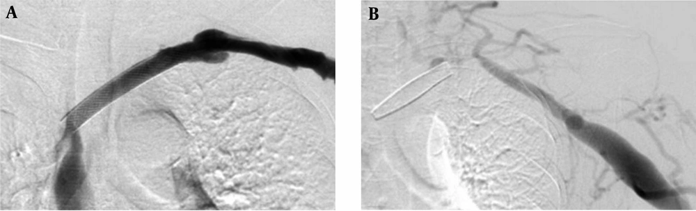
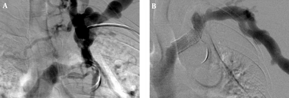
Figure 5).
A 46-year-old patient with brachio-basilic fistula. A, Fistulogram shows segmental stenosis with severe tortuosity and pronounced collateral flow at left innominate vein. B, Fistulogram shows restoration of flow and vanishing of collateral vessels after open-cell stent placement.
The Wallstent is constructed of Elgiloy, the free ends are sharp and capable of embedding into the vessel wall. Eccentric loading of the Wallstent such as that produced by a stenosis results in concentric narrowing beyond the point at which the load is applied, making it susceptible to reduced wall contact and decreased radial strength (
32). Poor or incomplete wall contact appears to be a risk factor for in-stent stenosis (
33,
34), and decreased radial strength (
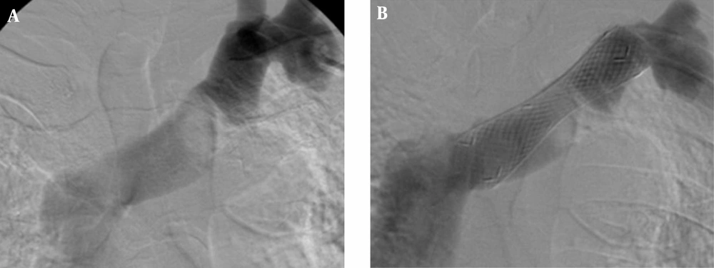
29). In this study, there was a case of incomplete wall contact in the closed-cell stent group for stenosis at the left innominate vein (
Figure 6). Closed-cell stent has also been observed to migrate and foreshorten in central venous segments (
Figure 4) (
8,
10,
11,
13). In addition, two cases of central stent migration were observed in patients with closed-cell stent in this study.
A 71-year-old-man with radio-cephalic fistula and left upper arm swelling. A, Fistulogram shows stenosis in left innominate vein. B, Fistulogram obtained after closed-cell stent placement shows incomplete contact of the stent to the vessel wall.
Open-cell stents are structured to allow for minimal foreshortening. Open-cell stents have become standard practice over closed-cell stents, mainly due to easier and more precise placement, non-shortening of the open-cell stents, and a longer vascular patency. The superelasticity and shape-memory characteristics of the nitinol stent improve flexibility, which also improve opposition to the vessel wall (
Figure 5).
Depending on the density of the bridges between the different rings, nitinol stents can be classified into stents with a closed-cell or an open-cell configuration. Flexibility and scaffolding are key characteristics derived from stent design (
15). Flexibility depends on the stent’s ability to conform the vessel tortuosity in the deployed state. Closed-cell stents are rigid, less flexible and may develop kinks and incomplete deployment in the tortuous vessel. Conversely, stents with an open-cell configuration conform best to angulated vessels or tortuous anatomy. Stents with a flexible and conformable open-cell configuration are preferred in vessels that are angulated or have a tortuous anatomy.
Patients in this study group did not experience any major morbidity or mortality, except for two cases of stent migration and minor complications.
There are several limitations to the present study. First, this study was retrospective and had mixed group patients among those with or without a history of hypertensio, diabetes mellitus, peripheral vascular disease, and use of tobacco, so their possible effects on patency rates could not be assessed. Although a history of diabetes, hypertension, duration of chronic renal failure and history of previous catheter insertion is more common in the open-cell stents group, the open-cell stent is more effective for treatment of central vein stenosis or occlusion in hemodialysis patients.
Second, differences in baseline and lesion characteristics may have influenced the observed results. It is unknown what role concurrent stenosis along the access circuit may have on patency following intervention for central venous occlusion.
In conclusion, the open-cell stent is effective for the treatment of central vein stenosis or occlusion in hemodialysis patients who have incomplete PTA results. These results suggest the use of open-cell stent for the treatment of central vein stenosis or occlusion in hemodialysis patients.