1. Background
Cerebral palsy (CP) is a major childhood neurodevelopmental disorder characterized by permanent, nonprogressive deficits in movement, posture, and coordination. It is attributed to injury to or abnormal development of the immature brain during fetal, neonatal, or early postnatal life. Associated cognitive, speech, sensory, or behavioral problems may further affect children, families, and healthcare systems (1, 2).
Globally, CP occurs in approximately 2 - 3 per 1,000 live births (3). It is reported more frequently among children born preterm or with low birth weight (4, 5). Its relatively stable prevalence despite improved neonatal care suggests multifactorial pathways (5). Iranian estimates range from 1.5 to 2.5 per 1,000 births; however, limited data from southeastern regions remain a barrier to context-specific planning for screening, referral, rehabilitation, and supportive care services.
Several maternal, perinatal, and neonatal characteristics, including infection, placental problems, hypoxic events, seizures, and hyperbilirubinemia, have been documented in children with CP (6-8). However, CP often lacks a single clear cause, consistent with the complex and often overlapping nature of documented clinical exposures and developmental vulnerability during brain development (9, 10). This complexity supports an integrated descriptive evaluation and cautious interpretation of maternal, perinatal, neonatal, clinical, and radiological findings without assuming causal pathways or independent effects.
MRI helps define patterns of brain lesions and the possible timing of injury in CP (10-13). Reported abnormalities include periventricular leukomalacia, cystic necrosis, basal ganglia injury, and cortical atrophy (14, 15). Lesion burden may vary across clinical phenotypes and levels of functional involvement and may guide clinical planning. However, clinical-radiological patterns should be interpreted cautiously in descriptive cross-sectional studies that do not establish independent or causal relationships.
Most studies examine maternal and perinatal characteristics, imaging, or clinical features separately, and integrated regional data remain limited, particularly in southeastern Iran. Assessing observed co-occurrence patterns among clinical phenotypes, documented maternal and perinatal characteristics, and MRI findings may support care planning in underserved settings. In descriptive studies, such information should be interpreted as a summary of coexisting clinical, historical, and radiological features within a defined cohort rather than as evidence of causal, independent, or prognostic relationships. Therefore, the present study was designed to provide a descriptive profile rather than to test etiological, predictive, or prognostic hypotheses.
2. Objectives
Ali Asghar Hospital, a tertiary referral center in southeastern Iran, served as the study setting. The primary objective was to describe clinical phenotypes, associated comorbidities, documented maternal and perinatal characteristics, and brain MRI findings among children with CP referred to the center. The primary descriptive outcomes were CP motor types, topographical patterns, and brain MRI findings; secondary descriptive variables included developmental comorbidities and documented maternal, prenatal, perinatal, and neonatal characteristics. No causal, prognostic, or independent association objective was specified.
3. Methods
3.1. Study Design and Setting
This descriptive cross-sectional study included 103 children with CP who were referred to Ali Asghar Hospital, a tertiary referral center in southeastern Iran, during 2020 - 2021. Motor and topographical clinical patterns, documented maternal and perinatal characteristics, comorbidities, and MRI findings were summarized. The study was not designed to assess causal, prognostic, or independent associations. A census sampling approach was used, and all children who met the eligibility criteria during the study period were enrolled. Reporting of this observational study was guided by relevant STROBE recommendations (16).
3.2. Participants and Eligibility
Eligibility criteria included a confirmed clinical diagnosis of CP by a pediatric neurologist, available and interpretable brain MRI reports, sufficiently documented medical records for the study variables, and written informed consent from a parent or legal guardian. Exclusion criteria were progressive neurological or neurodegenerative disorders, major congenital anomalies unrelated to CP, progressive genetic or metabolic disorders, incomplete key clinical data, or unavailable or uninterpretable MRI reports. Children with suspected CP but insufficient documentation to confirm the diagnosis or classify the main clinical and radiological variables were not included.
3.3. Data Collection and Variables
Information was obtained from medical records, MRI reports, and parent interviews using a researcher-designed checklist. The form was reviewed and approved for content validity by a pediatric neurologist and a neuroradiologist and included demographic, clinical, perinatal, and imaging items. When discrepancies or ambiguities were identified, data were cross-checked against hospital records, clinical notes, discharge summaries, MRI reports, and parental reports. Variables were recorded only when sufficient supporting information was available.
Recorded variables included age, sex, maternal and obstetric history, neonatal events, neonatal intensive care unit (NICU) admission, hypoxia/asphyxia, seizures, jaundice requiring treatment, developmental delay, comorbidities, CP subtype, and MRI abnormalities. These variables were considered documented clinical characteristics rather than confirmed causal risk factors. Perinatal hypoxia/asphyxia was defined as documented birth asphyxia, neonatal resuscitation, a low Apgar score when available, or physician-documented hypoxic-ischemic events. Neonatal seizures were defined as clinically documented seizures during the neonatal period, with or without electroencephalographic confirmation. Neonatal jaundice was recorded when hyperbilirubinemia requiring phototherapy or exchange transfusion was documented. Developmental delay was recorded when a delay in one or more developmental domains was documented by the treating physician or in the clinical record, with parental reports used as supplementary information when available; no standardized developmental assessment tool was used.
CP was classified by motor type and topographical pattern. Motor types included spastic, dyskinetic, ataxic, hypotonic, and mixed forms, whereas topographical patterns included hemiplegia, diplegia, and quadriplegia where applicable. Comorbidities, including motor, speech, cognitive/developmental delay, epilepsy, and visual or hearing problems, were obtained from documentation and parent reports. Functional severity scales such as the Gross Motor Function Classification System (GMFCS) were not consistently available and therefore were not analyzed.
MRI data were extracted from official routine radiology reports. No study-specific MRI acquisition or blinded reinterpretation was performed. Reported abnormalities were recorded when available, including periventricular leukomalacia, cystic encephalomalacia/porencephaly, basal ganglia involvement, cerebral atrophy, vascular lesions, ventriculomegaly, and gliosis. When more than one abnormality was reported, all documented abnormalities were recorded when available; therefore, MRI categories were not mutually exclusive. Because MRI categorization was based on routine reports rather than standardized independent rereading, MRI findings were interpreted descriptively.
3.4. Statistical Analysis
SPSS version 26 was used. Continuous data were presented as the mean ± standard deviation, and categorical data as frequency and percentage. Demographic characteristics, developmental comorbidities, documented maternal, prenatal, perinatal, and neonatal characteristics, motor types of CP, topographical patterns, and brain MRI findings were summarized descriptively. No formal inferential tests, P values, causal, prognostic, independent association, or multivariable analyses were performed. All findings were interpreted as descriptive summaries of documented clinical, historical, and radiological features within this referral-center cohort.
3.5. Ethical Considerations
Ethics approval was obtained from Zahedan University of Medical Sciences (Ethics Code: IR.ZAUMS.REC.1400.329), and written informed consent was obtained from parents or legal guardians. All collected data were anonymized and handled confidentially in accordance with ethical research standards.
4. Results
4.1. Demographic and Developmental Characteristics
A total of 103 children with CP who were referred to Ali Asghar Hospital during 2020 - 2021 were assessed and included in the final descriptive analysis. The mean age was 2.7 ± 3.5 years, with an age range of 21 days to 13 years. The cohort comprised 70 boys (68.0%) and 33 girls (32.0%). Cesarean and vaginal deliveries were recorded in 53 (51.5%) and 50 (48.5%) children, respectively (Table 1). These findings describe the composition of the referred cohort and were not interpreted as independent determinants of the CP phenotype.
Table 1.
Demographic and Selected Baseline Characteristics of Children with Cerebral Palsy (N = 103) a
| Variables | Value |
|---|---|
| Age (y), mean ± SD | 2.7 ± 3.5 |
| Age (y), median (IQR) | 1.8 (0.6 - 4.5) |
| Age range | 21 (d) –13 (y) |
| Male sex | 70 (68.0%) |
| Female sex | 33 (32.0%) |
| Cesarean delivery | 53 (51.5%) |
| Vaginal delivery | 50 (48.5%) |
a Values are expressed as mean ± SD, median (IQR) or No. (%) unless otherwise indicated. The age distribution was skewed toward younger ages due to the predominance of infants and young children.
Neurodevelopmental delay, motor developmental delay, and speech developmental delay were documented in 39 (37.9%), 33 (32.0%), and 24 (23.3%) children, respectively. The mean birth weight was 2443.6 ± 547.7 g, and the mean head circumference at birth was 33.9 ± 1.9 cm (Table 2). Developmental impairments were recorded as associated clinical features based on available clinical records and, where applicable, parental reports.
Table 2.
Developmental Characteristics and Birth Anthropometric Measures (N = 103) a
| Indicator | Value |
|---|---|
| Neurodevelopmental delay | 39 (37.9) |
| Motor developmental delay | 33 (32.0) |
| Speech developmental delay | 24 (23.3) |
| Birth weight (g) | 2443.6 ± 547.7 |
| Head circumference at birth (cm) | 33.9 ± 1.9 |
a Values are expressed as No. (%) or mean ± SD. Developmental disorders were defined based on clinical neurological assessment and medical record review.
4.2. Prenatal, Perinatal, Neonatal, and Maternal Characteristics
Documented perinatal and neonatal features included preterm birth in 19 children (18.4%), birth asphyxia in 31 (30.1%), neonatal seizures in 31 (30.1%), and NICU admission for more than 3 days in 36 (35.0%). Neonatal jaundice, infection, and small-for-gestational-age status were noted in 25.2%, 12.6%, and 4.9% of cases, respectively (Table 3). These variables were summarized descriptively within the cohort rather than as confirmed causal risk factors.
Table 3.
Documented Prenatal, Perinatal, and Neonatal Characteristics (N = 103)
| Characteristic | No. (%) |
|---|---|
| Preterm birth | 19 (18.4) |
| Birth asphyxia | 31 (30.1) |
| NICU admission > 3 (d) | 36 (35.0) |
| Neonatal seizures | 31 (30.1) |
| Neonatal jaundice requiring treatment | 26 (25.2) |
| Neonatal infection | 13 (12.6) |
| Small for gestational age (SGA) | 5 (4.9) |
Consanguinity was reported in 69 cases (67.0%). Gestational diabetes mellitus and thyroid disorders were the most frequently recorded maternal conditions, affecting 14.6% and 8.7% of cases, respectively. Multiple pregnancy was reported in 12.6% of cases, and previous abortion in 6.8% (Table 4). Maternal and familial characteristics were presented descriptively, and no independent maternal predictors were inferred.
Table 4.
Maternal and Socio-Familial Characteristics (N = 103)
| Variables | No. (%) |
|---|---|
| Consanguineous marriage | 69 (67.0) |
| Gestational diabetes | 15 (14.6) |
| Thyroid disorders | 9 (8.7) |
| Multiple pregnancy | 13 (12.6) |
| History of abortion | 7 (6.8) |
4.3. MRI Findings and Cerebral Palsy Phenotypes
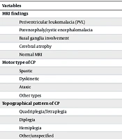
Periventricular leukomalacia was the most common MRI finding, reported in 35.0% of children, followed by porencephaly or cystic encephalomalacia (32.0%), basal ganglia involvement (13.6%), and cerebral atrophy (6.8%). Normal MRI findings were reported in 1.9%. Spastic CP was the predominant motor type, accounting for 74.8% of cases, and quadriplegia was the most frequent topographical pattern (Table 5). MRI findings were extracted from routine clinical radiology reports; when multiple abnormalities were documented, categories were recorded as non-mutually exclusive descriptive neuroimaging patterns. The observed frequencies of spastic CP, quadriplegia, and periventricular leukomalacia describe the clinical-radiological profile of this referral-center cohort and should not be interpreted as evidence of causal, prognostic, or independent relationships.
Table 5.
MRI Findings, Motor Types, and Topographical Patterns of Cerebral Palsy (N = 103) a
| Variables | No. (%) |
|---|---|
| MRI findings | |
| Periventricular leukomalacia (PVL) | 36 (35.0) |
| Porencephaly/cystic encephalomalacia | 33 (32.0) |
| Basal ganglia involvement | 14 (13.6) |
| Cerebral atrophy | 7 (6.8) |
| Normal MRI | 2 (1.9) |
| Motor type of CP | |
| Spastic | 77 (74.8) |
| Dyskinetic | 14 (13.6) |
| Ataxic | 4 (3.9) |
| Other types | 8 (7.7) |
| Topographical pattern of CP | |
| Quadriplegia/Tetraplegia | 33 (32.0) |
| Diplegia | 25 (24.3) |
| Hemiplegia | 19 (18.4) |
| Other/unspecified | 26 (25.2) |
a Values are expressed as No. (%). MRI categories were not mutually exclusive; therefore, children could have more than one documented MRI finding. MRI findings were based on routine reports. Other motor types included mixed, hypotonic, extrapyramidal, and dyskinetic-mixed forms. Other/unspecified topographical patterns included unclassified forms.
Overall, these findings provide a descriptive summary of demographic characteristics, developmental impairments, documented prenatal, perinatal, and neonatal characteristics, maternal and socio-familial characteristics, CP motor types, topographical patterns, and brain MRI findings in this referral-center cohort. No missing values were identified for variables entered into the final analysis, and all results are reported descriptively without causal, independent, or prognostic inference.
5. Discussion
Cerebral palsy is a leading cause of motor disability in childhood and involves nonprogressive but heterogeneous disturbances affecting motor, cognitive, communication, social, and family-related domains (1, 17). By jointly examining clinical features, documented maternal and perinatal characteristics, MRI patterns, and topographical subtypes, the present study provides an integrated descriptive profile of children with CP referred to a tertiary center in southeastern Iran. In this referral-center cohort, CP was accompanied by developmental impairments, neonatal events, maternal conditions, and diverse neuroimaging abnormalities. Given the cross-sectional design and the absence of a comparison group, these findings should be interpreted as descriptive observations rather than as evidence of causal pathways, prognostic relationships, or independent associations.
The broad age range at evaluation may reflect variability in recognition, diagnosis, and referral, which may be influenced by parental awareness, service accessibility, and referral systems in underserved areas (18, 19). This pattern underscores the importance of systematic developmental screening, provider education, and more efficient referral networks to facilitate earlier multidisciplinary intervention.
The male predominance in this cohort is consistent with several previous reports. Although increased male vulnerability to hypoxic-ischemic or inflammatory injury has been suggested, the present study was not designed to investigate sex-specific mechanisms or determine whether sex was independently associated with clinical phenotype or MRI findings. Therefore, sex distribution should be considered a demographic observation, while early neurodevelopmental monitoring remains important for all high-risk infants within intervention programs (20, 21).
Associated developmental problems were common, including motor, neurological/developmental, and speech impairments, reflecting broad neurodevelopmental involvement (1, 22). Their frequency may partly reflect the referral of more complex cases to this center. Such comorbidities can reduce child and family quality of life, increase care demands (5, 22), and underscore the need for coordinated multidisciplinary services (23). Nurses are central to this approach through monitoring, family education, care coordination, and support for long-term follow-up and continuity of care.
Several perinatal and neonatal events were documented, including prolonged NICU admission, neonatal seizures, jaundice requiring treatment, and birth asphyxia (24, 25). Their frequency in this cohort may be related to the tertiary referral setting and the concentration of children with complicated neonatal histories. Although these variables are clinically important for maternal and neonatal care, the present findings should not be interpreted as demonstrating that the documented neonatal characteristics caused CP in the included children. These observations support strengthening perinatal and neonatal services and improving early recognition of warning signs by healthcare and nursing teams.
Consanguineous marriage was frequent in this population and represents an important contextual feature (26). Although CP is not generally classified as a single-gene disorder, inherited susceptibility or genetic conditions may contribute to neurodevelopmental vulnerability in selected cases. However, this study did not include genetic testing and was not designed to determine whether consanguinity contributed directly to CP occurrence, clinical phenotype, or severity. Accordingly, this finding should be interpreted with caution and may primarily indicate the need for culturally appropriate education and genetic counseling in settings where consanguinity is common.
Maternal conditions such as gestational diabetes and thyroid disease were documented in some children (20). Their frequency may reflect local population characteristics, referral patterns, and the completeness of available records. Although previous studies have linked maternal disorders to adverse pregnancy and neurodevelopmental outcomes through placental, metabolic, or inflammatory pathways, the present study provides no direct evidence for these mechanisms and cannot determine whether maternal conditions were independently associated with CP phenotypes or MRI abnormalities. These findings are consistent with the broader literature emphasizing prenatal and perinatal monitoring in high-risk pregnancies (9, 25).
MRI provided useful descriptive information on structural brain abnormalities in this cohort (2, 27). Periventricular leukomalacia was the most frequent pattern, possibly reflecting immature white-matter vulnerability and the referral of clinically complicated cases to a tertiary center (12, 28). Additional abnormalities, including basal ganglia involvement, porencephaly/cystic encephalomalacia, cerebral atrophy, vascular lesions, ventriculomegaly, and gliosis, illustrate the diversity of brain injury patterns in CP. In this study, MRI findings were summarized descriptively from routine clinical radiology reports; when multiple abnormalities were documented, MRI categories were not treated as mutually exclusive, and no formal phenotype-imaging correlation analysis or blinded MRI reinterpretation was conducted (27).
Recent Iranian rehabilitation studies have also highlighted the value of broader multidisciplinary and family-centered care for children with CP and their families (29). Exercise-based rehabilitation approaches have been evaluated for functional balance in children with spastic diplegic CP (30), and culturally adapted tools for assessing family functioning have been developed for Iranian rehabilitation settings (31). These findings support the importance of context-sensitive rehabilitation planning and family assessment in pediatric CP care.
Topographically, quadriplegia was the most frequent pattern, which may reflect the referral of more severe or complex cases to this tertiary center. This finding aligns with the high frequency of developmental comorbidities observed in the cohort and highlights the potential need for sustained rehabilitation services tailored to functional needs. However, because standardized functional severity scales such as the GMFCS were not consistently available, the study could not formally quantify functional severity.
5.1. Study Limitations
Several limitations should be noted. The cross-sectional, single-center design limits temporal and causal interpretation, and the lack of a control group precludes relative-risk estimation. Because the study was conducted in a tertiary referral hospital, more severe or complex cases may have been overrepresented. This referral pattern may have contributed to the relatively high frequency of quadriplegia and other severe clinical phenotypes observed in the cohort. Retrospective data extraction from records and parental reports may also have introduced information bias. In addition, the absence of standardized functional tools such as the GMFCS, together with reliance on archived MRI reports without uniform rereading or inter-rater assessment, may reduce comparability with other studies. Potential confounding by prematurity, age at assessment, referral severity, comorbidity burden, and overlapping neonatal exposures could not be addressed because multivariable analyses were not performed. Therefore, the generalizability of the findings to the broader population of children with CP should be considered limited.
Despite these limitations, the simultaneous assessment of clinical features, documented maternal and perinatal characteristics, and neuroimaging patterns is a strength. The study adds context-specific data from an underserved region of southeastern Iran and may support local service planning, multidisciplinary organization, and research prioritization. Future multicenter longitudinal studies using standardized functional classification, harmonized MRI interpretation, and, when indicated, genetic or metabolic evaluation are needed to clarify developmental courses and phenotype-imaging patterns in children with CP. Such studies would also allow more robust assessment of temporal relationships and potential contributing clinical factors.
5.2. Conclusions
In summary, this study provides a regional descriptive overview of children with CP referred to a tertiary center in southeastern Iran. The distribution of clinical manifestations, documented maternal and perinatal characteristics, and MRI findings may help inform local assessment, rehabilitation planning, and family-centered care. However, these findings should be interpreted within the limitations of a single-center descriptive study and should not be considered evidence of causal, prognostic, or independent relationships. Spastic CP, quadriplegia/tetraplegia, and periventricular leukomalacia were prominent, delineating the main clinical and imaging profile. The findings may support early screening, timely referral, multidisciplinary management, and family-centered follow-up. Larger multicenter longitudinal studies using standardized functional and MRI protocols are recommended to validate these findings and improve regional planning and resource allocation.