Cover image of the article: Design, Fabrication, and Evaluation of Solid Lipid Nanoparticles Containing Ferula assa-foetida Essential Oil as an Antimicrobial Agent Against Human Pathogens

Image Credit:Zahedan J Res Med Sci

Design, Fabrication, and Evaluation of Solid Lipid Nanoparticles Containing Ferula assa-foetida Essential Oil as an Antimicrobial Agent Against Human Pathogens

Authors

Mehdi JahantighMehdi Jahantigh ORCID1, Saeide SaeidiSaeide Saeidi ORCID2,*, Masoomeh Rezaei3
1Department of Clinical Sciences, Faculty of Veterinary Medicine, University of Zabol, Zabol, Iran
2Biotechnology Research Institute, National University of Zabol, Zabol, Iran
3Cellular and Molecular Research Institute, Zahedan University of Medical Sciences, Zahedan, Iran
*Corresponding Author: Biotechnology Research Institute, National University of Zabol, Zabol, Iran. Email: s.saeedi12@yahoo.com

Zahedan Journal of Research in Medical Sciences:Vol. 28, issue 4; e174906
Published online:Oct 04, 2026
Article type:Research Article
Received:Sep 05, 2026
Accepted:Sep 30, 2026
How to Cite:Jahantigh M, Saeidi S, Rezaei M. Design, Fabrication, and Evaluation of Solid Lipid Nanoparticles Containing Ferula assa-foetida Essential Oil as an Antimicrobial Agent Against Human Pathogens. Zahedan J Res Med Sci. 2026;28(4):e174906. doi: https://doi.org/10.5812/zjrms-174906

Abstract

Background:

Ferula assa-foetida essential oil has demonstrated antimicrobial activity, making it a promising candidate for plant-derived antimicrobial formulations. However, poor aqueous solubility, volatility, and instability may limit the application of essential oils. Encapsulation in solid lipid nanoparticles (SLNs) may help overcome these limitations and improve antimicrobial delivery. This study aimed to formulate and characterize SLNs containing F. assa-foetida essential oil and to evaluate their antibacterial activity against foodborne pathogens.

Objectives:

This study aimed to design, fabricate, and evaluate solid lipid nanoparticles containing F. assa-foetida essential oil as an antimicrobial agent against foodborne pathogens.

Methods:

Lipids were heated to 5 - 10 °C above their melting point. Lipophilic substances were added to the lipid phase, and hydrophilic substances were added to the aqueous phase. Both phases were maintained at the same temperature in a water bath. The aqueous phase was added to the lipid phase to form a pre-emulsion, which was homogenized using an Ultratorex homogenizer and then reduced to fine particles using an ultrasound device. SLN formation at room temperature required approximately 4 - 12 hours and, in some cases, up to 24 hours. Fourier transform infrared spectroscopy (FTIR) and electron microscopy analyses were then performed, and incubation rates were assessed. The antimicrobial properties of the SLNs were assessed using antibacterial methods.

Results:

The FTIR spectrum of SLNs containing F. assa-foetida essential oil showed functional groups associated with the lipid matrix and the active compounds of the essential oil, including phenolic and alcoholic compounds and hydrogen-bonding interactions. The differential thermal analysis (DTA) plot showed three main thermal events. A small change at approximately 25.5 °C, with a negative heat change, was attributed to the release of moisture or volatile compounds. The event at 262.2 °C could reflect the onset of decomposition of light organic compounds and plant components of the F. assa-foetida essential oil. The main broad peak began at 485.5 °C and peaked at approximately 560 - 570 °C, corresponding to major decomposition of organic compounds and the lipid matrix. Overall, DTA indicated relative stability up to intermediate temperatures and major degradation at high temperatures. The SLNs produced inhibition halos and showed good inhibitory activity against foodborne pathogenic bacteria.

Conclusions:

Physicochemical evaluation of the nanosystem after loading F. assa-foetida essential oil into SLNs showed that the system was anionic and exhibited slow-release behavior. Its particle size, appropriate morphology, and acceptable essential oil uptake rate indicated its suitability as a carrier for F. assa-foetida essential oil, thereby increasing the essential oil’s stability and solubility and consequently improving its performance.

1. Background

Medicinal plants have been used since ancient times to treat diseases. Their use worldwide, particularly in developing countries such as India, contributes substantially to primary health care. Herbal medicines have considerable potential that has not been adequately explored. Knowledge of medicinal plants is sometimes considered the only source of treatment for certain communities and ethnic groups.
Essential oils contain volatile compounds, including terpenoids, phenols, and aliphatic components, and are widely used in the food and pharmaceutical industries (1).
Because essential oils are highly volatile, encapsulation methods can increase their aqueous solubility, reduce volatility, and ultimately improve their effectiveness (2).
Nanocarriers can be produced using a wide range of materials and designs (3). Liposomes are bilayer vesicles formed by the assembly of amphiphilic lipids (4-5).
New drug delivery methods using nanotechnology encapsulate drugs in carrier systems to increase drug stability and solubility (6-7).
Newer methods use lipid viscogels, including flexible liposomes with shape-changing properties, and liposomes containing propylene glycol or polysorbate (8).
The genus Ferula belongs to the family Apiaceae (9). Ferula species exhibit analgesic, anti-inflammatory, antipyretic, antispasmodic, and anticonvulsant effects (10-11).
Various studies have demonstrated the antimicrobial and antifungal activities of these plants (12).
Previous studies have also shown that the essential oil of this plant contains sulfur compounds and monoterpenes (13).

2. Objectives

This study aimed to design, fabricate, and evaluate solid lipid nanoparticles containing Ferula assa-foetida essential oil as an antimicrobial agent against food-borne pathogens.

3. Methods

3.1. Formulation Design and Data Analysis

Formulations were designed using Design Expert software and the response surface method. This software was used to select the optimal formulation while reducing time and cost. A central composite design was used to design the formulations. The variables included the percentage of lipid (stearic acid), surfactant (Poloxamer 188 and Span 80), and essential oil, as well as the Poloxamer 188:Span 80 ratio. Variable ranges were selected based on published studies: 3% - 5% for stearic acid, 1% - 3% for F. assa-foetida essential oil, 1% - 2% for surfactant, and 0.5 - 2 for the dimensionless Poloxamer 188:Span 80 ratio.

3.2. Preparation of Formulations by Hot Homogenization and Ultrasound

Lipids were heated to 5 - 10 °C above their melting point. Lipophilic substances were added to the lipid phase, and hydrophilic substances were added to the aqueous phase. Both phases were maintained at the same temperature in a water bath. The aqueous phase was added to the lipid phase to form a pre-emulsion, which was homogenized using an Ultratorex homogenizer and then reduced to small particles using an ultrasound device. The formation of solid lipid nanoparticles (SLNs) at room temperature required approximately 4 - 12 hours and, in some cases, up to 24 hours.

3.3. Nanoparticle Size

Electron microscopy and a particle size analyzer were used to assess the size of the prepared SLNs. Particle size and zeta potential were measured.

3.4. Physical Stability

Color and odor were observed and recorded after 1 month and 6 months at room temperature.

3.5. Nanoparticle Morphology

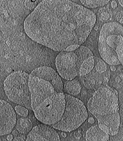
Nanoparticle morphology was examined using scanning electron microscopy (SEM) and transmission electron microscopy (TEM). Samples were diluted with double-distilled water and placed on a carbon-coated copper grid. They were negatively stained with 1% phosphotungstic acid, dried at room temperature, and observed using a microscope (14).

3.6. Fourier Transform Infrared Spectroscopy

Fourier transform infrared spectroscopy (FTIR) was used to observe infrared peaks arising from changes in molecular vibrational energy levels. First, 10 mg of the sample was placed in a mortar with 50 times its amount of potassium bromide (KBr) and ground counterclockwise with a pestle until a uniform mixture was obtained. The pestle was then placed inside the die through the transparent part. The uniform powder was placed inside the die and smoothed with a pestle. Another pestle was placed on top, and the steel cylinder was placed inside the die. The assembly was pressed using a press machine at a load of 10 tons for 2 minutes. After the die was opened, the resulting disk was placed on its base and transferred to the instrument. Results were recorded using Spectrum software.

3.7. Minimum Inhibitory and Minimum Bactericidal Concentrations

Gentamicin is often used as a reference antibiotic in microbial tests because it is a broad-spectrum antibiotic effective against both gram-positive and gram-negative bacteria.
Bacterial strains were obtained from a standard laboratory. The antibacterial activity of the SLNs was investigated against Staphylococcus aureus ATCC 1189, Listeria monocytogenes ATCC 1298, and Bacillus cereus PTCC 1015. The minimum inhibitory concentration (MIC) and minimum bactericidal concentration (MBC) were evaluated.

3.8. Statistical Analysis

Data were subjected to one-way analysis of variance (ANOVA) using SPSS version 17.

4. Results

4.1. Fourier Transform Infrared Spectroscopy

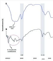
The FTIR spectrum of SLNs containing F. assa-foetida essential oil showed functional groups associated with the lipid matrix and the active compounds of the essential oil. The broad band at 3200 - 3600 cm-1 was attributed to O–H stretching vibrations, which may arise from phenolic and alcoholic compounds in the essential oil, as well as hydrogen-bonding interactions. Peaks at approximately 2920 and 2850 cm-1 corresponded to C–H stretching vibrations in lipid aliphatic chains, confirming the presence of a lipid phase in the nanoparticle structure. The band at 1700 - 1750 cm-1 corresponded to stretching vibrations of the carbonyl group (C=O), indicating acidic or ester compounds in the formulation. Bands at 1600 - 1500 cm-1 were attributed to C=C vibrations, and those at 1000 - 1300 cm-1 to C–O and C–O–C vibrations, indicating oxygenated and bioactive compounds in F. assa-foetida essential oil. Overall, the FTIR results indicated successful loading of the essential oil into the lipid nanoparticle matrix. The absence of new peaks or significant band shifts indicated that interactions between the essential oil and lipids were mainly physical, with no significant chemical reaction (Figure 1).
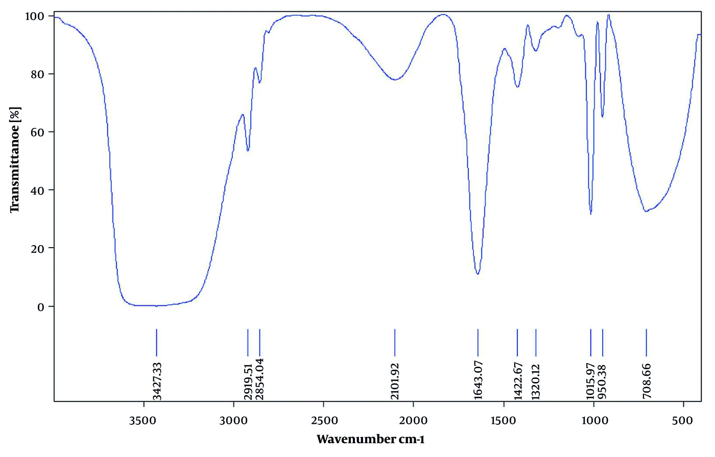
Fourier transform infrared spectroscopy (FTIR) of solid lipid nanoparticles
Figure 1.
Fourier transform infrared spectroscopy (FTIR) of solid lipid nanoparticles

4.2. Differential Thermal Analysis

The differential thermal analysis (DTA) plot of SLNs containing F. assa-foetida essential oil showed three main thermal events. A small change at approximately 25.5 °C, with a negative heat change, was attributed to the release of moisture or volatile compounds. The event at 262.2 °C may reflect the onset of decomposition of light organic compounds and plant components of F. assa-foetida essential oil. The main broad peak began at 485.5 °C and peaked at approximately 560 - 570 °C, corresponding to major decomposition of organic compounds and the lipid matrix. Overall, DTA showed relative stability up to intermediate temperatures and major degradation at high temperatures (Figure 2).
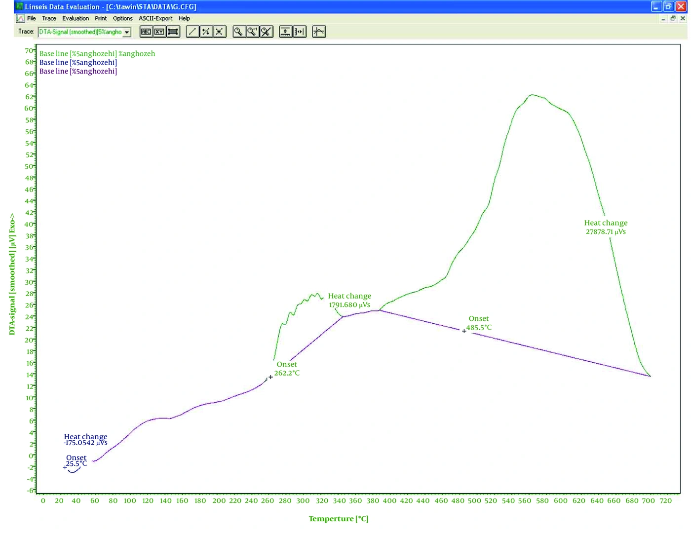
Differential thermal analysis (DTA) of solid lipid nanoparticles
Figure 2.
Differential thermal analysis (DTA) of solid lipid nanoparticles

4.3. Thermogravimetric Analysis

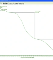
The thermogravimetric analysis (TGA) curve of SLNs containing F. assa-foetida essential oil showed thermal degradation in multiple stages. Initial weight loss at low temperatures was attributed to evaporation of absorbed moisture and the release of volatile compounds. Subsequent weight loss at intermediate temperatures may reflect decomposition of light organic compounds and bioactive components in the essential oil. The main weight loss at higher temperatures corresponded to major degradation of the lipid matrix and decomposition of organic chains. Residual weight at the end of the test was also likely attributable to ash, minerals, or thermally stable components. Overall, TGA showed relative thermal stability up to intermediate temperatures and main degradation at higher temperatures (Figure 3).
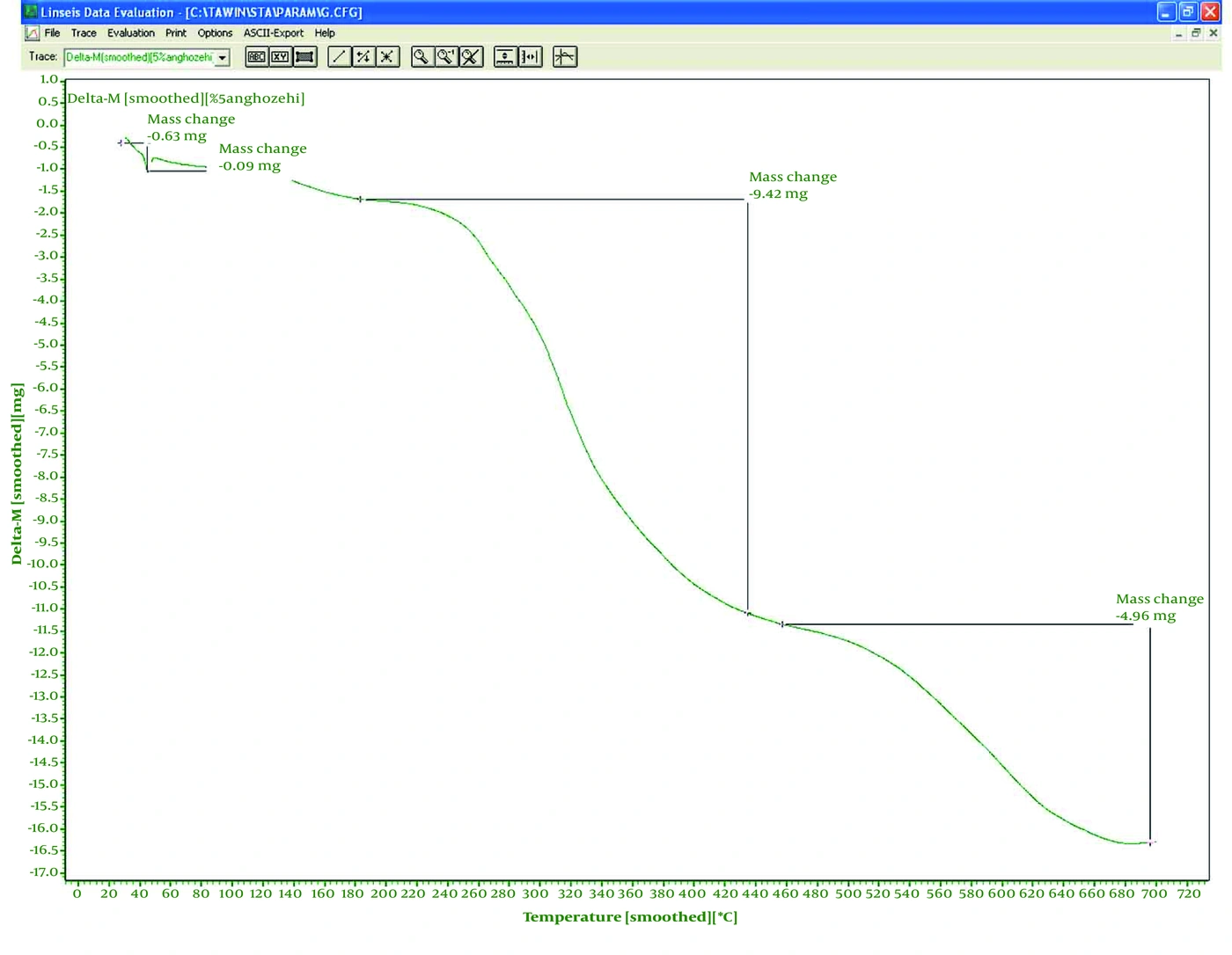
Thermogravimetric analysis (TGA) of solid lipid nanoparticles
Figure 3.
Thermogravimetric analysis (TGA) of solid lipid nanoparticles

4.4. Antibacterial Activity

Antimicrobial testing showed that the lowest inhibitory concentration against S. aureus was 50 mg/mL, whereas the highest inhibitory concentration against L. monocytogenes was 100 mg/mL.
The lowest inhibitory concentration against S. aureus was 50 mg/mL. The highest inhibitory concentration was 100 mg/mL, which inhibited L. monocytogenes and B. cereus strains (Table 1).
Table 1.
Minimum Inhibitory Concentration (MIC) and Minimum Bactericidal Concentration (MBC) of Solid Lipid Nanoparticles (mg/mL)
Bacterial speciesMICMBC
Staphylococcus aureus 50100
Listeria monocytogenes 100200
Bacillus cereus 100200

5. Discussion

In a study examining the antimicrobial properties of F. assa-foetida, the hydroalcoholic extract inhibited S. aureus and Bacillus subtilis (15).
Majdizadeh et al. synthesized nanoliposomes containing peppermint essential oil and investigated their stability and antimicrobial, antioxidant, and antiproliferative properties. Formulation F1 was selected, with an encapsulation efficiency of 62.12%, a nanoparticle size of 121 nm, and a zeta potential of -21.8 mV (16).
Faraji et al. investigated the encapsulation of Mentha pulegium and Ferula gummosa essential oils using nanoliposome technology as a safe botanical pesticide. The mean (± standard deviation) particle sizes of nanoliposomes containing M. pulegium and F. gummosa essential oils were 345 ± 3.2 and 309 ± 1.67 nm, respectively, and their encapsulation efficiencies were 99.38 ± 0.24% and 96.41 ± 0.26%, respectively. The type of essential oil did not significantly affect the physicochemical properties of the nanoparticles. After 24 hours, essential oil release from M. pulegium and F. gummosa nanoliposomes was 46% and 33%, respectively. The estimated LC50 values for nanoliposomes and crude M. pulegium essential oil against T. castaneum were 36.53 and 75.23 μL/L air, respectively (17).
In a study preparing nanoliposomes with rosemary essential oil, the particle size was 162 nm, and the particles were spherical (18).
A study examining the combined activity of several essential oils against Candida albicans reported an inhibition halo diameter of 9 mm (19).
The effect of a chitosan nanoparticle biofilm containing F. assa-foetida on the healing of full-thickness wounds infected with methicillin-resistant S. aureus was investigated. Microbiological, planimetric, biomechanical, histological, and quantitative morphometric studies, along with the determination of hydroxyproline levels, showed a significant difference between animals in group 4 and those in the other groups (20).
A study investigating the antimicrobial activity of several medicinal plants against Escherichia coli showed that P. harmala extract had the highest antimicrobial activity, with a MIC of 2.5 mg/mL (21).
In a study investigating the synthesis of silver nanoparticles using Sesamum indicum, the minimum inhibitory concentration against E. coli was 200 ppm (22).
Studies synthesizing silver nanoparticles using Plantago ovata via a green method reported a nanoparticle size of 13 nm. The highest and lowest inhibitory concentrations were 100 and 12.5 mg/mL, respectively (23).

5.1. Conclusions

The results indicated that F. assa-foetida essential oil in SLNs had a long shelf life at 4 °C and exhibited good antimicrobial properties.
Acknowledgments

Footnotes

  • AI Use Disclosure:The authors declare that no generative AI tools were used in the creation of this article.

  • Authors' Contribution:Study Concept and Design: M. J. and S. S.; Analysis and Interpretation of Data: M. R.

  • Conflict of Interests Statement:The authors declared no conflict of interest.

  • Data Availability:The dataset presented in the study is available on request from the corresponding author during submission or after publication.

  • Ethical Approval:IR- RIOZ-GR-9027.

  • Funding/Support:This research was funded by Zabol University of Medical Sciences.

References

  • 1.
    Bilia AR, Guccione C, Isacchi B, Righeschi C, Firenzuoli F, Bergonzi MC. Essential oils loaded in nanosystems: a developing strategy for a successful therapeutic approach. Evid Based Complement Alternat Med. 2014;2014(1). 651593. [PubMed ID: 24971152]. [PubMed Central ID: PMC4058161]. https://doi.org/10.1155/2014/651593.
  • 2.
    Sherry M, Charcosset C, Fessi H, Greige-Gerges H. Essential oils encapsulated in liposomes: a review. Journal of Liposome Research. 2013;23(4):268-75. [PubMed ID: 23879218]. https://doi.org/10.3109/08982104.2013.819888.
  • 3.
    Ajazuddin, Saraf S. Applications of novel drug delivery system for herbal formulations. Fitoterapia. 2010;81(7):680-9. [PubMed ID: 20471457]. https://doi.org/10.1016/j.fitote.2010.05.001.
  • 4.
    Moghimipour E, Aghel N, Zarei Mahmoudabadi A, Ramezani Z, Handali S. Preparation and characterization of liposomes containing essential oil of Eucalyptus camaldulensis leaf. Jundishapur Journal of Natural Pharmaceutical Products. 2012;7(3):117-22. https://doi.org/10.17795/jjnpp-5261.
  • 5.
    Mozafari MR. Nanoliposomes: preparation and analysis. Liposomes: Methods and Protocols, Volume 1: Pharmaceutical Nanocarriers. 2010;605:29-50. [PubMed ID: 20072871]. https://doi.org/10.1007/978-1-60327-360-2_2.
  • 6.
    Suryani A, Laksitorini MD, Sulaiman TNS. Ferrous fumarate nanoliposomes: formulation, characterization, and diffusion profiles. J Appl Pharm Sci. 2024;14(5):157-65. https://doi.org/10.7324/JAPS.2024.154580.
  • 7.
    Chabib L, Suryani A, Pangestu MI, Hidayat AMUJ, Trianloka AMB. IAI CONFERENCE: The development of Origanum vulgare L. into nanoparticles in dosage forms. Pharm Educ. 2021;21(2):205-9. https://doi.org/10.46542/pe.2021.212.205209.
  • 8.
    Guillot AJ, Martínez-Navarrete M, Garrigues TM, Melero A. Skin drug delivery using lipid vesicles: a starting guideline for their development. J Control Release. 2023;355:624-54. [PubMed ID: 36775245]. https://doi.org/10.1016/j.jconrel.2023.02.006.
  • 9.
    Mozaffarian V. A dictionary of Iranian plant names. Tehran: Farhang Mo'aser; 1996.
  • 10.
    Valencia E, Feria M, Díaz J, González A, Bermejo J. Antinociceptive, anti-inflammatory and antipyretic effects of lapidin, a bicyclic sesquiterpene. Planta Med. 1994;60(5):395-399. [PubMed ID: 7997463]. https://doi.org/10.1055/s-2006-959517.
  • 11.
    Aqel MB, Al-Khalil S, Afifi F. Effects of a Ferula sinaica root extract on the uterine smooth muscle of rat and guinea pig. J Ethnopharmacol. 1991;31(3):291-7. [PubMed ID: 2056757]. https://doi.org/10.1016/0378-8741(91)90014-5.
  • 12.
    Abedi D, Jalali M, Asghari G, Sadeghi N. Composition and antimicrobial activity of oleogumresin of Ferula gumosa Bioss. essential oil using Alamar Blue. Res Pharm Sci. 2008;3(1):41-5.
  • 13.
    Khajeh M, Yamini Y, Bahramifar N, Sefidkon F, Reza Pirmoradei M. Comparison of essential oils compositions of Ferula assa-foetida obtained by supercritical carbon dioxide extraction and hydrodistillation methods. Food Chem. 2005;91(4):639-644. https://doi.org/10.1016/j.foodchem.2004.06.033.
  • 14.
    Daneshmand S, Jaafari MR, Movaffagh J, Malaekeh-Nikouei B, Iranshahi M, Seyedian Moghaddam A, et al. Preparation, characterization, and optimization of auraptene-loaded solid lipid nanoparticles as a natural anti-inflammatory agent: in vivo and in vitro evaluations. Colloids and Surfaces B: Biointerfaces. 2018;164:332-339. [PubMed ID: 29413613]. https://doi.org/10.1016/j.colsurfb.2018.01.054.
  • 15.
    Jalili B, Saeidi-sar S, Masoudian N, Zarban A, Namaei MH. Evaluation of antimicrobial activity of hydroalcoholic extracts from different parts of Ferula assafoetida L. in three regions of South Khorasan Province. Journal of Chemical Health Risks. 2022;12(4):621-34. https://doi.org/10.22034/jchr.2022.1938969.1397.
  • 16.
    Majdizadeh M, Akbarzadeh S, H Al-Turnachy H, Hemati M, Akhlaghi M, Haghiralsadat BF, et al. Synthesis of nano-liposomes containing Mentha piperita essential oil and investigating their stability, antimicrobial, antioxidant, and antiproliferative properties. Nanomed J. 2025;12(1):122-39. https://doi.org/10.22038/nmj.2024.79435.1960.
  • 17.
    Faraji Z, Shakarami J, Varshosaz J, Jafari S. Encapsulation of essential oils of Mentha pulegium and Ferula gummosa using nanoliposome technology as a safe botanical pesticide. Journal of Applied Biotechnology Reports. 2020;7(4):237-42. https://doi.org/10.30491/jabr.2020.121704.
  • 18.
    Arabi MH, Chabok H, Mirzapour A, Ardestani MS, Mostafa M. Preparation of nanoliposomes containing Rosmarinus officinalis L essential oil: a comparative study. Biosc Biotech Res Comm. 2017;10(1):103-108. https://doi.org/10.21786/bbrc/10.1/15.
  • 19.
    Iyer MS, Sheshanna SH, Rao RN, Puttegowda D, Ramu R. Antibiofilm and antimicrobial potential of Trigonella foenum-graecum and Ferula asafoetida essential oils against oral pathogens: synergistic effects and in silico insights. The Microbe. 2026;10. 100667. https://doi.org/10.1016/j.microb.2026.100667.
  • 20.
    Sadeghi MA, Kalantari S, Gharib FZ, Faed-Maleki F, Yousefi A. Influence of Ferula assa-foetida loaded chitosan nanoparticle biofilm on wound healing in full-thickness wounds infected with methicillin resistant Staphylococcus aureus. Iran J Vet Surg. 2020;15(1):42-52. https://doi.org/10.30500/ivsa.2019.201547.1200.
  • 21.
    Saeidi S, Amini Boroujeni N, Ahmadi H, Hassanshahian M. Antibacterial activity of some plant extracts against extended-spectrum beta-lactamase producing Escherichia coli isolates. Jundishapur J Microbiol. 2015;8(2). e15434. [PubMed ID: 25793093]. [PubMed Central ID: PMC4353063]. https://doi.org/10.5812/jjm.15434.
  • 22.
    Bokaeian M, Sheikh M, Hassanshahian M, Saeidi S, Sahraei S. The antibacterial activity of silver nanoparticles produced in the plant Sesamum indicum seed extract: a green method against multi-drug resistant Escherichia coli. International Journal of Enteric Pathogens. 2014;2(2). e17928. https://doi.org/10.17795/ijep17928.
  • 23.
    Bokaeian M, Fakheri BA, Mohasseli T, Saeidi S. Antibacterial activity of silver nanoparticles produced by Plantago ovata seed extract against antibiotic resistant Staphylococcus aureus. Int J Infect. 2015;2(1). e22854. https://doi.org/10.17795/iji-22854.

Copyright

Copyright © 2026, Jahantigh et al. This open-access article is available under the Creative Commons Attribution 4.0 (CC BY 4.0) International License (https://creativecommons.org/licenses/by/4.0/), which allows for unrestricted use, distribution, and reproduction in any medium, provided that the original work is properly cited.

Similar Articles

25
Jul
2015
Fabrication of Gelatin Nano-Capsules Incorporate Ferula assa-foetida Essential Oil With Antibacterial and Antioxidant

Fabrication of Gelatin Nano-Capsules Incorporate Ferula assa-foetida Essential Oil With Antibacterial and Antioxidant

Somayeh Jahani,
Amin Shakiba,
Leila Jahani

Jahani S, Shakiba A, Jahani L. Fabrication of Gelatin Nano-Capsules Incorporate Ferula assa-foetida Essential Oil With Antibacterial and Antioxidant. Zahedan J Res Med Sci. 2015;17(7):-. doi: https://doi.org/10.17795/zjrms1018

16
Jun
2013
Essential Oil Composition of Ferula Assa-Foetida L. Fruits from Western Iran

Essential Oil Composition of Ferula Assa-Foetida L. Fruits from Western Iran

Gholamreza Bahrami,
Reza Soltani,
Sayed-Ebrahim Sajjadi,
Mohammad-Reza Kanani,
Reza Naderi,
Nastaran Ghiasvand
,et al.

Bahrami G, Soltani R, Sajjadi S, Kanani M, Naderi R, et al. Essential Oil Composition of Ferula Assa-Foetida L. Fruits from Western Iran. J Rep Pharm Sci. 2013;2(2):e147736. doi:

23
Aug
2026
Synthesis and Characterization of Nanostructured Lipid Carriers Containing Pomegranate Essential Oil and Grape Seed Oil Using Hot Homogenization and Ultrasound

Synthesis and Characterization of Nanostructured Lipid Carriers Containing Pomegranate Essential Oil and Grape Seed Oil Using Hot Homogenization and Ultrasound

Omolbanin Shahraki,
Farzane Nasiri,
Sara Daneshmand

Shahraki O, Nasiri F, Daneshmand S. Synthesis and Characterization of Nanostructured Lipid Carriers Containing Pomegranate Essential Oil and Grape Seed Oil Using Hot Homogenization and Ultrasound. Jundishapur J Nat Pharm Prod. 2026;21(3):e162314. doi: https://doi.org/10.5812/jjnpp-162314

11
Sep
2024
Development of Sesame Oil Loaded in Maltodextrin/Acacia Gum-Coated Nanoemulsion and Investigation of Its Physicochemical Properties

Development of Sesame Oil Loaded in Maltodextrin/Acacia Gum-Coated Nanoemulsion and Investigation of Its Physicochemical Properties

Malihe Karami,
Ali Fazlara,
Mehdi Zarei,
Behzad Sharif Makhmal Zadeh,
Mehdi Safdarian

Karami M, Fazlara A, Zarei M, Sharif Makhmal Zadeh B, Safdarian M. Development of Sesame Oil Loaded in Maltodextrin/Acacia Gum-Coated Nanoemulsion and Investigation of Its Physicochemical Properties. Jundishapur J Nat Pharm Prod. 2024;19(4):e149354. doi: https://doi.org/10.5812/jjnpp-149354

6
May
2023
Targeted Solid Lipid Nanoparticles Formulation for Colon Cancer Treatment and Cytotoxicity Assessment Using HT29 Cell Line

Targeted Solid Lipid Nanoparticles Formulation for Colon Cancer Treatment and Cytotoxicity Assessment Using HT29 Cell Line

Eskandar Moghimipour,
Maryam Abedini,
Somayeh Handali

Moghimipour E, Abedini M, Handali S. Targeted Solid Lipid Nanoparticles Formulation for Colon Cancer Treatment and Cytotoxicity Assessment Using HT29 Cell Line. Jundishapur J Nat Pharm Prod. 2023;18(2):e135987. doi: https://doi.org/10.5812/jjnpp-135987

More by these authors

Mehdi JahantighPubMedScholar
Saeide SaeidiPubMedScholar
Masoomeh RezaeiPubMedScholar
Share
Cited by
Metrics