1. Introduction
In the last months of 2019, the advent of a new virus called SARS-CoV-2 caused the spread of a pandemic, COVID-19. Among the peculiarities of patients who died of severe COVID-19 is the increase of highly pro-inflammatory cytokines (1). This phenomenon, called Cytokine Storm (CS), consists of a cascade of immune system responses that can dramatically worsen the prognosis. Endothelial damage with cytomixoid exudate is largely found in both lungs in COVID-19 patients. At the same time, a decrease in CD4 and CD8 cells is detected, which is inversely proportional to the increase in Th17 cells in COVID-19 patients (1). High levels of IL-6, IL-23, and TGF-β would induce the differentiation of helper T cells from type T0 into Th17 (2). In mild cases, the reduction of lymphocytes is not as evident as in severe cases. Even in the most severe cases, there is a simultaneous reduction of CD8 T cells compared to an increase in the serum levels of all inflammatory cytokines (IFN-γ, IL-2, IL-6, etc.) (3). In most severe COVID-19 patients, it is necessary to identify the cytokine storm to implement a therapeutic strategy to reduce serum IL-6 levels (3). To achieve this therapeutic strategy, the administration of monoclonal antibody (tocilizumab) capable of blocking IL-6 receptors (IL-6R) has been proposed. Tocilizumab produces a selective blockage of the IL-6 signal transduction pathway and has been successfully used in some preliminary trials and is currently one of the most widely used drugs for the treatment of severe COVID-19. A patient recently implanted in our Pain Department with a spinal cord stimulator for failed back surgery syndrome (FBSS), following SARS‐CoV‐2 infection, received Tocilizumab, which caused a severe pocket inflammatory device reaction and imposed a high risk of IPG rejection.
2. Case Presentation
We describe the case of a 63-year-old woman, affected by FBSS after L5 surgical arthrodesis, with chronic back and bilateral radicular pain, treated for several years with opioids, anti-inflammatory drugs, corticosteroids, and GABAergic drugs without clinical outcomes. She underwent two dorsal ganglion pulsed radiofrequency treatments with no durable results. In December 2019, spinal cord stimulation was performed on the patient with the implant of two electrodes. After three weeks of trial, a significant reduction in NRS (Numeric Rating scale), PCS (Pain Catastrophizing scale), and ODI (Oswestry Disability Index) allowed for an IPG permanent implant.
In the second decade of March, she had a fever, ageusia, anosmia, mild respiratory symptoms, dyspnea, and SatO2 95%; thus, she underwent a nasopharyngeal test that showed a positive SARS-CoV-2 infection. After 48 h of isolation therapy at home, only with oral hydroxychloroquine 200 mg/day, Azithromycin 500 mg/day, PEA 1200 mg/day, prednison 25 mg/die, and O2, her clinical conditions worsened, so she was recovered in a sub-intensive care unit where she was subjected to NIV-CPAP ventilation, with consequent optimization of respiratory parameters and improvement of SatO2 from 91% to 97%, and P/F ratio from 130 to 250 mmHg in four days. The most important variation in pharmacological therapy was the administration of Tocilizumab, 8 mg/kg EV bolus in 60 min, for IL-6 increasing in the blood (IL-6 48.3 pg/mL; normal values 0 - 5 pg/mL). After administration, she experienced mild hypertransaminasemia, which promptly returned to normal in four days. After five days, the patient continued therapy only with corticosteroids in isolation in a COVID-19 care unit. She never complained about any problems related to spinal cord stimulation, nor about her previous neuropathic pain, throughout the above-mentioned period.
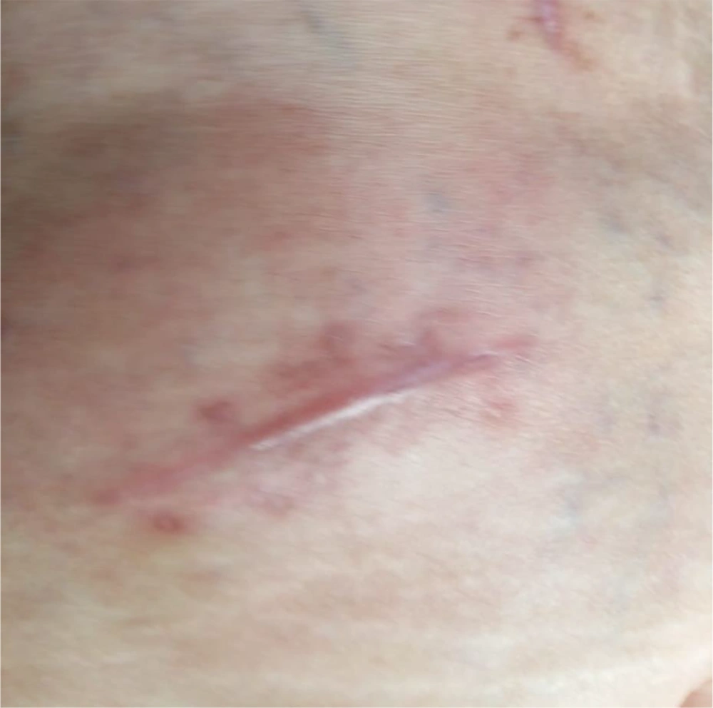
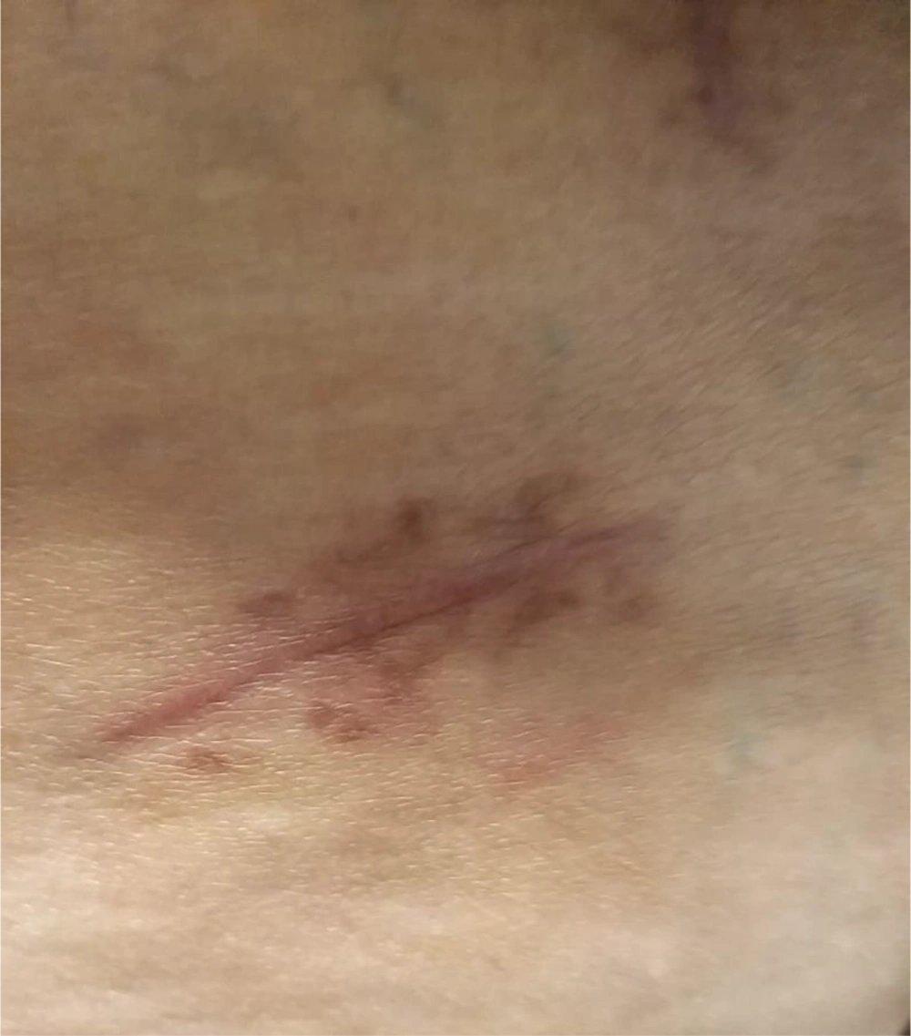
Just 10 days after tocilizumab administration, the patient reported a new problem on the surgical pocket for the IPG. The pocket was warm, painful, and red, with no fluid to the touch (Figure 1). After a sonographic evaluation, tissue edema without evident effusion was detected. An immunological evaluation was performed, and after evaluating the risks/benefits of performing a biopsy or microbiological sampling, we decided to follow a conservative approach, with laboratory and clinical evaluation of the inflammatory pattern and empirical antimicrobial therapy. She was treated with trimethoprim-sulfamethoxazole (800/160 mg.day) for seven days. Laboratory tests showed no serious alterations, except for these values: white blood cells (WBC) 12.06 103/µL, ESR (Erythrocyte Sedimentation Rate) 60 mm/h, IL-6 4.2 pg/mL, and Procalcitonin < 0.0200 ng/mL. After seven days of antibiotic therapy (17 days after tocilizumab therapy), the pocket area appeared clear and not painful (Figure 2). A new sonographic evaluation of the pocket did not show clear images possibly related to infections and/or fluids.
3. Discussion
As an important function, IL-6 influences the specific differentiation of naive CD4+ T cells by modulating a variation between the innate and the acquired immune response. We know that IL-6, together with transforming growth factor (TGF)-β, is essential for Th17 differentiation from naive CD4+ T cells (4). On the other hand, IL-6 can inhibit TGF-β-induced Treg differentiation (5). The upregulation of the Th17/Treg equilibrium can lower immunological tolerance and trigger the mechanisms of the development of chronic inflammatory autoimmune or chronic diseases (6). The same immunological pattern is emerging caused by COVID-19 immunological damage and IL-6 overexpression (7-9).
In this context, IL-6 is represented as a fundamental mediator to generate an alarm signal in adverse situations. When the release of this alarm signal occurs as a result of an infection, it reaches the entire organism, activating monocyte and macrophage cells rushing recognized by specific pathogen-recognition receptors (PRRs). Another function of IL-6 is to alert the body in the case of tissue damage. Damaged tissue cells, as occur in the case of trauma or infections, produce damage-associated molecular patterns (DAMPs), which is the direct or indirect cause of inflammation (10). This phenomenon is triggered in a similar way, even after sterile surgery. Thus, an increase in serum IL-6 levels is associated with an increase in the serum acute-phase protein concentration and often with a simultaneous rise in body temperature (11).
Regardless of the cause of the inflammation, which cannot be determined without a biopsy, the cytokine storm from COVID-19 would have had far more serious causes if it had not been contrasted with tocilizumab. We hypothesize that the activation of PRRs or DAMP receptors in scar tissue remodeling and both the presence of a prosthetic implant and the increased systemic levels of IL-6 due to COVID-19 were the triggers of inflammatory tissue reaction. Finally, it is well known that polymorphism of the IL-6 promoter region in position 174 is associated with severe autoimmune and autoinflammatory diseases (12); in the next step, we should research IL-6 polymorphisms in consideration of their prevalence in European people (13). We will also evaluate the administration of Tocilizumab in future patients with aggressive IL-6/174 polymorphism and post-surgical hyperimmune reaction.

