The Use of Intraoperative Transesophageal Echocardiography in Thoracic Aortic Dissection Due to Chronic Cocaine Abuse

authors:

avatar Ali Dabbagh ORCID 1 , * , avatar Mohammad Kazem Arabnia 2 , avatar Mahnoosh Foroughi 3 , avatar Mehran Shahzamani 4 , avatar Hassan Rahmian 5

MD, Professor of Cardiac Anesthesia, Anesthesiology Research Center, Shahid Beheshti University of Medical Sciences, Tehran, Iran
MD, Assistant Professor, Cardiac Surgery Department, Shahid Beheshti University of Medical Sciences, Tehran, Iran
MD, Associate Professor, Cardiovascular Research Center, Shahid Beheshti University of Medical Sciences, Tehran, Iran
MD, Assistant Professor, Cardiac Surgery Department, Isfahan University of Medical Sciences, Isfahan, Iran
MD, Cardiac Surgery Department, Shahid Beheshti University of Medical Sciences, Tehran, Iran

How To Cite Dabbagh A, Arabnia M K, Foroughi M, Shahzamani M, Rahmian H. The Use of Intraoperative Transesophageal Echocardiography in Thoracic Aortic Dissection Due to Chronic Cocaine Abuse. Anesth Pain Med. 2017;7(1):e35254. https://doi.org/10.5812/aapm.35254.

Abstract

Introduction:

Aortic dissection is a life threatening disease and is usually accompanied by a high rate of mortality and morbidity. Here we present a case report in which intraoperative tranesophageal echocardiography was used for intraoperative assessments of thoracic aortic dissection due to cocaine abuse.

Case Presentation:

A 45- year- old male was admitted to a university hospital due to severe chest pain. He was suffering from severe excruciating chest pain that had started after a psychological stress, leading to heavy cocaine abuse. He was admitted to the emergency department of the hospital, and was then transferred to the cardiac care unit to control the chest pain. The patient underwent emergent surgery. After induction of anesthesia, tranesophageal echocardiography probe was introduced gently and a full exam was done. The surgeon decided to perform a classic Bentall procedure. Cardiopulmonary bypass was started. Everything was acceptable, but bleeding was uncontrolled. The surgical team could not control the bleeding, and he passed away due to bleeding.

Conclusions:

This case report stresses the use of IOTEE as a means for more accurate diagnosis of the lesion under general anesthesia, especially when there is not time to do preoperative TEE, or when bedside echocardiography does not give us adequate data.

1. Introduction

Aortic dissection is a life threatening disease and is usually accompanied by a high rate of mortality and morbidity (1-5). Usually, most of these patients require acute medical and surgical treatment (5-9). Cocaine induced vascular injuries and ischemia is among the complications of cocaine abuse (10, 11). One of the rare complications of cocaine inhalation is dissection of the thoracic aorta. Although it is a rare complication, it is well recognized and may involve both the thoracic and abdominal aorta (12, 13).

Different methods are used to diagnose this disease entity. However, we could find only one citation in the Medline that had presented an atypical presentation of aortic dissection due to cocaine abuse (type A of dissection), which was diagnosed by transesophageal echocardiography (6). Here, we present a case report in which intraoperative tranesophageal echocardiography was used for intraoperative assessments of thoracic aortic dissection due to cocaine abuse.

2. Case Presentation

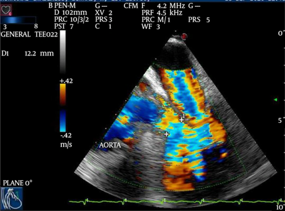
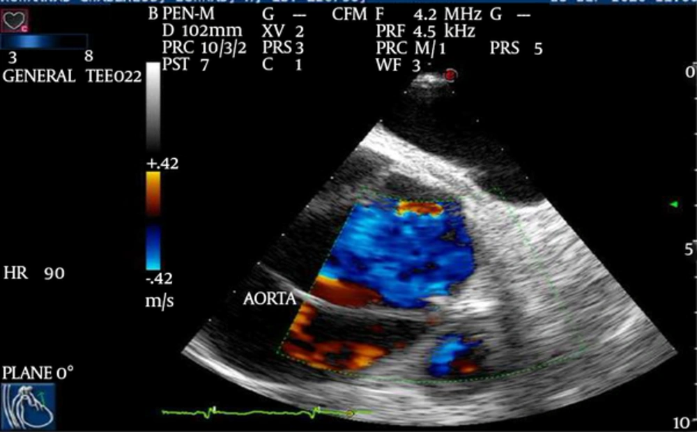
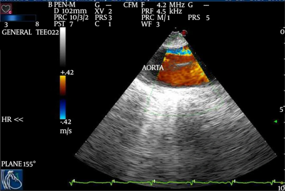
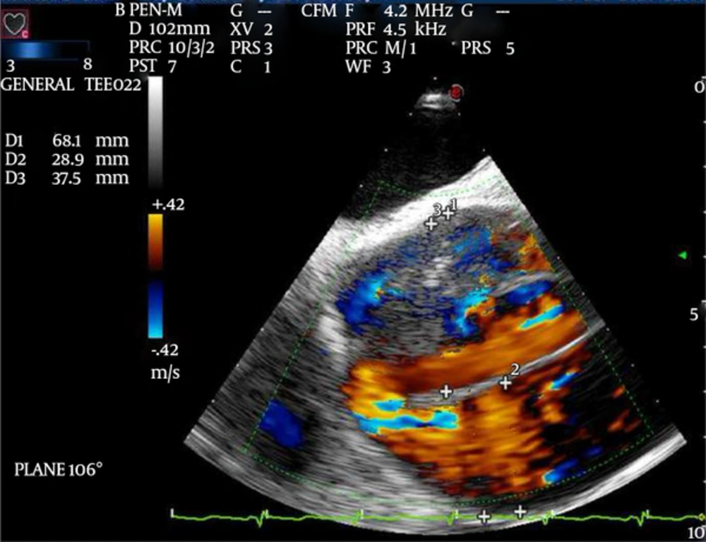
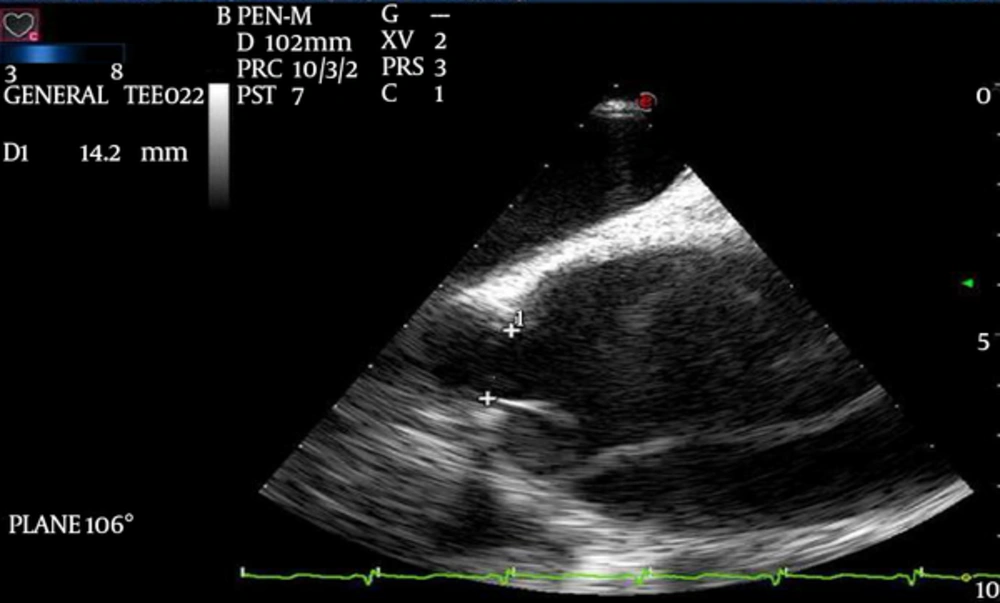
Consent for publication of this case was obtained from the patient. A 45-year- old male was admitted to a university hospital due to severe chest pain. He was suffering from severe excruciating chest pain that had started after a psychological stress, leading to heavy cocaine abuse. He was admitted to the emergency department of the hospital, and was then transferred to the cardiac care unit to control the chest pain. The primary therapeutic modalities could not completely suppress the chest pain while the patient underwent diagnostic evaluations including chest X-ray, echocardiography, and computed tomography angiography. These assessments revealed a dilated aneurysm involving the arch of aorta, the origin of the right common carotid artery, and the aortic valve. The patient had a history of habitual opium and severe cocaine abuse. In addition, he was a heavy smoker, hypertensive and very obese (120 Kg body weight and 179 cm height). The patient was transferred to the cardiac operation room for a Bentall procedure. He was monitored with standard monitoring including 5- lead electrocardiogram, pulse oximeter, and invasive blood pressure monitoring. Then anesthesia was induced using a combination of sufentanil, cisatracurium, and etomidate. The patient was intubated and a central venous catheter was inserted through the right internal jugular vein. Moreover, 3 large bore intravenous catheters (14 F) were prepared for him. The transesophageal echocardiography probe was introduced gently and a full exam was done. The results of the exam revealed the following results (Figures 1 - 5):

Midesophageal Long Axis (LAX) View of the Ascending Aorta With Color Doppler Demonstrating Flow Through the Intimal Layer
Midesophageal Long Axis (LAX) View of the Ascending Aorta With Color Doppler Demonstrating the Distances
Midesophageal 5- Chamber View, Demonstrating the Left Ventricular Outflow Tract (LVOT), the Aortic Valve, and the Proximal Part of the Ascending Aorta With Color Doppler Regurgitation Through the Aortic and Mitral Valves
Midesophageal 5- Chamber View Focusing on the Proximal Part of the Ascending Aorta, Demonstrating the Ascending Aorta With Color Doppler Flow through the Intimal Layer
Upper Esophageal Short Axis View, Demonstrating the Aortic Arch With Color Doppler and the Aortic Flap

• Dilated ascending aorta and the arch;

• A mobile flap undulating inside the proximal aorta extending to the arch and producing a lumen;

• Severe aortic insufficiency;

• Left ventricular hypertrophy;

• Good function of the LV.

After preparation and wide drape, the left femoral artery and vein, and right axillary artery were exposed and cannulated as routine. Then cardiopulmonary bypass (CPB) was started using femoral artery and vein only. At that point, sternotomy was done and pericardium was opened; only some serosanguinous liquid was in the pericardial cavity. Aorta was huge and from the outflow of the left ventricle to just near the innominate artery was taken off. Aorta appeared normal. Then the surgeon decided to perform a classic Bentall procedure. The aorta was clamped as distal as possible. Anterograde and then retrograde cold blood cardioplegia were used and repeated every 20 minutes. The aorta was opened, the dissection flap was exposed, and the aortic valve was resected. Afterwards, the left and the right buttons were prepared using the standard method. Aorta, the aortic valve, and the left ventricle outflow tract were assessed and found to be very fragile and thin. A tube graft with a number 25 accompanying valve and a number 28 tube, SJM, was inserted in place and sutured in the aortic valve site, and its distal end was sutured to the aorta just beneath the clamp and a felt strip (after suturing the intima to the wall of aorta with a thin felt inside the aorta). De-airing and cross clamp opening were done using routine manner. There was some bleeding from the proximal suture line at the site of left coronary leaflet, and the outflow of the left ventricular tract was very fragile there.

Cardiopulmonary bypass was started to be turned off and everything was acceptable, but bleeding continued. Therefore, CPB was started again, and aorta cross clamp over the tube was opened after infusion of the anterograde cold blood cardioplegia. The site of bleeding was sutured again and aorta was closed, de-airing and opening of the cross clamp were done again.

However, the bleeding continued. Weaning from CPB was done, protamine infusion was performed, and the site of bleeding was packed. Additional actions (including cryoprecipitate, fresh frozen plasma, and platelet) were administered, but no good results were achieved. The surgical team could not control the bleeding and the patient passed away due to bleeding.

3. Discussion

Such cases with acute dissection of the aorta are not common, though not rare. However, not a considerable number of these cases have been reported in the literature (having a history of cocaine abuse, leading to acute signs and symptoms leading to surgery) (1-5). On the other hand, in the new era of intraoperative echocardiography (IOTEE), the role of cardiac anesthesiologist in using perioperative echo is of utmost importance; therefore, discussing the comments of this patient is of importance and its presentation here is useful for other cardiac anesthesiologists. The authors could not find many cases like this one with acute aortic dissection mainly after cocaine abuse who had underwent IOTEE (11). However, there was another case report in which the aorta was not divided as an intimal flap and a double lumen was not created at any level of the aorta (7).

This case report stresses the use of IOTEE as a means for more accurate diagnosis of the lesion under general anesthesia, especially when there is not enough time for the physicians to perform preoperative TEE and when due to a number of problems like obesity, the bedside transthoracic echocardiography does not give us adequate data.
References

  • 1.

    Perron AD, Gibbs M. Thoracic aortic dissection secondary to crack cocaine ingestion. Am J Emerg Med. 1997;15(5):507-9. [PubMed ID: 9270392]. https://doi.org/10.1016/S0735-6757(97)90196-0.

  • 2.

    Restrepo CS, Rojas CA, Martinez S, Riascos R, Marmol-Velez A, Carrillo J, et al. Cardiovascular complications of cocaine: imaging findings. Emerg Radiol. 2009;16(1):11-9. [PubMed ID: 18773229]. https://doi.org/10.1007/s10140-008-0762-x.

  • 3.

    Vannemreddy P, Caldito G, Willis B, Nanda A. Influence of cocaine on ruptured intracranial aneurysms: a case control study of poor prognostic indicators. J Neurosurg. 2008;108(3):470-6. [PubMed ID: 18312093]. https://doi.org/10.3171/JNS/2008/108/3/0470.

  • 4.

    Majlesi N, Greller H, Su M. Cocaine and aneurysms. J Neurosurg. 2008;109(4):779. [PubMed ID: 18826369]. https://doi.org/10.3171/JNS/2008/109/10/0779.

  • 5.

    Chang RA, Rossi NF. Intermittent cocaine use associated with recurrent dissection of the thoracic and abdominal aorta. Chest. 1995;108(6):1758-62. [PubMed ID: 7497801]. https://doi.org/10.1378/chest.108.6.1758.

  • 6.

    Riaz K, Forker AD, Garg M, McCullough PA. Atypical presentation of cocaine-induced Type A aortic dissection: a diagnosis made by transesophageal echocardiography. J Investigative Med. 2002;50(2):140-2.

  • 7.

    Neri E, Toscano T, Massetti M, Capannini G, Frati G, Sassi C. Cocaine-induced intramural hematoma of the ascending aorta. Tex Heart Inst J. 2001;28(3):218-9. [PubMed ID: 11678260].

  • 8.

    Johnson JA, Callison C, Miller AN. A 48-year-old man with paralysis and hypotension. Chest. 2008;134(4):862-5. [PubMed ID: 18842920]. https://doi.org/10.1378/chest.08-0253.

  • 9.

    Feldhendler M, May JA, Gandhi SD, Iqbal Z, Butler EG, Lilly RE, et al. A triple-lumen aorta: an unusual manifestation of complex aortic pathology associated with crack cocaine abuse. J Cardiothorac Vasc Anesth. 2007;21(3):462-4. [PubMed ID: 17544910]. https://doi.org/10.1053/j.jvca.2006.09.007.

  • 10.

    Swalwell CI, Davis GG. Methamphetamine as a risk factor for acute aortic dissection. J Forensic Sci. 1999;44(1):23-6. [PubMed ID: 9987866]. https://doi.org/10.1520/JFS14407J.

  • 11.

    Fisher A, Holroyd BR. Cocaine-associated dissection of the thoracic aorta. J Emerg Med. 1992;10(6):723-7. [PubMed ID: 1491155]. https://doi.org/10.1016/0736-4679(92)90532-X.

  • 12.

    Bacharach JM, Colville DS, Lie JT. Accelerated atherosclerosis, aneurysmal disease, and aortitis: possible pathogenetic association with cocaine abuse. Int Angiol. 1992;11(1):83-6.

  • 13.

    Gadaleta D, Hall MH, Nelson RL. Cocaine-induced acute aortic dissection. Chest. 1989;96(5):1203-5. [PubMed ID: 2805854]. https://doi.org/10.1378/chest.96.5.1203.