1. Introduction
Difficult intubation is one of the most significant issues anesthesiologists deal with, occasionally. Among these, mandibulofacial deformities face the biggest challenge in intubation and make an anticipated difficult airway (1), due to short horizontal length of mandible (HLM), short thyromental distance (TMD), short inter-incisors gap (IIG) and high grade modified Mallampati test (MMT) (2).
There are several strategies to approach these patientsand each technique has unique benefits that should be used on experience. Sitting endotracheal intubation is a useful technique for airway control, in patients with difficult airway or in patients in whom maintenance of the upright posture is beneficial (3), such as patients with tracheal compression (4), or superior vena cava syndrome (5, 6). The technique consists of advancing the endotracheal tube to vocal cords of patient in face-to-face position, when patient is in the sitting or semi-sitting position (7).
2. Case Presentation
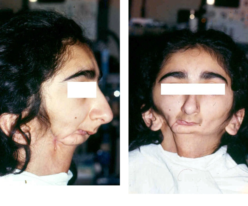
The patient was a 23-year-old girl who weighed 40 kilograms and had 157 cm in height (body mass index: 16.22). She was diagnosed as a mandible osteosarcoma in her childhood and received radiotherapy that led to underdevelopment of the mandible. In 2011, drainage of pus accumulation was performed and biopsies were obtained. She underwent surgery to remove half of the mandible because of osteonecrosis and osteomyelitis, in 2013 (Figure 1). The correction surgery was not performed; therefore, her chin deviated to the opposite side and her mouth and pharynx remained small, which resulted in an obvious asymmetry of her face. On the other hand,the previous radiotherapy sessions resulted in severe fibrosis in the mouth and neck area, which caused troubles of deglutition, therefore also diagnosing her case as adifficult airway (2).
In 2014, she was scheduled for reconstructive surgery of the mandible, with free-flap and free fibula. In this procedure, after prelevating flap and creating anastomosis to the external carotid artery and jugular vein, skin and muscles were used to reconstruct the mandible defect. The initial surveys, including routine laboratory tests, urine analysis and spirometry, had normal results. Six bags of packed red blood cell (RBC) and four units of fresh frozen plasma (FFP) were reserved. Awake intubation was planned in consultation with the ear, nose and throat (ENT) specialist. The chest X-ray revealed no tracheal deviation or any other important upper airway pathology. She explained recent night snoring, after her last surgery. The patient was examined by an expert anesthesiologist and her Mallampati class was IV, while the TMD was imprecise due to mandible removal in last surgery. Echocardiography, pulse oximetry and non-invasive blood pressure (NIBP) monitoring, were performed, before anesthesia. The patient position was supine. All future events, which were undertaken in awake fiberoptic intubation, were explained to the patient.
Facemask preoxygenation was done for 3 minutes. Thepremedications, which were used for awake intubation, were atropine (0.01 mg/kg), midazolam (0.05 mg/kg), fentanyl (4 µ/kg) and lidocaine (1.5 mg/kg), which was injected titrated, over 5 minutes. Mask ventilation was tested and showed no difficulties. Nasal drops of phenylephrine and the mesh dipped in epinephrine were used, before nasal intubation. Transtracheal block was performed by a green catheter (18 G), which entered into trachea between the 2nd and 3rd tracheal cartilages and after removing the mandarin out and pushing the catheter toward vocal cords, 4 mL of lidocaine 4% was injected. During injection, the patient was asked to cough voluntary. The cricothyroid membrane was not recognized, because of fibrosis. At the end of block, the catheter was preserved. Then, the patient gargled 5 mL of lidocaine 4%, in semi-sitting position, for 5 minutes. The position of the patient was changed to supine and then, 10 mg ketamine were injected intravenously. Finally, to improve intubation maneuvers, a shoulder roll and a head ring were placed. Intubation was tried by blind awake nasal intubation and it was failed; then, fiberoptic oral intubation was tried, which failed, also. After failure of fiberoptic oral intubation, awake fiberoptic nasal intubation was tempted, also resulting in failure. Finally, after 15 minutes, the patient, who was conscious and cooperating, adopted a sitting position and awake nasal intubation in sitting position, using fiberoptic led to success. Tracheal intubation was performed using flexible-spiral tracheal tube, with 7.0-mm internal diameter. All the time, an ENT specialist was available to perform possible jet ventilation through the catheter already placed.
After intubation and auscultation of apex and base of lungs and epigastric zone, a chest X-ray was performed to ensure of optimal endotracheal tube positioning and, also, to observe any complications and then, the tracheal tube was sutured to nasal septum.
The anesthesia was maintained with isoflurane and ventilated by controlled mechanical ventilation. Her blood loss was 1200 mL and her urine output was about 2300 mL. The operation took 18 hours and the patient received six liters of isotonic serum, four units of packed RBC and two units of FFP. Her urine output was 3.1 mL/kg/hr.
Three arterial blood gas (ABG) samples were sent to laboratory, 1) after 4 hours from induction, which was in the normal range, 2) after 14 hours from induction, which showed metabolic acidosis (pH = 7.13, partial pressure of carbon dioxide: 36, base excess: -16) and was corrected by 100 cc of 8.4%sodium bicarbonate, and 3) near to the end of surgery, which was in the normal range. At the end of surgery, she was reversed with 0.02 mg/kg atropine and 0.04 mg/kg neostigmine, and after recovering consciousness, she was moved to recovery while intubated and then transferred to intensive care unit (ICU). She was extubated after 48 hours and kept at ICU for better monitoring. Two days later, she developed a complication with her airways, as a result of infection and edema of flap. Therefore, percutaneous dilatational tracheostomy was done bedside in the ICU. The mucosal defect led to severe microstomy from lips to pharynx. Hence, oral and pharyngeal mucosa dissection was performed in the operation room and she received general anesthesia and was ventilated through the tracheostomy, during operation. Her tracheostomy was removed after one week from surgery. Dysphagia and dysphonia occurred, as a result of pharyngeal fibrosis. Therefore, jejunostomy was inserted before release. She had jejunostomy for 4 months, after which thetube was removed. She is healthy now and she is ranked class II, based on New York Heart Association functional classification.
3. Discussion
A difficult airway is defined as difficulty with facemask ventilation, difficulty with tracheal intubation, or both (8). According to new updates on difficult airway management, by the American Society of Anesthesiologists, there are non-invasive and invasive interventions for the management of difficult airway. Non-invasive interventions include, without being limited to: awake intubation, video-assisted laryngoscopy, intubating stylets or tube-changers, supraglottic airway (SGA) for ventilation (e.g., LMA, laryngeal tube), SGA for intubation (e.g., ILMA), rigid laryngoscopic blades of variousdesign and size, fiberoptic-guided intubation, and lighted stylets or light wands, while invasive interventions include surgical or percutaneous airway, jet ventilation and retrograde intubation (8).
Sitting endotracheal intubation is more successful, compared to intubation at patient’s upper side for conventional intubation (9). To our knowledge, there arelimited case reports concerning sitting nasal endotracheal intubation in difficult airways, which werenot nasally intubated by fiberoptic and, in the majority of similar case reports, after fiberoptic failure, tracheostomy, and then retrograde intubation was chosen, which involved its specific aesthetic complications. Submental intubation is another alternative airway management in patients who require mandible or maxilla surgeries (1). Nevertheless, there were few cases of neck masses that were intubated nasally, in sitting position and with fiberoptic (4, 10).
In mandible interventions, in which oral intubation is not possible, sitting nasal intubation, as an alternative method, can be chosen, instead of nasal supine fiberoptic, especially when the difficult airway is anticipated. Sitting position also facilitates visualization by gravity drainage of saliva and blood (4). Bouaggad et al. revealed that a significantly increased incidence of difficulty in endotracheal intubation is seen in patients with Mallampati class III/IV airway, neck mobility of <90°, tracheal compression, tracheal deviation and presence of dyspnea (11).
Fiberoptic assisted intubation is one of the new procedures that has representeda major revolution in difficult cases. Although the percentage of successful intubations with fiberoptic is remarkable and has the least side-effects, in comparison with previous procedures, it does however sometimes fail. In this case, after failure of fiberoptic intubation, awake direct laryngoscopy and blind nasal intubation, the awake nasal intubation in sitting position, using fiberoptic has led to success. It should be mentioned that, if this method also fails, the anesthesiologist shouldnot give up and by using the catheter already inserted in the trachea, the patient canbe intubated retrogradely.
