1. Introduction
Pulmonary collapse after intubation is common and is caused by a variety of factors (1-3). We treated a young healthy male who developed severe hypoxemia and generalized wheezing over both lung fields immediately after intubation. Diagnostic and treatment bronchoscopy showed that a yellow sticky mucus had been secreted into the right main bronchus. Plausible causes are discussed.
2. Case Presentation
A 21-year-old man, 74 kg in weight and 177 cm in height, presented at our operation room to undergo an appendectomy. Except for a history of pain localized to the right iliac fossa for two days and cigarette smoking (> 1.5 pack/day) for 5 years, his history was negative. He was forced to stop smoking at admission, one day before the operation. There were no previous anesthetics or drug allergies, recent respiratory infection or airway disease. Except for findings related to his appendicitis, his physical examination was unremarkable and his chest abdominal radiograph and computed tomography scans were clear. The SPO2 was above 97% in room air.
While the patient was preoxygenated for 3 minutes, routine intraoperative monitoring devices (an electrocardiogram, non-invasive blood pressure monitor, and pulse oximeter) were placed on the subject. Anesthesia was then induced with 100% oxygen and 1.5% sevoflurane, remifentanil infusion at 0.4 mcg/kg/minute, and 150 mg of propofol. Neuromuscular block was obtained with 50 mg of rocuronium. We normally use lidocaine gel for the lubrication of tracheal tube, but there was no lidocaine gel prepared in the operation room; instead, 3 minutes after neuromuscular block, the tip and cuff of an 8.5 mm tracheal tube were lubricated with 3 mL of a topical metered-dose of 8% Lidocaine pump spray. A clear view of the larynx was obtained with a laryngoscope, so the trachea was intubated and the tracheal tube was secured at 21 cm at the lips. On immediate inspection, both side of the chest appeared to inflate; auscultation revealed bilateral normal breathing sounds, and capnography revealed a normal waveform by bag ventilation, so we confirmed that the tube position was appropriate. Thereafter, anesthesia was maintained with 1.5% sevoflurane and oxygen and air (FiO2 = 0.5), remifentanil at 0.4 mcg/kg/minute, and propofol at 100 mg/hour. Mechanical ventilation was set with a tidal volume of 650 mL and a respiratory rate of 12 breaths/minutes.
Within 3 minutes after intubation, the SPO2 suddenly decreased from 99% to 86%. The inspired oxygen fraction was increased to 1. The peak inspiratory pressure increased up to 30 - 40 cm H2O and the tidal volume decreased to around 200 mL. The left chest was expanding and breathing sounds were vesicular with no wheezing. On the right chest, slight expansion and marked wheezing were observed. The chest X-ray taken at this juncture revealed right upper lobe atelectasis, and the tip of the tracheal tube was found to be 3 cm above the carina. Fiber optic bronchoscopy showed that a large amount of yellow sticky mucus had been secreted into the right main bronchus, so the secretion was removed using a suction port of the bronchoscopy; thereafter, manual ventilation with an O2 bag was applied for 5 minutes for the re-expansion of the collapsed lung. We also administered 125 mg of methylprednisolone intravenously to reduce the secretion of sticky mucus. Since the SPO2 had increased to 95% and the patient was now stable, the operation commenced. Mechanical ventilation was set with a tidal volume of 560 mL, a PEEP of 3 cmH2O, and a respiratory rate of 12 breaths/minutes.
After the operation was completed, a chest X-ray revealed right upper lobe atelectasis, and the SPO2 was 96%. Then, fiber optic bronchoscopy showed that a small amount of yellow sticky mucus had been secreted into the right and left main bronchus, so the secretion was removed using a suction port of the bronchoscopy again. Muscle relaxation was reversed with 200 mg of sugammadex, and then sevoflurane, remifentanil, and propofol were discontinued. The patient responded well; the SPO2 was 96%, while spontaneously breathing 100% oxygen without any sign of respiratory distress. We administered 125 mg of methylprednisolone intravenously again for postoperative management. Afterward, the trachea was extubated. Following this, 10 L/minutes of oxygen was applied via a face mask, the SPO2 was maintained above 93%, and the patient did not complain of any discomfort.
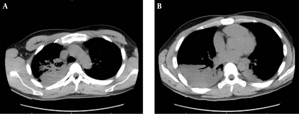
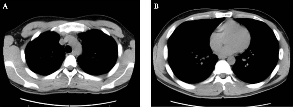
On the following day, computed tomography scans revealed right upper and lower lobe and left lower lobe consolidation (Figure 1A and B), the SPO2 was maintained above 93% with 4 L/min of oxygen applied via a face mask and the patient did not complain of dyspnea. On day 5 following the operation, the patient showed no fever or dyspnea in room air and displayed normal breathing sounds; in addition, computed tomography scans showed an improvement of the lungs (Figure 2A and B).
3. Discussion
Pulmonary collapse may be caused by such factors as endobronchial intubation, mucus secretion plug, and bronchospasm (1, 3, 4). The most likely cause is endobronchial intubation, but it is unlikely that the tip of the tracheal tube was beyond the carina in this case because we checked its position via a chest X-ray.
Bronchial secretion is known to be a cause of atelectasis, and segmental or lobar collapse results from bronchial obstruction by secretion (3). In our case, fiber optic bronchoscopy showed that a large amount of sticky mucus had been secreted, so we postulated that the sticky mucus might have caused the lobar collapse, thereby reducing the SPO2. It is possible that aspiration occurred. However, the oral cavity was clean at intubation and there were no gastric contents or foreign bodies apart from the sticky secretion shown by the bronchoscopy.
Unfortunately, we did not sample and analyze the removed secretion, so the etiology of the acutely induced airway secretion is not clear. We usually employ lidocaine gel for the lubrication of tracheal tube, but in the present case, the tip and cuff of a 8.5 mm tracheal tube were lubricated with 3 mL of a topical metered-dose of 8% Lidocaine pump spray instead. The dose of 8% Lidocaine pump spray used for the lubrication of the tracheal tube might have triggered excessive mucus production. The aerosolized products used for 8% Lidocaine pump spray in Japan contain menthol and ethanol as additives (5, 6). Menthol and ethanol can irritate airway mucosa (6). For example, they can induce a sore throat. Furthermore, menthol-type cigarette smoking can cause pulmonary disease (7, 8). We thus speculate that these additives, especially menthol, might have led to excessive mucus production, although we did not analyze the mucus secretion in this case. Secretion management in the mechanically ventilated patient includes secretion removal and manual hyperinflation (9), which we think worked effectively in this case.
Smokers tend to require deep general anesthesia not only for its induction but also for maintenance (10). Thus, in the present case, we have to consider the possibility that inadequate anesthetic medication might have induced not only the increased secretion but also severe bronchospasm at first, which could have precipitated atelectasis.
In conclusion, we have reported a case of severe pulmonary collapse after intubation related to general anesthesia. When sudden hypoxia occurs, the anesthesiologist should check for endotracheal tube malposition and breathing sounds. In such cases, it is helpful to conduct a chest X-ray and diagnostic bronchoscopy.

