1. Background
When etomidate was first introduced in 1973, it was known as an agent with hypnotic properties that possessed minimal cardiovascular and pulmonary complications (1). Etomidate maintains hemodynamic stability and eliminates histamine release more profoundly than other drugs (2). Research has shown that using etomidate at a dose of 0.3 mg/kg does not cause significant changes in the cardiac stroke volume, heart rate, right atrial pressure, cardiac index, systemic and pulmonary vascular pressures, leading to the increasing use of this medication (3, 4). Despite all of these benefits, there are two major complications associated with etomidate use. The first one is pain on injection, which can be prevented to some extent with a new lipophilic combination (5). The second major problem with etomidate is the occurrence of myoclonus, which is still one of the difficulties of its use among anesthesiologists.
Various studies have reported the incidence of myoclonus in up to 80% of the times when it was administered. Myoclonus can be detrimental to full-stomach patients undergoing emergency surgery, patients with an open eye injury, and those with a history of cardiovascular disease (2, 6). Although the mechanism of myoclonus is not fully understood, various medications have been used to prevent the occurrence of myoclonus (1), including fentanyl (2), benzodiazepines (7), rocuronium (8), opioids (9), dexmedetomidine (10), and etomidate (at pre-treatment dose of 0.03 mg/kg) (11).
Midazolam is a drug from the benzodiazepine group. Concerning the mechanism of action, this group of drugs is attached to their specific GABA receptors in CNS synapses, preventing the opening of GABA chloride channels, thus creating an increase in brain hyper-repolarization. The most desirable feature of these drugs is anxiolytic and anterograde amnesia effects. They are clinically used for acute anxiety disorders, panic attacks, insomnia, other sleep disorders, skeletal muscle relaxation, seizure disorders, and anesthesia induction.
2. Objectives
The present study assessed premedication with low-dose midazolam (0.015 mg/kg) on 132 patients. Midazolam caused a decrease in the incidence of myoclonus. The goal of the study was to compare the effectiveness of low-dose etomidate with midazolam in the prevention of myoclonus induced by etomidate, which is regularly used as an anesthetic drug for ECT. If one of these drugs is found to be superior to the other, we can propose it to be used to prevent myoclonus during ECT.
3. Methods
This study was a randomized double-blind clinical trial performed on 132 patients who referred to the electroconvulsive therapy ward at Alzahra Hospital affiliated to the Isfahan University of Medical Sciences in 2017 - 18.
3.1. Inclusion Criteria
They included age between 6 and 25, no previous history of renal or hepatic diseases, no decompensated history of cardiovascular diseases, no history of chronic respiratory disease, and no history of allergy to midazolam or etomidate.
3.2. Exclusion Criteria
They included patient refusal to participate in the study, patient mortality, and no possibility of intubation during anesthesia.
The study was approved by the Ethics Committee of the University (IR.MUI.REC.1396.3.442) and informed consent was obtained from the patients/legal guardians. This clinical trial was registered at www.irct.ir with identification code IRCT2016092826950N2. A Thymatron IV device (Somatics, LLC. Lake Bluff, IL, USA) was used for ECT.
Moreover, patients were randomly allocated into three groups of control, midazolam, and etomidate. Normal saline solution was administered to the control or no pre-treatment group while 0.015 mg/kg of midazolam (Midazolam, Caspian Tamin Pharmaceutical Co. Rasht, Iran) was administered to the midazolam group and 0.03 mg/kg of etomidate (12, 13) (Etomidate, B.Braun Medical S.R.L, Romania) to the etomidate group.
Patients received isotonic saline solution (5 mL/kg) over 10 min at room temperature and were preoxygenated with 100% oxygen. Then, the medication was injected over 30 s. Ninety seconds after pre-treatment with the medication, etomidate (0.15 mg/kg) was intravenously injected for all the three groups for anesthesia induction within 20 s.
After the administration of the above-mentioned medications in control, midazolam, and etomidate groups, general anesthesia was induced using 0.15 mg/kg of etomidate. After the induction of anesthesia using etomidate, the presence and intensity of myoclonus were assessed, recorded, and scored as follows: score zero for no myoclonus, score one for mild myoclonus (short movement in one part of the body), score two for moderate myoclonus (mild movements in two different muscles), and score three for severe myoclonus (severe clonic movements in two or more muscles) (14).
The sample size included 44 patients per group to achieve a 0.05 level of significance and 80% power in detecting the myoclonus improvement at least with a 50% difference between the control group and any of the other two groups.
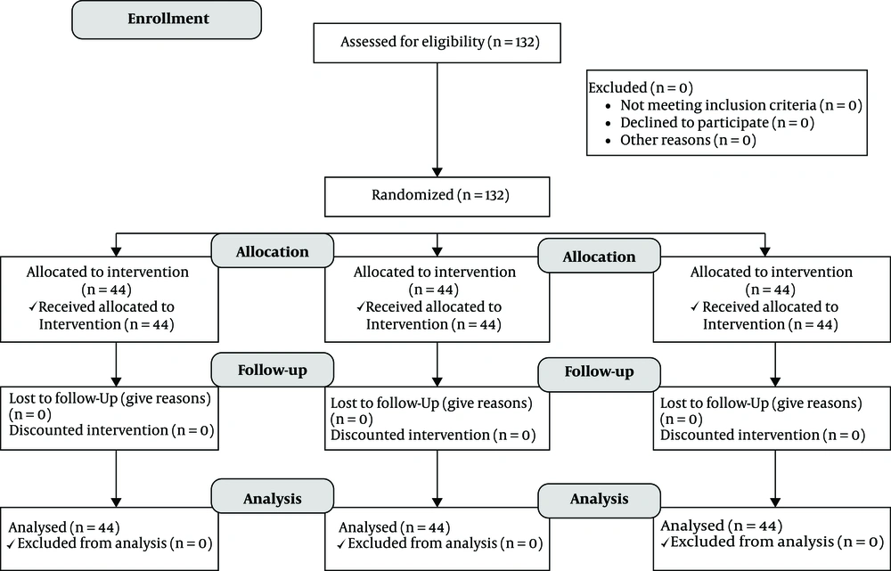
The data were entered into SPSS version 20 software (IBM, USA). Descriptive data were reported as means and percentages. One-way analysis of variance, chi-square test, Kruskal-Wallis test, Mann-Whitney test, and Tukey Post Hoc test were performed for data analysis. A P value of less than 0.05 was considered significant (Figure 1).
4. Results
The study was done on 132 patients in three groups of 44 patients administered with placebo, midazolam, and etomidate. The age of the patients was 10 to 16 years. The demographic data including age (P value = 0.87), height (P value = 0.80), weight (P value = 0.94), and body mass index (BMI) (P value= 0.93) did not show any significant differences. Table 1 compares the data in these three groups (Table 1).
| Variables | Unit | Control | Midazolam | Etomidate | P Valuea |
|---|---|---|---|---|---|
| Age | yrs | 13.8 ± 1.4 | 13.9 ± 1.36 | 14 ± 1.4 | 0.87 |
| Height | cm | 159.3 ± 7.2 | 160.3 ± 7.9 | 159.8 ± 6.9 | 0.80 |
| Weight | kg | 58.6 ± 10.6 | 59.1 ± 10.5 | 59.3 ± 9.6 | 0.94 |
| BMI | kg/m2 | 23.1 ± 4.1 | 23.03 ± 4.1 | 23.3 ± 4.2 | 0.93 |
aOne-way ANOVA test
According to Table 2, the most prevalent psychiatric disorder in this study was bipolar disease among others including major depression, psychosis, schizophrenia, and autism. The three groups did not have significant differences in gender or the type of psychiatric disease (P value > 0.05) (Table 2).
| Variables | Frequency (%) | P Valuea | |||
|---|---|---|---|---|---|
| Control | Midazolam | Etomidate | Total | ||
| Gender | 0.69 | ||||
| Female | 22 (50) | 20 (15.9) | 24 (45.5) | 66 (50) | |
| Male | 22 (50) | 24 (15.9) | 20 (54.5) | 66 (50) | |
| Type of diseases | 0.99 | ||||
| BD II | 30 (68.2) | 33 (75) | 31 (70.5) | 94 (71.2) | |
| BD I | 3 (6.8) | 4 (9.1) | 3 (6.8) | 10 (7.5) | |
| MD | 5 (11.4) | 2 (4.5) | 3 (6.8) | 10 (7.5) | |
| PSY | 2 (4.5) | 2 (4.5) | 2 (4.5) | 6 (4.5) | |
| SCZ | 2 (4.5) | 2 (4.5) | 2 (4.5) | 6 (4.5) | |
| ASD | 2 (4.5) | 1 2.3) | 3 (6.8) | 6 (4.5) | |
Abbreviations: ASD, autism; BD, bipolar disorder; MD, major depression; PSY, psychosis; SCZ, schizophrenia
aIndependent t-test
Table 3 compares the mean values of systolic blood pressure (SBP), Diastolic Blood Pressure (DBP), and Mean Arterial Pressure (MAP) at various measurement times. The independent t-test showed no differences between mean SBP, mean DBP, mean MAP, heart rate (HR), apnea time, recovery time, and arterial oxygen saturation (SaO2) between the three groups. However, there were significant differences in the seizure and recovery time among the three groups. The mean seizure duration in the three groups was not equal (P value < 0.001). The seizure duration was significantly shorter in the midazolam group than in the placebo and etomidate groups (P value < 0.001) but it showed no difference between the placebo and etomidate groups (P value = 0.71) (Table 3).
| Variables | Unit | Control | Midazolam | Etomidate | P Valuea |
|---|---|---|---|---|---|
| SBP | mmHg | 123.8 ± 11.9 | 124.2 ± 11.03 | 125.9 ± 13.1 | 0.69 |
| DBP | mmHg | 74.5 ± 8.9 | 72.1 ± 9.1 | 70.9 ± 9.1 | 0.17 |
| MAP | mmHg | 90.9 ± 8.7 | 89.5 ± 8.5 | 89.3 ± | 0.62 |
| HR | beat/min | 95 ± 11.5 | 96 ± 11.7 | 96.4 ± 10.1 | 0.84 |
| SpO2 | % | 96.9 ± 1.7 | 97.4 ± 1.4 | 97.6 ± 1.5 | 0.12 |
| SD | sec | 32.5 ± 6.5 | 25.7 ± 5.7 | 33.6 ± 7.04 | < 0.001 |
| BS | sec | 24.4 ± 4.1 | 24.136.6 ± 7.94.9 | 24.8 ± 3.4 | 0.76 |
| RD | min | 32.1 ± 8.4 | 36.6 ± 7.9 | 34.7 ± 9.7 | 0.07 |
Abbreviations: BS, breathe spontaneously; DBP, diastolic blood pressure; HR, heart rate; MAP, mean arterial pressure; RD, recovery duration; SBP, systolic blood pressure; SD, seizure duration; SpO2, saturation of peripheral oxygen
aIndependent t-test
The findings of the study demonstrated significant differences in the incidence of myoclonic movements during general anesthesia (P value < 0.001). The prevalence of such movements was lower in the midazolam group (n = 10; 22.7%) than in the placebo groups (n = 29; 69.9%) (P value < 0.001), but with no difference between the etomidate (n = 34; 77.3%) and placebo groups. Also, the intensity of myoclonic movements during general anesthesia was significantly higher in the midazolam group than in the placebo and etomidate groups (P value < 0.001), whereas there was no difference between the placebo and etomidate groups (P value = 0.092) (Table 4).
| Variables | Number (%) | P Value | ||
|---|---|---|---|---|
| Control | Midazolam | Etomidate | ||
| Headache | 6 (13.6) | 2 (4.5) | 14 (31.8) | F = 0.646 |
| Nausea and vomiting | 7 (15.9) | 0 (0) | 13 (29.5) | F = 0.754 |
| Muscular pain | 7 (15.9) | 4 (9.1) | 17 (38.6) | F = 0.026 |
| Myoclonus | 29 (69.9) | 10 (22.7) | 34 (77.3) | 0.001 |
| 0 | 13 (29.5) | 32 (72.3) | 18 (40.9) | 0.001 |
| 1 | 14 (31.8) | 6 (13.6) | 5 (11.4) | |
| Myoclonus intensity | ||||
| 2 | 8 (18.2) | 4 (9.1) | 10 (22.7) | |
| 3 | 7 (15.1) | 0 (0) | 9 (20.4) | |
5. Discussion
Recovery after anesthesia for ECT is of utmost importance since ECT is an effective treatment from both psychiatrists and patients’ perspectives. Decreasing the complications of ECT, which also include the side effects of anesthesia, increases the compliance and satisfaction of patients (10).
Myoclonus following the administration of etomidate is one of the complications of anesthesia used for ECT. This compound is a desirable drug used by anesthesiologists since it has no cardiovascular and hemodynamic changes; however, myoclonus is the only complication still challenging anesthesiologists (15).
It seems that myoclonus caused by etomidate is the result of subcortical dis-inhibition in the brain. It appears that using etomidate can decline cortical activity and the decline occurs prior to the control of subcortical activity. Therefore, the myoclonus is created. Hence, various medications are utilized to prevent the asynchrony of brain activity caused by etomidate to control the center responsible for creating myoclonus even before it starts (14, 16, 17).
The present study compared the prevalence and intensity of myoclonus after the administration of 15 mg/kg of etomidate during anesthesia induction for ECT in groups given placebo, midazolam, and etomidate. The three groups did not have any differences in age, height, BMI, gender distribution, and primary psychiatric disorders requiring ECT. The confounding variables that could negatively affect the study results were similar in these groups.
Our study findings indicated that the occurrence and intensity of myoclonus were significantly lower in the group treated with midazolam. However, the comparison of the prevalence and intensity of myoclonus in the etomidate and placebo groups did not show any significant differences.
Because of the high prevalence of myoclonus following etomidate injection, various studies have been performed to reduce and control such an event. It is important to use a drug that has the least complications not adding to myoclonus of etomidate; for example, many studies have shown that administering an opioid can control myoclonus effectively but may unexpectedly cause side effects such as apnea, respiratory failure, nausea, and vomiting. Therefore, in using opioids, benefits should be weighed against disadvantages (18, 19).
Mullick et al. studied the use and non-use of etomidate pre-treatment with a bolus dose followed by a therapeutic dose of etomidate while the third group was pretreated with a slow injection of etomidate followed by a therapeutic dose of etomidate. It was shown that myoclonus occurred at a significantly lower rate in patients who received a bolus pre-treatment dose followed by a therapeutic dose (11). Also, Salim et al. showed that the intensity of myoclonus was higher in patients not receiving pre-treatment than in the other two groups but no significant difference was observed between the two groups receiving etomidate pre-treatment (20). In our study, we tried an initial bolus dose of pre-treatment, but did not study the bolus and infusion administration of the drug.
Other studies focused more on comparing etomidate premedication with a control group. Aissaoui et al. and Doenicke et al. emphasized the benefits of premedication with etomidate compared to not using etomidate during anesthesia induction using etomidate (11, 16, 21). In another study performed in 2016, Sedighinejad et al. studied the effects of low-dose midazolam, magnesium sulfate, remifentanil, and low-dose etomidate on the prevention of myoclonus caused by etomidate for orthopedic surgery. They showed conflicting findings with our study. The results of statistical analysis showed the benefits of using low-dose etomidate in the decrease of prevalence and intensity of etomidate-induced myoclonus (22).
In the current study, 0.015 mg per kg of midazolam was used, which is quite different than the dose used in other studies. Myoclonus was observed in only 23.8% of patients under study. This is in contrast to Wasinwong et al.’s study that used a double dose of midazolam and observed myoclonus in 60% of patients (23). The rate of myoclonus was up to 75% in the study by Sadighinejad et al. who used the same dose of midazolam as used in our study (22). Myoclonus occurred in only 10% of patients in a study by Huter et al. who used the same dose of midazolam as used in our study (7).
In addition, Salim et al. used 0.05 mg per kg of midazolam and reported the myoclonus rate of 5.6% in patients, with 1.6% showing severe signs that appeared to be due to a difference in methodology and patient selection (19).
The study by Zhang et al. compared the effects of midazolam, butorphanol, and a combination of both drugs on the prevention of myoclonus caused by etomidate. The results of the study demonstrated the effectiveness of using midazolam in decreasing the occurrence and intensity of myoclonus compared to the lack of using a premedication. Indeed, there was no difference between midazolam and butorphanol, but combination therapy was superior to single therapy (24).
Our study also assessed the vital signs, seizure duration, recovery time, and occurrence of apnea in the groups. We showed no significant differences, except for seizure duration that was significantly shorter in the midazolam group. Various other studies assessed these complications while using different kinds of drug compounds. They have mostly focused on using one anesthetic drug rather than using premedication with a drug while using etomidate. For example, Singh et al. showed that seizure duration was significantly shorter when using etomidate than using propofol and thiopental. But in this study, there was no special mention or emphasis on myoclonus as the main side effect of etomidate (25). Hoyer et al. compared etomidate, thiopental, ketamine, and propofol and showed the superiority of ketamine and etomidate, with ketamine being superior to etomidate (26). Tan and Lee also studied etomidate in controlling seizure during ECT; they stressed the high efficacy of this drug in this regard and demonstrated its priority to propofol (27). In a further study by Nazemroaya et al. to assess the effects of premedication on ECT complications, it was specified that using midazolam in ECT could decrease headaches and pain (28). Additionally, Mizrak et al. performed one of a few studies on the effects of premedication on ECT complications. Their study assessed the effects of using midazolam, dexmedetomidine, and placebo prior to the principal anesthetic drug, propofol. The results did not indicate any difference between midazolam and dexmedetomidine, yet both drugs were preferable to placebo (29).
5.1. Limitations
The sample of our study was small. Further studies are required to compare different doses of etomidate and determine other adverse effects.
5.2. Conclusions
According to the findings of our study, using 0.015 mg/kg of midazolam as premedication prior to anesthesia induction using etomidate for ECT significantly decreased the prevalence and intensity of myoclonus compared to placebo. More studies are recommended in this regard considering the conflicting results of various studies.
