1. Background
The shoulder is the most mobile human joint, and shoulder motion needs the coordinated effort of muscles, tendons, ligaments, and bones (1). Rotator cuff muscles play a key role in the shoulder function as the most critical component (2). The tendon of the subscapularis muscle is in the anterior portion of the shoulder (3). It is the main internal rotator of the humerus and acts as the dynamic anterior stabilizer of the glenohumeral joint (4). One of the common causes of shoulder pain and dysfunction is rotator cuff tears (5), and subscapularis tendon tear was known as an uncommon disorder. However, the development of arthroscopic diagnostics indicated the higher prevalence of subscapularis tendon tears (6) as it is diagnosed in about 30% of all arthroscopic shoulder procedures and approximately 50% of rotator cuff tendon repairs (4). The most common signs and symptoms of subscapularis tendon tear include shoulder pain, decreased range of motion, and shoulder weakness (7). Some previous investigations reported positive clinical test results, which were 20% and 31% for the belly-press and lift-off tests after open repair, respectively (8, 9). Moreover, the prevalence ratio of isolated rupture to total injuries involving the subscapularis muscle has been estimated to be about 10.1%. The isolated rupture of the subscapularis muscle was first introduced by Smith (10).
Arthroscopy is considered the gold standard in diagnosing rotator cuff tears because of direct vision and relatively high diagnostic power (11). Although magnetic resonance imaging (MRI) has a high accuracy for diagnosing rotator cuff tear due to the specific insertion site of the subscapularis tendon, the accuracy of MRI for diagnosing subscapularis tendon tear is about 70% (12). Nowadays, shoulder arthroscopy with a direct vision of the subscapularis tendon is the preferred method for diagnosing such lesions. Over the last decade, arthroscopy surgery was improved for shoulder treatments (13). There have been many studies on the outcomes of the arthroscopic repair of a rotator cuff tear, while few studies investigated the outcomes of isolated subscapularis tendon tear.
2. Objectives
Consequently, the present study aimed to evaluate the medium-term clinical outcomes of the arthroscopic repair of an isolated subscapularis tendon tear in a mean follow-up time of 4 years. We hypothesized that patients with isolated subscapularis tendon tears benefit from arthroscopic repair in the medium-term clinical evaluation, and its outcome is comparable to open surgery.
3. Methods
This prospective cohort study was conducted on patients referred to our shoulder clinic in Besat Hospital, Hamadan, Iran, during April 2011 - March 2017. All patients with shoulder pain who were diagnosed as isolated subscapularis tendon tear according to MRI and had an indication for surgical repair were included in the current study and were treated by arthroscopic repair. Cases with any other rotator cuff tear in MRI or arthroscopic evaluation were excluded from the study. Patients who had type 1 or type 5 subscapularis tendon tears were also excluded from the study as they have no indication for surgical repair. In addition, reluctance and not cooperating in the examinations were considered exclusion criteria.
3.1. Surgical Technique
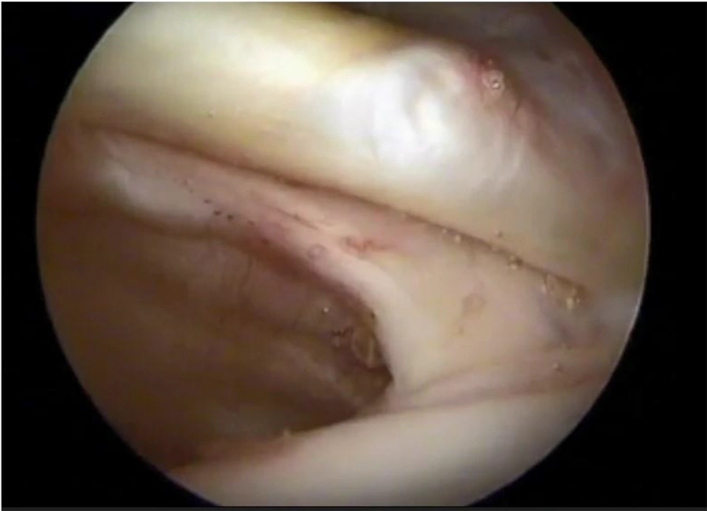
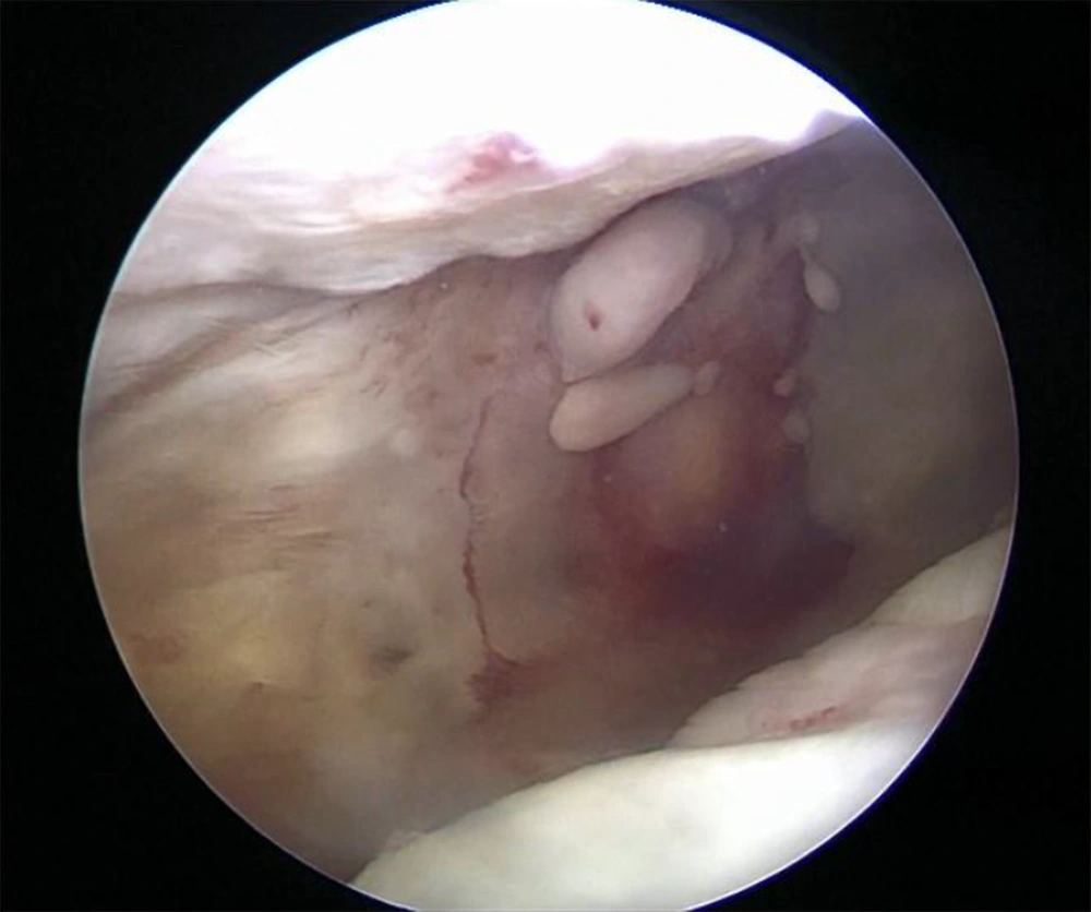
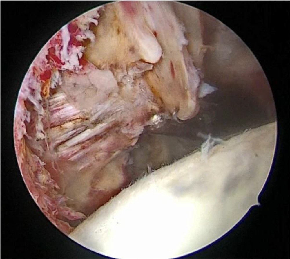
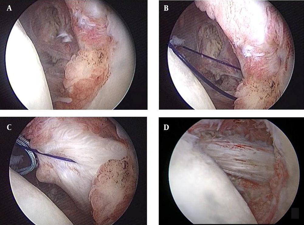
Under general anesthesia, the patient was in a beach chair position with the arm in 60° forward flexion and 30° abduction. We evaluated the glenohumeral joint for any pathology, including subscapularis or other rotator cuff tears, pulley integrity, and LHB tendon, using a 30° scope from the posterior portal. It is important to see all the profiles of subscapularis insertion by internal rotation of the arm while looking at the insertion site with a 30° or 70° arthroscopic lens (Figure 1). If there was a type 3 subscapularis tendon tear (Figure 2), we performed tendon repair by using an anchor suture from the anterosuperior portal while looking at the joint from the posterior portal. If there was a type 4 subscapularis tendon tear (Figure 3), we switched the scope to subacromial space and performed out-of-the-box release and tendon repair using an anterolateral portal. We used a nylon suture strand during tendon release to be aware of the efficacy of tendon release to reach the footprint in all cases (Figure 4). Video of arthroscopic procedure was recorded for future evaluation in follow-up. The individuals were followed for at least 3 years. The modified UCLA, Quick DASH, and visual analogue scale (VAS) were measured, and the belly-press and lift-off tests were performed in the examination.
3.2. Data Collection Tools
Data collection tools in this study were as follow:
(1) Patient registration profile questionnaire: It included age at the time of surgery, gender, occupation and description, date and type of surgery, a history of previous examinations, manner and time of injury, the interval between injury and surgery, records regarding sports activities.
(2) Modified UCLA score: The result of this numerical criterion is in the range of 0 - 35, which is interpreted as weak, medium, good, and excellent when the score is < 21, 21 - 27, 28 - 33, and 34 - 35, respectively.
(3) VAS: The result is numerically in the range of 0 - 10, with 10 indicating the most severe pain the patient has ever had and 0 representing no pain.
(4) Quick DASH Questionnaire: It is an abbreviated version of the DASH Outcome measure that examines 11 upper limb physical and clinical functions in people with musculoskeletal problems instead of 30. The result of this criterion is numerically in the range of 0 - 100, with a higher score showing greater disability.
(5) Belly-press Test: The test result is binary (positive/negative), with a positive result being the sign of damage to the subscapularis muscle.
(6) Lift-off Test (Garber’s Test): This test is also binary (positive/negative), with a negative result being the sign of a lesion in the subscapularis muscle.
3.3. Statistical Analysis
The collected data were analyzed by the SPSS software version 24. Frequency and percentage for qualitative variables and mean and standard deviation for quantitative variables were used to describe the data. The mean scores of modified UCLA, Quick DASH, and VAS before and after surgery in patients were compared using the paired t-test. P-value < 0.05 was considered significant.
4. Results
Out of 11 patients who met our inclusion criteria, three were female (27.3%), and seven were male (72.7%). The mean age of patients was 58.5 ± 8.12 years (range: 52 - 70 years), and the mean interval between injury and surgery was 5.28 ± 3.9 months. One of the patients was a professional athlete, and the rest did not have regular exercise. The cause of injury was degenerative in two patients (18.2%) and trauma in 9 cases (81.8%). The mean follow-up time was 5.3 years (range: 3 - 9 years). Nine patients were completely satisfied with the surgery outcome, two cases were relatively satisfied with the operation outcome, and there was no dissatisfaction.
All seven patients were asked whether they would choose surgery again if they went back in time?" and they answered affirmatively. All 11 patients returned to their daily activities after recovering. The belly-press test was negative in ten patients and positive in one patient. Furthermore, the lift-off test was positive in ten patients and negative in the same patient who had a positive belly-press test. As shown in Table 1, the mean of UCLA score augmented significantly after surgery (33.28 ± 2.92 vs. 10.71 ± 3.4, P < 0.001). The mean Quick DASH declined from 38.26 ± 27.94 preop to 7.56 ± 16.43 postop (P = 0.003). Moreover, the mean VAS score significantly reduced (0.57 ± 1.51 post-intervention compared to 4.57 ± 1.21 pre-intervention, P < 0.001).
| Index | Pre-intervention | Last Follow-up Post-intervention | P-Value |
|---|---|---|---|
| UCLA score | 10.71 ± 3.40 | 33.28 ± 2.92 | < 0.001 |
| Quick DASH | 38.26 ± 27.94 | 7.56 ± 16.43 | 0.003 |
| VAS score | 4.57 ± 1.21 | 0.57 ± 1.51 | < 0.001 |
a Values are expressed as Mean ± SD.
5. Discussion
The subscapularis is the largest and most powerful rotator cuff muscle and has an important role in shoulder movement and stability (14). Detecting subscapularis injury by physical examination is difficult, and the assessment of subscapularis integrity by MRI may have limitations (12, 15). As a result, arthroscopic evaluation and repair could be more valuable. In the present study, the medium-term clinical outcome of the arthroscopic repair of isolated subscapularis tendon tear was evaluated in Besat Hospital of Hamadan, Iran. We observed excellent results following the arthroscopic repair of isolated subscapularis tears. Our findings showed that out of eleven patients, nine were completely satisfied, only two cases were relatively satisfied with operation outcome, and no dissatisfaction. Edwards et al. similarly demonstrated that the open repair of isolated subscapularis tears resulted in an acceptable improvement in shoulder function (16).
Burkhart and Tehrany reported a series of eight cases of isolated subscapularis tears in which the UCLA scores significantly improved from a preoperative average of 10 to a postoperative average of 32.8 (P < 0.002) (17). In another follow-up of 20 years conducted by Collin et al. (18), satisfactory results were achieved for patients after repairing isolated supraspinatus tendon tears, and the clinical outcome significantly improved. Saltzman et al. (19), in a systematic review of eight studies, showed that arthroscopic subscapularis repair seemed to be a reasonable option for treating isolated tears of subscapularis to obtain successful functional and patient-reported clinical outcomes. Yoon et al. (20) compared the single-row and double-row repairs of the isolated subscapularis tendon. These authors indicated that the mean UCLA score rose significantly two years after surgery. It was found that both arthroscopic single-row and double-row suture-bridge repairs performed for isolated subscapularis tears of total thickness resulted in satisfactory clinical outcomes and suitable structural integrity. It was also noted that patients with good muscle quality had no significant difference.
Nove-Josserand et al., in a retrospective study of 25 patients, found that the arthroscopic repair of isolated subscapularis tears is associated with improved shoulder function and enhanced clinical results (21). Lafosse et al. studied 17 patients after arthroscopic repair and observed that the arthroscopic repair of an isolated subscapularis tear could yield remarkable improvements in shoulder function and significantly reduce pain, resulting in a durable structural repair (22). Arthroscopic repair of isolated subscapularis tendon tears caused an obvious enhancement in the shoulder function and a low re-rupture rate.
In addition, the mean VAS score was significantly reduced. Katthagen et al. demonstrated that improved function and decreased pain were associated with high patient satisfaction (23). In the research by Lafosse et al. (22), the mean UCLA score, pain score, forward flexion, and external rotation improved (P < 0.05). There are few studies regarding the evolution of Quick DASH, and similar findings were previously reported by Eren et al. (24), indicating that the mean of Quick DASH score significantly declined (P < 0.05). Seppel et al. (8) evaluated three patients (17.6%) and showed persistent positive clinical test results (belly-press/lift-off). However, in our study, the belly-press test was negative in all patients, and the lift-off test was positive in all participants. Other studies have reported satisfactory clinical outcomes after repairing isolated subscapularis tears (22). The current investigation, for the first time, presents quantitative and reproducible medium-term data after the arthroscopic repair of isolated subscapularis tendon tear of types 3 and 4 in Laffose classification.
5.1. Conclusions
Considering the satisfaction rate, functional outcome, and the rate of returning to daily activities in the medium-term follow-up of the arthroscopic repair of isolated subscapularis tendon tear, this technique can be highly recommended as an alternative method for the open repair of lesions. All patients returned to their daily activities after recovering at a medium-term follow-up.


