Effects of Peripheral Cold Application and Antipyretics on Hemodynamic Parameters in Febrile ICU Patients Diagnosed with Hospital-Acquired Infections

authors:

avatar Hossein Asgarpour 1 , * , avatar Rahşan Çevik Akyil 2 , avatar Serdal Öğüt 3

Department of Surgical Nursing, Çanakkale Onsekiz Mart University, Çanakkale, Turkey
Department of Internal Nursing, Adnan Menderes University, Aydın, Turkey
Department of Nutrition and Dietetic, Adnan Menderes University, Aydın, Turkey

how to cite: Asgarpour H, Çevik Akyil R, Öğüt S. Effects of Peripheral Cold Application and Antipyretics on Hemodynamic Parameters in Febrile ICU Patients Diagnosed with Hospital-Acquired Infections. Ann Mil Health Sci Res. 2018;16(3):e85116. https://doi.org/10.5812/amh.85116.

Abstract

Background:

Accurately assessing hemodynamic parameters plays an important role in preventing complications during and after interventions related to a fever in febrile patients. There are not enough studies related on the effects of peripheral cold application (PCA) and antipyretics (APs) on hemodynamic parameters in febrile patients.

Objectives:

The aim was to determine the effects of PCA and APs on hemodynamic parameters in febrile ICU patients diagnosed with hospital-acquired infections (HAIs).

Methods:

In this retrospective study, the archive documents of the 346 adult ICU patients diagnosed with HAIs of a university hospital in Turkey were evalauted. Body temperature (BT), systolic/diastolic blood pressure (SBP/DBP), pulse rate (PR), respiratory rate (RR), and arterial O2Sat (aO2sat) changes one hour before, during, and one hour after application of PCA or APs were evalauted.

Results:

The incidence of HIAs was 4.16%. A total of 46% of the patients had a bacteremia infection and causative microorganisms in 16.8% were Acinetobacter spp. BT, DBS, PR, and aO2Sat changes one hour before, during, and one hour after PCA were significantly different. In addition, BT, RR, and aO2Sat changes one hour before, during, and one hour after APs were also significantly different (P < 0.05). More decrease in SBP and DBP were observed between during and one hour after APs compare with PCA.

Conclusions:

BT and hemodynamic parameters, especially SBP and DBP changes, were higher in APs compared with PCA. Knowledge regarding the effects of APs on BT and hemodynamic parameters will be of benefit to the ICU staff in terms of quality of care in febrile patients. The ICU staff should be proactive in ensuring that they have an evidence-based management plan for the prevention of HIAs and management of fever that conforms to the relevant clinical guidelines and integrates multidisciplinary care.

1. Background

Fever is defined as an increase in the core body temperature ≥ 38.3°C (1). The incidence of fever ranges between 28% and 75% in critically ill patients (2, 3). Fever depends on a complex physiologic response to infectious agents and other conditions such as pulmonary embolism, gastrointestinal bleeding, drug reactions, trauma, surgery, and etc. (2, 4, 5). During a febrile episode, an increase of oxygen consumption and energy expenditure occurs (6). Subsequent increases in oxygen consumption, respiratory quotient, and cardiac output add a considerable burden to febrile patients who might be unable to compensate for the increased metabolic demand (7, 8). With the increasing of oxygen demand in cell levels during fever, easily separated bunding oxygen on Hb in the cells occur (6, 9). Based on this information, blood gas values are directly affected by fever (10, 11).

Fever is a manifestation of cytokine release in response to a variety of stimuli. Cytokines may be released in response to exogenous pyrogens such as microorganisms (12). Cytokines stimulate the release of prostaglandins from the anterior hypothalamus and the prostaglandins are then responsible for acting to reset the thermoregulatory center, thereby, causing a rise in the temperature set point that manifests as a fever (13). Besides evaluating the febrile patient and initiating a workup based on the clinical evaluation, it is common for the patients to receive either pharmacologic or mechanical antipyretic therapy (14, 15). Clinicians have controlled fevers by using external cooling methods and antipyretics (APs) (16, 17). Studies showed that the use of APs to diminish fevers correlates with negatively affecting patient outcomes (18). APs act mainly by the inhibation of prostaglandin synthesis or decrease prostaglandin E2 levels during fever (5, 19). Furthermore, APs have some adverse effects such as hypotension, renal, and hepatic toxicity (20). In the study of Vera et al. the effects of different APs on hemodynamic parameters decrease in systolic blood pressure were observed (16). Although, non-pharmacological and pharmacological methods are used to reduce core BT in febrile patients, little research related to the effects of these methods on BT and hemodynamic parameters have been performed (21-23). In the present study we aimed to determine the effects of peripheral cold application (PCA) and APs on BT and heamodynamic parameters (systolic/diastolic and mean arterial blood pressure, pulse rate, repiratory rate and arterial O2 saturation) in febrile ICU adult patients diagnosed with hospital-acquired infections (HAIs).

2. Methods

This retrospective study was conducted by evaluating the documents of the patients diagnosed with HAIs in the internal-surgical ICUs at a university hospital in the west of Turkey between 2014 - 2015. It was determined that in total, 9547 patients stayed in the adult and pediatrics ICUs, and 448 of these patients were diagnosed with a HAIs. A total of 346 of the 448 patients that stayed in the adults ICUs had HAIs and related documents were evaluated. In order to be able to carry out the study, written permissions from Adnan Menderes University Non-Invasive Ethics Committee, Chief Physician of Adnan Menderes University Hospital, and the Head of the Department of Infectious Diseases were received. One hour before, during, and one hour after PCA or APs interventions related to febrile episodes, BT, SBD, DBS, PR, RR, and aO2Sat values were recorded. For statistical analysis, SPSS version 17 (SPSS Inc., Chicago, IL) was used. Shapiro Wilk test was used to check the normality of distribution of continuous variables. As appropriate, data are presented as percent and mean (SD). To compare mean values, paired t test and one-way ANOVA with repeated measures test were used. Statistical significance was set at P < 0.05, as appropriate.

3. Results

The mean age of the patients was 67.91 ± 14.76, mean length of stay in the ICU was 27.02 ± 25.68 days. A total of 46% and 31.8% of the patients had bacteremia and VIP, respectively; 16.8% of the causative microorganisms were Acinetobacter spp.

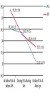
A decrease of 0.17°C (P = 0.004) and 0.44°C (P < 0.001) of mean BT was observed during and one hour after PCA compared with one hour before, respectively. A decrease of 0.16°C (P = 0.004) and 0.65°C (P < 0.001) was observed during and one hour after APs compared with one hour before, respectively (Figure 1).

Mean body temperature before, during, and after PCA and APs

A decrease of 5.19 mmHg (P = 0.002) and 5.06 mmHg (P = 0.009) of mean SBP was observed during and one hour after PCA compared with one hour before, respectively. A decrease of 3.55 mmHg (P = 0.121) and 5.73 mmHg (P = 0.010) was observed during and one hour after AP compared with one hour before, respectively (Figure 2).

Mean systolic blood pressure before, during, and after PCA and APs

A decrease of 1.18 mmHg (P = 0.249) and 2.76 mmHg (P = 0.049) of mean DBP was observed during and one hour after PCA compared with one hour before, respectively. A decrease of 2.69 mmHg (P = 0.039) and 5.33 mmHg (P < 0.001) was observed during and one hour after AP compared with one hour before, respectively (Figure 3).

Mean diastolic blood pressure before, during, and after PCA and APs

A decrease of 3.85 beats/min (P = 0.003) and 5.20 beats/min (P < 0.001) of mean PR was observed during and one hour after PCA compared with one hour before, respectively. A decrease of 4.63 beats/min (P < 0.001) and 5.69 beats/min (P < 0.001) was observed during and one hour after AP compare with one hour before, respectively (Figure 4).

Mean pulse rate before, during, and after PCA and APs

A decrease of 0.11 breaths/min (P = 0.850) and 0.46 breaths/min (P = 0.585) of mean RR was observed during and one hour after PCA compared with one hour before, respectively. A decrease of 0.10 breaths/min (P = 0.887) and 1.12 breaths/min (P = 0.209) was observed during and one hour after AP compared with one hour before, respectively (Figure 5).

Mean respiratory rate before, during, and after PCA and APs

An increase of 0.37% (P = 0.134) and a decrease of 0.02% (P = 931) of mean aO2Sat was observed during and one hour after PCA compared with one hour before, respectively. An increase of 0.68% (P = 0.139) and 0.86 % (P = 0.020) was observed during and one hour after AP compared with one hour before, respectively (Figure 6).

Mean arterial O2 saturation before, during, and after PCA and APs

Comparing BT and hemodynamic parameters over time, from one hour before, during, and one hour after, PCA, BT, DBP, PR, and aO2Sat parameters did significantly change (P < 0.05), while SBP and RR parameters did not significantly changes (P > 0.05). Comparing BT and hemodynamic parameters over time, from one hour before, during, and one hour after APs, BT, RR, and aO2Sat parameters did significantly change (P < 0.05), while SBP, DBP, and PR parameters did not significantly changes (P > 0.05) (Table 1).

Table 1.

BT and Hemodynamic Parameters Changes 1 Hour Before, During, and 1 Hour After PCA and APs

Variables1 Hour Before PCADuring PCA1 Hour After PCAP Value1 Hour Before APsDuring APs1 Hour After APsP Value
BT,°C38.51 ± 0.3738.33 ± 0.5738.07 ± 0.76 < 0.001a39.35 ± 0.9038.25 ± 0.6037.73 ± 0.60 < 0.001a
SBP, mmHg129.61 ± 3.23124.41 ± 3.16124.54 ± 3.190.250124.06 ± 3.67119.84 ± 3.39117.19 ± 3.240.865
DBP, mmHg65.92 ± 1.8764.80 ± 1.8263.16 ± 1.970.002a59.32 ± 1.5457.40 ± 1.5854.69 ± 1.280.492
PR, beats/min113.38 ± 1.65108.73 ± 1.68108.52 ± 1.680.003a117.58 ± 1.80112.80 ± 1.56111.88 ± 1.940.700
RR, breaths/min23.57 ± 1.0123.45 ± 0.8623.11 ± 0.930.08026.10 ± 1.0325.56 ± 0.9424.79 ± 0.92 < 0.001a
O2Sat, %96.86 ± 2.5697.22 ± 1.9896.84 ± 2.580.004a95.53 ± 2.1696.25 ± 2.7496.39 ± 2.38 < 0.001a

4. Discussion

In the present study, the incidence of the HIAs was 4.16%, and 46% of the patients had a bacteremia infection, and the most causative microorganism was Acinetobacter spp. In the O'Shea et al. (24) study 28.8% of the ICU patients developed an infection and the responsible organisms included Pseudomonas, Acinetobacter, E. coli, ect. (18). In the Erman et al. (25) study 6.2% of neurosurgery patients developed an infection and the predominantly isolated microorganisms were SA, Acinetobacter baumanii, and Staphylococcus epidermidis (19). In the Sturm (26) study the majority of microorganisms in craniotomy wounds were gram-positive (20). In the Celik et al. study Acinetobacter and Pseudomonas were the most common causes of nosocomial infections in ICU patients (4). Most studies stated that the most frequent HIAs in ICU patients were urinary tract infections (24-27). Erbay et al. (28), Meric et al. (29), Akhtar, (30) Baghaei et al. (31), Pradhan et al. (32) and Khan et al. (33) stated that the most frequent HIAs in ICU patients were respiratory tract infections (28-30, 34-36). In the study of Dereli et al. (37), the most frequent HIAs were bloodstream, soft tissue, and skin infections (31). In the study of Farzianpour et al. (36) and Amazian et al. (38) E. coli was the most isolated microorganism in HIAs (27, 32). In the study of de Oliveira et al. (34), Pradhan et al. (32), Dereli et al. (37), Alatrouny and Luna, Acinetobacter baumanii was the most isolated microorganism (25, 29, 31, 33, 37). In the Erbay et al. (28), Akhtar, Baghaei et al. (31) and Khan et al. (33) study, Pseudomonas aeruginosa, and in the Kolgeliler study, MRSA were the most isolated microorganisms (28, 30, 34, 36, 38).

Fever is a complex physiologic response triggered by infectious or aseptic stimuli. Elevations in body temperature occur when concentrations of prostaglandin E2 increase within certain areas of the brain (22). The induction and maintenance of fever during infection involves the tightly coordinated interplay between the innate immune system and neuronal circuitry within the central and peripheral nervous systems (23). At the persent study, BT, DBS, PR, and aO2Sat changes one hour before, during, an done hour after PCA were significantly different, and BT, RR, and aO2Sat changes one hour before, during, and one hour after APs were significantly different. More decrease in SBP and DBP were observed between during and one hour after APs compared with PCA.

In the studies of Kiekkas et al. (6) and Asgar Pour and Yavuz (9) fever episodes were followed by changes in hemodynamic parameters. In the present study, BT and hemodynamic parameters changes were higher in AP compared with PCA. In the study by Polderman et al. (39), on neurology, febrile patients, to avoid myocardial and neurological damage mild to moderate hypotermia, applied cold water, drafted a blanket, and applied cold normal salin infusion. The MAP increased 15 mmHg, this parameter was seen higher in patients without hemodynamic instability. The study results showed that hypothermia is achieved safely and rapidly with cold water, drafted blanket, and cold normal salin infusion together (39). Cormio and Citero study results to assess two different APs effects on MAP and ICP and evaluation their efficacy in fever control on patients with brain injury showed that the CPP and MAP were found high in the diclofenac applied patient group (40). In the study by Hata et al. results reducing the core BT effects oxygen consumption in acute brain injury (7).

In conclusion, as the rate of the HAIs is the most important indicator in the quality of care, it becomes important for the ICU clinicians to use their roles as a caregiver and as an educator in the prevention of the HAIs. Moreover, knowledge regarding the effects of APs on BT and hemodynamic parameters will be of a benefit to clinicians in terms of quality of care in ICU patients.

References

  • 1.

    Laupland KB, Shahpori R, Kirkpatrick AW, Ross T, Gregson DB, Stelfox HT. Occurrence and outcome of fever in critically ill adults. Crit Care Med. 2008;36(5):1531-5. [PubMed ID: 18434882]. https://doi.org/10.1097/CCM.0b013e318170efd3.

  • 2.

    Henker R, Rogers S, Kramer DJ, Kelso L, Kerr M, Sereika S. Comparison of fever treatments in the critically ill: A pilot study. Am J Crit Care. 2001;10(4):276-80. [PubMed ID: 11432215].

  • 3.

    Fernandez A, Schmidt JM, Claassen J, Pavlicova M, Huddleston D, Kreiter KT, et al. Fever after subarachnoid hemorrhage: Risk factors and impact on outcome. Neurology. 2007;68(13):1013-9. [PubMed ID: 17314332]. https://doi.org/10.1212/01.wnl.0000258543.45879.f5.

  • 4.

    Celik S, Yildirim I, Arslan I, Yildirim S, Erdal F, Yandi YE. Hemodynamic and oxygenation changes in surgical intensive care unit patients with fever and fever lowering nursing interventions. Int J Nurs Pract. 2011;17(6):556-61. [PubMed ID: 22103821]. https://doi.org/10.1111/j.1440-172X.2011.01973.x.

  • 5.

    Lee JJ, Simmons DL. Antipyretic therapy: Clinical pharmacology. Handb Clin Neurol. 2018;157:869-81. [PubMed ID: 30459046]. https://doi.org/10.1016/B978-0-444-64074-1.00054-9.

  • 6.

    Kiekkas P, Brokalaki H, Manolis E, Askotiri P, Karga M, Baltopoulos GI. Fever and standard monitoring parameters of ICU patients: A descriptive study. Intensive Crit Care Nurs. 2007;23(5):281-8. [PubMed ID: 17531490]. https://doi.org/10.1016/j.iccn.2007.04.001.

  • 7.

    Hata JS, Shelsky CR, Hindman BJ, Smith TC, Simmons JS, Todd MM. A prospective, observational clinical trial of fever reduction to reduce systemic oxygen consumption in the setting of acute brain injury. Neurocrit Care. 2008;9(1):37-44. [PubMed ID: 17985248]. https://doi.org/10.1007/s12028-007-9015-1.

  • 8.

    Asgar Pour H, Yavuz M. [Effect of body temperature height (fever) on the hemodynamic parameters: Literature review]. J Maltepe Univ School Nurs. 2010;3:73-9. Turkish.

  • 9.

    Asgar Pour H, Yavuz M. Effects of fever on haemodynamic parameters in neurosurgical intensive care unit patients. Intensive Crit Care Nurs. 2014;30(6):325-32. [PubMed ID: 25242210]. https://doi.org/10.1016/j.iccn.2014.07.001.

  • 10.

    Yenen OS, Altunay H. Ateş ve nedeni bilinmeyen ateş. In: Willke Topçu A, Söyletir G, Doğanay M, editors. Enfeksiyon hastalıkları ve mikrobiyolojisi. 2nd ed. Istanbul, Turkey: Nobel Tıp Kitapevleri; 2002. p. 388-98. Turkish.

  • 11.

    Laws C, Jallo J. Fever and infection in the neurosurgical intensive care unit. JHNJ. 2010;5(2):5. https://doi.org/10.29046/JHNJ.005.2.005.

  • 12.

    Rudra A, Pal S, Acharjee A. Postoperative fever. Indian J Crit Care Med. 2006;10(4):264. https://doi.org/10.4103/0972-5229.29848.

  • 13.

    Perlino CA. Postoperative fever. Med Clin North Am. 2001;85(5):1141-9. [PubMed ID: 11565491]. https://doi.org/10.1016/S0025-7125(05)70369-2.

  • 14.

    Hedenstierna G, Mancebo J, Brochard L, Pinsky MR. Applied physiology in intensive care medicine, bacher A. Effects of body temperature on blood gases. Springer; 2009. p. 49-52. https://doi.org/10.1007/978-3-642-01769-8.

  • 15.

    Ryan M, Levy MM. Clinical review: Fever in intensive care unit patients. Crit Care. 2003;7(3):221-5. [PubMed ID: 12793871]. [PubMed Central ID: PMC270667]. https://doi.org/10.1186/cc1879.

  • 16.

    Vera P, Zapata L, Gich I, Mancebo J, Betbese AJ. [Hemodynamic and antipyretic effects of paracetamol, metamizol and dexketoprofen in critical patients]. Med Intensiva. 2012;36(9):619-25. Spanish. [PubMed ID: 22425338]. https://doi.org/10.1016/j.medin.2012.02.003.

  • 17.

    Lee JH, Wei L, Gu X, Won S, Wei ZZ, Dix TA, et al. Improved therapeutic benefits by combining physical cooling with pharmacological hypothermia after severe stroke in rats. Stroke. 2016;47(7):1907-13. [PubMed ID: 27301934]. [PubMed Central ID: PMC4927220]. https://doi.org/10.1161/STROKEAHA.116.013061.

  • 18.

    Karadakovan A, Aslan EF. Dahili-Cerrahi Hastaliklarda Hemsirelik Bakimi. Izmir, Turkey: Akademisyen Tıp Kitapevi; 2009. Turkish.

  • 19.

    Basoglu OK, Bacakoglu F, Ersin S, Erikoglu M, Kose T. Üst karın cerrahisinde postoperatif solunumsal komplikasyon riskinin preoperatif parametrelerle ilişkisi. Thorax J. 2000;2:17-22. Turkish.

  • 20.

    Kiekkas P, Aretha D, Bakalis N, Karpouhtsi I, Marneras C, Baltopoulos GI. Fever effects and treatment in critical care: Literature review. Aust Crit Care. 2013;26(3):130-5. [PubMed ID: 23199670]. https://doi.org/10.1016/j.aucc.2012.10.004.

  • 21.

    Asgar Pour H, Yavuz M. Effects of peripheral cold application on core body temperature and haemodynamic parameters in febrile patients. Int J Nurs Pract. 2014;20(2):156-63. [PubMed ID: 24713012]. https://doi.org/10.1111/ijn.12151.

  • 22.

    Aronoff DM, Neilson EG. Antipyretics: Mechanisms of action and clinical use in fever suppression. Am J Med. 2001;111(4):304-15. [PubMed ID: 11566461]. https://doi.org/10.1016/S0002-9343(01)00834-8.

  • 23.

    Evans SS, Repasky EA, Fisher DT. Fever and the thermal regulation of immunity: The immune system feels the heat. Nat Rev Immunol. 2015;15(6):335-49. [PubMed ID: 25976513]. [PubMed Central ID: PMC4786079]. https://doi.org/10.1038/nri3843.

  • 24.

    O'Shea M, Crandon I, Harding H, Donaldson G, Bruce C, Ehikhametalor K. Infections in neurosurgical patients admitted to the Intensive Care Unit at the University Hospital of the West Indies. West Indian Med J. 2004;53(3):159-63. [PubMed ID: 15352744].

  • 25.

    Erman T, Demirhindi H, Gocer AI, Tuna M, Ildan F, Boyar B. Risk factors for surgical site infections in neurosurgery patients with antibiotic prophylaxis. Surg Neurol. 2005;63(2):107-12. discussion 112-3. [PubMed ID: 15680644]. https://doi.org/10.1016/j.surneu.2004.04.024.

  • 26.

    Sturm L. Neurosurgical surgical site infection–review and prevention strategies. Int J Infect Control. 2009;5(2). https://doi.org/10.1016/j.surneu.2004.04.024.

  • 27.

    Shaikh JM, Devrajani BR, Shah SZ, Akhund T, Bibi I. Frequency, pattern and etiology of nosocomial infection in intensive care unit: An experience at a tertiary care hospital. J Ayub Med Coll Abbottabad. 2008;20(4):37-40. [PubMed ID: 19999200].

  • 28.

    Erbay H, Yalcin AN, Serin S, Turgut H, Tomatir E, Cetin B, et al. Nosocomial infections in intensive care unit in a Turkish university hospital: A 2-year survey. Intensive Care Med. 2003;29(9):1482-8. [PubMed ID: 12898002]. https://doi.org/10.1007/s00134-003-1788-x.

  • 29.

    Meric M, Willke A, Caglayan C, Toker K. Intensive care unit-acquired infections: Incidence, risk factors and associated mortality in a Turkish university hospital. Jpn J Infect Dis. 2005;58(5):297-302. [PubMed ID: 16249625].

  • 30.

    Akhtar N. Hospital acquired infections in a medical intensive care unit. J Coll Physicians Surg Pak. 2010;20(6):386-90. [PubMed ID: 20642968].

  • 31.

    Baghaei R, Mikaili P, Nourani D, Khalkhali HR. An epidemiological study of nosocomial infections in the patients admitted in the Intensive Care Unit of Urmia Imam Reza Hospital: An etiological investigation. Ann Biolog Res. 2011;2:172-8.

  • 32.

    Pradhan NP, Bhat SM, Ghadage DP. Nosocomial infections in the medical ICU: A retrospective study highlighting their prevalence, microbiological profile and impact on ICU stay and mortality. J Assoc Physicians India. 2014;62(10):18-21. [PubMed ID: 25906516].

  • 33.

    Khan MS, Kundra P, Cherian A, Noyal MJ, Sistla S. Epidemiology of nosocomial infections in an intensive care unit at a tertiary care hospital in India: A retrospective study. Int J Infect Control. 2015;11:1-5. https://doi.org/10.3396/IJIC.v11i2.016.15.

  • 34.

    de Oliveira AC, Kovner CT, da Silva RS. Nosocomial infection in an intensive care unit in a Brazilian university hospital. Rev Lat Am Enfermagem. 2010;18(2):233-9. [PubMed ID: 20549123]. https://doi.org/10.1590/S0104-11692010000200014.

  • 35.

    Ozer B, Tatman-Otkun M, Memis D, Otkun M. Nosocomial infections and risk factors in intensive care unit of a university hospital in Turkey. Open Med. 2010;5(2):203-8. https://doi.org/10.2478/s11536-009-0095-5.

  • 36.

    Farzianpour F, Sokhanvar M, Ashrafi E, Khalaj G. The 3-year trend of nosocomial infections in intensive care unit: A case Study in Iran. Indian J Appl Res. 2015;5(3):152-4.

  • 37.

    Dereli N, Ozayar E, Degerli S, Sahin S, Koc F. Three-year evaluation of nosocomial infection rates of the ICU. Braz J Anesthesiol. 2013;63(1):73-8. [PubMed ID: 24565091]. https://doi.org/10.1016/j.bjane.2012.03.004.

  • 38.

    Amazian K, Rossello J, Castella A, Sekkat S, Terzaki S, Dhidah L, et al. [Prevalence of nosocomial infections in 27 hospitals in the Mediterranean region]. East Mediterr Health J. 2010;16(10):1070-8. French. [PubMed ID: 21226344]. https://doi.org/10.26719/2010.16.10.1070.

  • 39.

    Polderman KH, Rijnsburger ER, Peerdeman SM, Girbes AR. Induction of hypothermia in patients with various types of neurologic injury with use of large volumes of ice-cold intravenous fluid. Crit Care Med. 2005;33(12):2744-51. [PubMed ID: 16352954]. https://doi.org/10.1097/01.CCM.0000190427.88735.19.

  • 40.

    Cormio M, Citerio G. Continuous low dose diclofenac sodium infusion to control fever in neurosurgical critical care. Neurocrit Care. 2007;6(2):82-9. [PubMed ID: 17522789]. https://doi.org/10.1007/s12028-007-0002-3.