Prevalence of Subarachnoid Hemorrhage in Traumatic Brain Injury Patients: A Cross-Sectional Study

authors:

avatar Hassan Reza Mohammadi 1 , avatar Sohrab Sadeghi 1 , * , avatar Masoud Hatefi ORCID 2 , avatar Aryoobarzan Rahmatian 3

Department of Neurosurgery, Imam Hossein Hospital, School of Medicine, Shahid Beheshti University of Medical Sciences, Tehran, Iran
Department of Neurosurgery, Ilam University of Medical Sciences, Ilam, Iran
Shahid Mostafa Khomaeini Hospital, School of Medicine, Ilam University of Medical Sciences, Ilam, Iran

how to cite: Mohammadi H R, Sadeghi S, Hatefi M, Rahmatian A. Prevalence of Subarachnoid Hemorrhage in Traumatic Brain Injury Patients: A Cross-Sectional Study. Arch Neurosci. 2023;10(2):e136402. https://doi.org/10.5812/ans-136402.

Abstract

Background:

One of the clinical manifestations and complications of traumatic brain injury patients is traumatic intracranial hemorrhages, divided into primary and secondary hemorrhages.

Objectives:

The present study was conducted to determine the prevalence of subarachnoid hemorrhage (SAH) in traumatic brain injury (TBI) patients.

Methods:

The present cross-sectional study was conducted on all TBI patients with SAH for one year. Data collection tools include a demographic profile form and a researcher-made checklist. The severity of TBI is divided according to the Glasgow Coma Scale (GCS) score. The patient’s history and clinical examinations were considered when admitting to the hospital. The consciousness level was measured at 6-to-24-hour intervals, a computed tomography (CT) scan was performed, and any abnormal SAH-related clinical findings and symptoms were recorded. If the patient had other hemorrhages besides SAH, the hematoma volume was recorded. The collected data were entered into and analyzed by SPSS version 16 software.

Results:

A total of 534 patients were investigated, of whom 84 (15.3%) had intracranial hemorrhage. Out of 84 patients with intracranial hemorrhage, 12 (2.2%) had SAH, of whom ten were male and 2 were female. Also, SAH occurred to traffic accidents, falls, and other related reasons in 7 (58.3%), 4 (33.3%), and 1 (8.3%) patients, respectively. It was also shown that 1 (8.3%), 2 (16.6%), and 9 (75%) patients with SAH had mild, moderate, and severe consciousness, respectively. Regarding the frequency of SAH-related diseases, it was shown that 2 (16.6%) and 10 (82.3%) patients were diabetic and non-diabetic, 4 (33.3%) and 8(66.6%) patients were hypertensive and non-hypertensive, and 7 (58.3%) and 5 (41.6%) patients were with and without a history of skull fractures, respectively, 12 (100%) of them had a history of coagulation disorders.

Conclusions:

The prevalence of intracranial hemorrhage and SAH in TBI patients is significantly high, which should be taken into consideration when performing diagnostic and therapeutic procedures for these patients.

1. Background

Traumas are among the serious life-threatening events that can have direct and serious adverse effects on patient mortality. There are various types of trauma, and the prevalence of each type affects the outcome of these patients. Traumatic brain injuries (TBIs) include a variety of mild reversible lesions to severe life-threatening lesions (1-3). Trauma affects all age groups and causes a lot of economic and psychological burdens for patients. This disease decreases patients’ quality of life and mental health (4-6), affecting all aspects of health and leading to short-term and long-term complications (7, 8).

One of the clinical manifestations and complications of these patients is traumatic intracranial hemorrhages, divided into primary and secondary hemorrhages. In primary hemorrhages, the patient's computed tomography (CT) scan showed hemorrhage in the first 6 hours after the trauma, while no evidence of hemorrhage was found in the initial CT scan in secondary hemorrhages. Also, new symptoms may be created by reperforming a CT scan (6 hours after the initial CT scan) (9-11).

A subarachnoid hemorrhage (SAH) is a type of hemorrhagic stroke that usually occurs in the sixth decade of life and has the worst prognosis in the subgroup of strokes. Subarachnoid hemorrhage is a serious disease with high morbidity and mortality. This disease affects about 30,000 to 40,000 people in the United States and 60,000 people in the world (12-14). Patients with SAH may suffer from various complications, such as systemic inflammation, myocardial damage, or cardiac dysfunction (15, 16). Various factors contribute to the development of SAH, including arteriovenous abnormalities, intracranial aneurysm rupture, and traumatic events, so intracranial aneurysm rupture accounts for about 85% of SAH cases (17, 18).

Various risk factors are known for SAH, including smoking, family history, heavy drinking, and hypertension, each of which can affect the mortality rate of these patients (19, 20). More than 30% of patients with SAH die within the first days to the first weeks after SAH. Also, if these patients survive, most of them suffer from cognitive disorders or disability (14, 21). This disease has significant complications and mortality, and knowing the prognostic predictors and investigating related factors are of special importance. One of these factors is electrolyte imbalances, which are observed in the acute stage of the disease (22, 23).

2. Objectives

Considering the importance of traumatic hemorrhages, particularly SAH, the present study was conducted to determine the prevalence of SAH in TBI patients.

3. Methods

3.1. Study Design

The present cross-sectional study was conducted on all TBI patients with SAH for one year.

3.2. Inclusion and Exclusion Criteria

Inclusion criteria included being hospitalized in Ilam and providing informed consent by companions to participate in the study. In order to comply with the ethics of the research, permissions were obtained from the Research Ethics Committee of the university. The sampling began in accordance with the university’s guidelines, including the confidentiality of the information and imposing no costs on the patient.

3.3. Data Collection

Data collection tools include a demographic profile form and a researcher-made checklist containing questions on age, marriage, gender, level of education, body mass index (BMI), history of diabetes, hypertension, coagulation disorders, and skull fracture status. The severity of TBI is divided according to the Glasgow Coma Scale (GCS) score. Scores of 13 - 15, 9 - 12, and 3 - 8 indicate mild, moderate, and severe TBI, respectively (24).

3.4. Procedure

The patient’s history and clinical examinations were considered when admitting to the hospital. The consciousness level was measured at 6-to-24-hour intervals, a CT scan was performed, and any abnormal SAH-related clinical findings and symptoms were recorded. If the patient had other hemorrhages besides SAH, the hematoma volume was recorded. The need for surgery was determined by a neurosurgeon based on clinical examinations, radiological and paraclinical findings, and consultation with other specialists.

This study was conducted in accordance with the university’s ethical research guidelines, and all patient information was kept confidential.

3.5. Data Analysis

The collected data were entered into and analyzed by SPSS version 16 software (descriptive statistics, including frequency and percentage).

4. Results

A total of 534 patients were investigated, of whom 84 (15.3%) had intracranial hemorrhage. Out of 84 patients with intracranial hemorrhage, 12 (2.2%) had SAH, of whom ten were male and 2 were female. Also, SAH occurred to traffic accidents, falls, and other related reasons in 7 (58.3%), 4 (33.3%), and 1 (8.3%) patients, respectively. It was also shown that 1 (8.3%), 2 (16.6%), and 9 (75%) patients with SAH had mild, moderate, and severe consciousness, respectively (Table 1).

Table 1.

Investigating the Relationship Between Subarachnoid Hemorrhage and Demographic Variables a

VariablesTotal Patients with TBI (N = 534)Types of Cerebral Hemorrhages (N = 84)SAH (N = 12)
Age
< 50313 (57.1)54 (64.3)6 (50)
> 50235 (42.9)30 (35.7)6 (50)
Gender
Male413 (75.4)62 (73.8)10 (83.3)
Female135 (24.6)22 (26.2)2 (16.7)
Education
Illiterate143 (26.1)14 (16.7)2 (16.7)
Cycle212 (38.7)28 (33.3)6 (50)
Diploma155 (28.3)31 (36.9)3 (25)
Bachelor's degree and higher38 (6.9)11 (13.1)1 (8.3)
Marital status
Single306 (55.8)26 (31)2 (16.7)
Married242 (44.2)58 (69)10 (83.3)
BMI
22 and lower353 (64.4)37 (44)5 (41.7)
More than 22195 (35.6)47 (56)7 (58.3)
History of blood pressure
Yes246 (44.9)22 (26.2)4 (33.3)
No302 (55.1)62 (73.8)8 (66.7)
Coagulation disorders
Yes269 (49.1)2 (2.4)0 (0)
No279 (50.9)82 (97.6)12 (100)

Regarding the causes of trauma in patients, it was shown that 228 patients due to traffic accidents, 169 patients due to sports injuries, 103 patients due to falls, 44 patients due to invasion, and four patients due to other causes had TBI (Table 2).

Table 2.

Distribution of Traumatic Brain Injury Patients According to Season and Type of Trauma a

SeasonsInvasionTraffic AccidentsSportsFallsOther CausesTotal
Spring35336110103
Summer165979341189
Fall157334483173
Winter10432010083
Total442281691034548

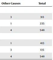
Concerning the relationship between patients’ age and the type of trauma, it was shown that most injuries leading to TBI in individuals under 50 years of age were related to traffic accidents. Also, in terms of male gender, 174 people were hospitalized due to traffic accidents, and 125 were hospitalized due to sports injuries (Table 3).

Table 3.

Investigating the Relationship Between Patients’ Age and Gender with the Mechanism of Trauma a

VariablesInvasionTraffic AccidentsSportsFallsOther CausesTotal
Age
< 502314886533313
> 50218083501235
Total442281691034548
Gender
Male33174125801413
Female115444233135
Total442281691034548

5. Discussion

According to the results, most patients (n = 413, 75.4) were male. According to Perel et al.’s study in England and Wales, 10,229 (73.3%) males and 3,733 (26.7%) females had intracranial hemorrhages, respectively. Concerning the type of hemorrhage, it was shown that the prevalence of intraparenchymal hemorrhage (IPH), epidural hematoma (EDH), SAH, and subdural hematoma (SDH) were equal to 2187 (73.1%) and 803 (26.9%), 2352 (74.9%) and 788 (25.1%), 2257 (74.6%) and 768 (25.4%), and 3050 (72.5%) and 1154 (27.5%) for males and females, respectively (25), which is consistent with the results of the present study.

Regarding the causes of trauma, it was shown that most causes of trauma were related to traffic accidents with 228 patients, sports accidents with 169 patients, falls with 103 patients, and invasions with 44 patients, respectively. In Rahimi-Movaghar et al.’s study (26) in Tehran, 7110 patients were examined, and TBI was observed in only 21 patients. Also, out of 21 patients with TBI, the causes of TBI included traffic accidents in 10 patients, falls in 8 patients, and sports accidents in 2 patients. In Reihanian et al.’s study (27) in the north of Iran, out of 166 patients with TBI, traffic accidents were observed in 99 patients, falls in 53 patients, and sports accidents in 4 patients. In Alavi et al.’s study (28) in Bandar Abbas, traffic accidents in 30 patients and falls in 13 patients were observed as causes of TBI.

According to the results, most TBIs occurred due to traffic accidents. In this regard, it was shown in studies by Rahimi-Movaghar et al. (26) in Tehran, Alavi et al. (28) in Bandar Abbas, and Zandi and Seyed Hoseini (29) in Hamedan that traffic accidents accounted for 10 (47.6%), 30 (55.6%), and 90 (29.8%) of TBIs, respectively, which is consistent with the results of the present study.

One of the symptoms in TBI patients may include hemorrhage. In this regard, Shahhosseini et al. showed that CT scan changes were observed in only 20 out of 1395 patients referring to the hospital due to TBI. Also, 40%, 35%, and 25% of these 20 studied patients had decreased hemorrhage, increased hemorrhage, and new hemorrhage (11), respectively, indicating the presence of hemorrhage in TBI patients. Moustafa et al. also found intracranial hemorrhage in 26 (8.9%) out of 993 TBI patients under investigation (30). Perel et al. also reported that approximately 46% of 13,962 TBI patients were in the intracranial hemorrhage stage. Also, subdural hemorrhage was observed in about 30% of these patients, and the risk of death increased with increasing intracranial hemorrhage (25).

According to the findings, most patients had low GCS. In different studies, TBI led to patients decreased GCS. In Sadegh et al. (31), Samadi Rad et al. (32), and Chabok et al.’s (33) studies, TBI led to decreased GCS in patients, which is consistent with the results of this study. Also, regarding the GCS status and mortality rate, it was shown in Hayati et al.’s study that in patients with GCS scores of 3 to 8, the mortality rate was equal to 28 (66.70%), and in patients with GCS scores of 9 to 12, the mortality rate equal to 7 (18.40%) was reported, which is consistent with the results of this study (34).

5.1. Conclusions

The prevalence of intracranial hemorrhage and SAH in TBI patients is significantly high, which should be taken into consideration when performing diagnostic and therapeutic procedures for these patients.

Acknowledgements

References

  • 1.

    Karimian M, Okhli A, Noormohammadi-Dehbalaee A, Gholami A, Abdi A, Salimi E, et al. Prevalence of Vascular Trauma and Related Factors in Iran: A Systematic Review. Int J Med Toxicol Forensic Med. 2021;11(2):31441.1-8. https://doi.org/10.32598/ijmtfm.v11i2.31441.

  • 2.

    Shokri M, Nayyeri S, Salimi N, Nourmohammadi A, Tarjoman A, Borji M, et al. Prevalence of Neonatal Birth Trauma in Iran: a Systematic Review and Meta-analysis. Int J Pediatr. 2021;9(10):14520-31. https://doi.org/10.22038/ijp.2020.50652.4032.

  • 3.

    Maleki MM, Afsharloo S, Tarjoman A, Borji M, Mahdikhani S, Shokri M. Prevalence of Trauma and Related Factors in Iranian Children and Adolescents: Systematic Review. Int J Pediatr. 2021;9(9):14293-306. https://doi.org/10.22038/ijp.2020.49720.3975.

  • 4.

    Sutton L, Rowe S, Hammerton G, Billings J. The contribution of organisational factors to vicarious trauma in mental health professionals: a systematic review and narrative synthesis. Eur J Psychotraumatol. 2022;13(1):2022278. [PubMed ID: 35140879]. [PubMed Central ID: PMC8820814]. https://doi.org/10.1080/20008198.2021.2022278.

  • 5.

    Sevillano L, Wood B, Franklin C. Mental Health Interventions That Address Historical Trauma among Indigenous Peoples: A Systematic Review. Soc Work Res. 2022;46(2):127-40. https://doi.org/10.1093/swr/svac007.

  • 6.

    Karbasfrushan A, Karimiyarandi H. Role of vitamin D on knee osteoarthritis pain: a systematic review. Eurasian Chem Commun. 2022;4(12):1241-50. https://doi.org/10.22034/ecc.2022.351411.1505.

  • 7.

    Taghinejad H, Otaghi M, Nikan A, Beiranvand R. Investigating the effects of nurse post-discharge follow-up phone calls on the self-efficacy of patients suffering from stroke. Bali Med J. 2018;7(3). https://doi.org/10.15562/bmj.v7i3.1008.

  • 8.

    Otaghi M, Hasanvand F, Mozafari M, Khorshidi A. Effects of home-based walking program on fatigue in patients with cance. Rev Latinoam Hipertens. 2019;14(3):284-9.

  • 9.

    Scantling D, Fischer C, Gruner R, Teichman A, McCracken B, Eakins J. The role of delayed head CT in evaluation of elderly blunt head trauma victims taking antithrombotic therapy. Eur J Trauma Emerg Surg. 2017;43(6):741-6. [PubMed ID: 28439613]. https://doi.org/10.1007/s00068-017-0793-7.

  • 10.

    Marincowitz C, Lecky FE, Townend W, Borakati A, Fabbri A, Sheldon TA. The Risk of Deterioration in GCS13-15 Patients with Traumatic Brain Injury Identified by Computed Tomography Imaging: A Systematic Review and Meta-Analysis. J Neurotrauma. 2018;35(5):703-18. [PubMed ID: 29324173]. [PubMed Central ID: PMC5831640]. https://doi.org/10.1089/neu.2017.5259.

  • 11.

    Shahhosseini R, Ebrahimi Nejad A, Shahba M, Tajoddini S, Ghaedamini H, Farahbakhsh S, et al. [Investigation of the changes in CT scan findings in patients with head trauma referred to the emergency department of Shahid Bahonar Hospital in Kerman in 2020]. Qom Univ Med Sci J. 2021;15(2):130-9. Persian. https://doi.org/10.52547/qums.15.2.130.

  • 12.

    Virani SS, Alonso A, Aparicio HJ, Benjamin EJ, Bittencourt MS, Callaway CW, et al. Heart Disease and Stroke Statistics-2021 Update: A Report From the American Heart Association. Circulation. 2021;143(8):e254-743. [PubMed ID: 33501848]. https://doi.org/10.1161/CIR.0000000000000950.

  • 13.

    Suzuki H, Kawakita F, Asada R. Neuroelectric Mechanisms of Delayed Cerebral Ischemia after Aneurysmal Subarachnoid Hemorrhage. Int J Mol Sci. 2022;23(6):3102. [PubMed ID: 35328523]. [PubMed Central ID: PMC8951073]. https://doi.org/10.3390/ijms23063102.

  • 14.

    Dayyani M, Sadeghirad B, Grotta JC, Zabihyan S, Ahmadvand S, Wang Y, et al. Prophylactic Therapies for Morbidity and Mortality After Aneurysmal Subarachnoid Hemorrhage: A Systematic Review and Network Meta-Analysis of Randomized Trials. Stroke. 2022;53(6):1993-2005. [PubMed ID: 35354302]. https://doi.org/10.1161/STROKEAHA.121.035699.

  • 15.

    Chen S, Li Q, Wu H, Krafft PR, Wang Z, Zhang JH. The harmful effects of subarachnoid hemorrhage on extracerebral organs. Biomed Res Int. 2014;2014:858496. [PubMed ID: 25110700]. [PubMed Central ID: PMC4109109]. https://doi.org/10.1155/2014/858496.

  • 16.

    Lauzier DC, Jayaraman K, Yuan JY, Diwan D, Vellimana AK, Osbun JW, et al. Early Brain Injury After Subarachnoid Hemorrhage: Incidence and Mechanisms. Stroke. 2023;54(5):1426-40. [PubMed ID: 36866673]. [PubMed Central ID: PMC10243167]. https://doi.org/10.1161/STROKEAHA.122.040072.

  • 17.

    Suarez JI, Tarr RW, Selman WR. Aneurysmal subarachnoid hemorrhage. N Engl J Med. 2006;354(4):387-96. [PubMed ID: 16436770]. https://doi.org/10.1056/NEJMra052732.

  • 18.

    Darkwah Oppong M, Lohrer L, Wrede KH, Chihi M, Santos AN, Dammann P, et al. Reevaluation of risk factors for aneurysmal subarachnoid hemorrhage associated epilepsy. J Neurol Sci. 2023;444:120519. [PubMed ID: 36563606]. https://doi.org/10.1016/j.jns.2022.120519.

  • 19.

    Kreitschmann-Andermahr I, Hoff C, Saller B, Niggemeier S, Pruemper S, Hutter BO, et al. Prevalence of pituitary deficiency in patients after aneurysmal subarachnoid hemorrhage. J Clin Endocrinol Metab. 2004;89(10):4986-92. [PubMed ID: 15472195]. https://doi.org/10.1210/jc.2004-0146.

  • 20.

    D'Amato SA, Chang TR. Advances in Intracranial Hemorrhage: Subarachnoid Hemorrhage and Intracerebral Hemorrhage. Crit Care Clin. 2023;39(1):71-85. [PubMed ID: 36333038]. https://doi.org/10.1016/j.ccc.2022.06.003.

  • 21.

    Catapano JS, Rumalla K, Koester SW, Winkler EA, Rudy RF, Cole TS, et al. Incidence and Prediction of Chronic Depression Following Aneurysmal Subarachnoid Hemorrhage: A Single-Center 17-Year Experience. World Neurosurg. 2023;171:e206-12. [PubMed ID: 36455851]. https://doi.org/10.1016/j.wneu.2022.11.120.

  • 22.

    Sabouri M, Moradian Lotfi H, Norian SM. [Assay of Serum Sodium Balance Disturbance in Spontaneous Subarachnoid Hemorrhage and Relation to Prognosis of Disease]. Avicenna J Clin Med. 2006;13(2):5-9. Persian.

  • 23.

    Pan J, Bonow RH, Temkin N, Robinson EF, Sekhar LN, Levitt MR, et al. Incidence and Risk Model of Venous Thromboembolism in Patients with Aneurysmal Subarachnoid Hemorrhage. World Neurosurg. 2023;172:e418-27. [PubMed ID: 36657716]. https://doi.org/10.1016/j.wneu.2023.01.045.

  • 24.

    Jain S, Iverson LM. Glasgow Coma Scale. StatPearls. Treasure Island, FL: StatPearls Publishing; 2022.

  • 25.

    Perel P, Roberts I, Bouamra O, Woodford M, Mooney J, Lecky F. Intracranial bleeding in patients with traumatic brain injury: a prognostic study. BMC Emerg Med. 2009;9:15. [PubMed ID: 19650902]. [PubMed Central ID: PMC2735732]. https://doi.org/10.1186/1471-227X-9-15.

  • 26.

    Rahimi-Movaghar V, Saadat S, Rasouli MR, Ghahramani M, Eghbali A. The Incidence of Traumatic Brain Injury in Tehran, Iran: A Population Based Study. Am Surg. 2011;77(6):112-4. https://doi.org/10.1177/000313481107700608.

  • 27.

    Reihanian Z, Noori Roodsari N, Rimaz S, Asadi P, Khoshsima N, Rafiee Zadeh A, et al. Traumatic injuries in children during COVID-19 pandemic: a national report from northern Iran. Int J Burns Trauma. 2022;12(5):188-93. [PubMed ID: 36420100]. [PubMed Central ID: PMC9677227].

  • 28.

    Alavi SA, Taghaviasl M, Baghr Zadeh Homaii N. Prevalence of head trauma causes in Bandar Abbas-Iran. Life Sci J. 2013;10(12s):284-7.

  • 29.

    Zandi M, Seyed Hoseini SR. The relationship between head injury and facial trauma: a case-control study. Oral Maxillofac Surg. 2013;17(3):201-7. [PubMed ID: 23100036]. https://doi.org/10.1007/s10006-012-0368-z.

  • 30.

    Moustafa F, Roubin J, Pereira B, Barres A, Saint-Denis J, Perrier C, et al. Predictive factors of intracranial bleeding in head trauma patients receiving antiplatelet therapy admitted to an emergency department. Scand J Trauma Resusc Emerg Med. 2018;26(1):50. [PubMed ID: 29914560]. [PubMed Central ID: PMC6006553]. https://doi.org/10.1186/s13049-018-0515-0.

  • 31.

    Sadegh R, Karimialavijeh E, Shirani F, Payandemehr P, Bahramimotlagh H, Ramezani M. Head CT scan in Iranian minor head injury patients: evaluating current decision rules. Emerg Radiol. 2016;23(1):9-16. [PubMed ID: 26407978]. https://doi.org/10.1007/s10140-015-1349-y.

  • 32.

    Samadi Rad B, Zeinali A, Shakeri Bavil M, Ashrafian F, Daghighi MA. Autopsy Findings of Brainstem in Head Trauma in Comparison with CT Scan Findings in Brain Trauma Ward in Tabriz, Iran. Acta Med Iran. 2009;47(5):409-14.

  • 33.

    Chabok S, Yousefzadeh Chabok S, Ramezani S, Kouchakinejad L, Saneei Z. Epidemiology of Pediatric Head Trauma in Guilan. Arch Trauma Res. 2012;1(1):19-22. https://doi.org/10.5812/atr.5381.

  • 34.

    Hayati S, Yazdani R, Ghasemi A, Yousefi Kafshgari M, Tabibzadeh Dezfuli SA. Epidemiology and radiologic findings of patients with traumatic brain injuries in emergency department of Shahid Mohammadi hospital. Eurasian Chem Commun. 2020;2(12):1210-5. https://doi.org/10.22034/ecc.2020.253265.1086.