


1. Background
One of the most common complaints in the emergency department (ED) is loss of consciousness (LOC) (1). Consciousness refers to a person's awareness of themselves and their environment and the ability to respond to external stimuli. A disorder of consciousness means a reduced level of excitability and awareness of oneself and the environment (2).
Several common causes of LOC range from benign situations like standing up quickly to more serious conditions such as structural brain damage, seizures, infections, shock, respiratory failure, or psychiatric disorders (3). One of the most common causes of LOC is vasovagal syncope, which occurs when there is a sudden drop in blood pressure, leading to decreased blood flow to the brain. This can be triggered by factors such as standing for long periods, dehydration, or emotional stress (4). Neurological conditions such as epilepsy, migraines, and transient ischemic attacks (mini-strokes) can also cause LOC by causing sudden disruptions in brain function, leading to fainting episodes (5).
Infections can cause dehydration and electrolyte imbalances that affect brain function, leading to confusion and LOC (4, 5). Shock and respiratory failure decrease blood flow to the brain, causing LOC. Heart failure (HF) can lead to LOC through various mechanisms, including reduced cardiac output, dysrhythmias, medication side effects, and, in severe cases, pulmonary edema due to impaired gas exchange (6). Dysrhythmias, which are irregular heart rhythms, can disrupt the normal pumping of blood to the brain, leading to a LOC. Other cardiac conditions such as heart attacks or heart failure can also result in fainting episodes (7).
Some clinical conditions, such as stroke, seizures, and poisoning, are easily identifiable. However, other causes, such as metabolic issues, electrolyte disorders, or endocrine disorders, require further evaluation and investigation (8). Understanding the etiology of this phenomenon is crucial for developing effective strategies for the prevention and management of patients. Emergency management of patients with reduced LOC is critical to prevent irreversible brain damage (9). Differentiating between traumatic and non-traumatic causes is important but challenging without a detailed history (10). However, most trauma patients are referred to the trauma department based on their history and are separated from non-trauma patients by EMS during the initial triage (11).
After stabilizing the patient, it is important to differentiate between structural and non-structural causes, typically done using a CT scan (12). Further examination and a thorough history can help diagnose the cause of LOC (13, 14). Additionally, determining the cause can also be used to predict the patient's prognosis (15, 16). The mortality rate due to trauma has been reported to range from 25% to 87% (10). The one-month mortality rate of patients with LOC lasting more than six hours is 76%. However, the prognosis of patients varies depending on the cause of LOC (10).
2. Objectives
This study aimed to investigate the cause and prognosis of patients with non-traumatic LOC referred to two referral hospitals of Mashhad University.
3. Methods
3.1. Study Design
This prospective cohort study was conducted on 1000 patients presenting with complaints of LOC who were referred to Ghaem and Imam Reza hospitals in Mashhad in 2020. The study spanned one year, and patients were followed up for one month from the date of admission to assess their final prognosis. All patients with a decreased level of consciousness (GCS < 14) were included in the study after obtaining informed consent from a legal guardian. Inclusion criteria comprised patients over 18 years of age with non-traumatic LOC. The exclusion criteria included a history of head trauma within one month before admission.
Initial patient assessments included demographic information, vital signs upon arrival, the time interval between the patient's arrival and the first visit, past medical history, and medications used by the patient. This information was recorded by the Emergency Medicine Assistant. The Emergency Medicine Assistant also followed up with patients for one month from the date of hospitalization to evaluate their recovery or prognosis.
3.2. Sample Size
This study was conducted over one year using a simple sampling method and included 1000 patients.
3.3. Statistical Analysis
Descriptive and inferential statistics were performed using SPSS software version 23 for data analysis. Charts and statistical tables were utilized to describe the data. The chi-square test was used to analyze the qualitative data. The relationship between different study outcomes was assessed with multivariate binary logistic regression, with a 2-sided P < 0.05 considered significant.
4. Results
A total of 1037 patients participated in this study. However, 37 patients were excluded: 18 were not satisfied to participate, and 19 were unresponsive in the follow-up. The study included 500 cases from Imam Reza (AS) Hospital and 500 cases from Ghaem Hospital. Among the participants, 524 (52.4%) were male and 476 (47.6%) were female, with a mean age of 59.85 ± 16.06 years, ranging from 18 to 95 years.
The history of underlying conditions among the patients was as follows: 404 (40.4%) had hypertension (HTN), 576 (57.6%) had diabetes, 355 (35.5%) had ischemic heart disease, 296 (29.6%) had hyperlipidemia (HLP), and 158 (15.8%) had a history of cerebrovascular accident (CVA).
The average vital signs at admission were as follows: Systolic blood pressure (SBP) was 113.90 mmHg, diastolic blood pressure (DBP) was 73.07 mmHg, pulse rate (PR) was 107 beats per minute, respiratory rate (RR) was 24 breaths per minute, body temperature was 38.05 degrees Celsius, oxygen saturation (O2 sat) in room air was 89.55%, and blood sugar reading on glucometer was 172 mg/dL. The patients’ Glasgow Coma Scale (GCS) was assessed with a mean score of 12 (Table 1). The average time between the patient's arrival and the first visit by a doctor was 15.41 minutes.
| Variables | Average | Standard Deviation |
|---|---|---|
| Temperature | 38.05 | 0.98 |
| Heart rate | 107.57 | 25.27 |
| SBP | 113.90 | 25.27 |
| DBP | 73.07 | 14.99 |
| RR | 23.60 | 7.1 |
| O2 sat | 85.99 | 9.36 |
| Blood sugar (Glucometer) | 172.25 | 90.63 |
| GCS | 11.45 | 1.75 |
Abbreviations: SBP, systolic blood pressure; DBP, diastolic blood pressure; RR, respiratory rate; O2 sat, oxygen saturation; GCS, Glasgow Coma Scale.
The most common etiology of decreased level of consciousness was sepsis (38.3%), followed by stroke (9.2%), heart failure (8%), myocardial infarction (7.3%), meningitis (7.2%), hepatic encephalopathy (6.5%), and toxic alcohols (5.8%).
A gender-based comparative study of underlying etiology showed that alcohol consumption (100%) was more common in men, while heart failure (100%) was more common in women. Other etiologies such as sepsis, meningitis, uremia, cholangitis, shock, and diabetic ketoacidosis were more prevalent in men. In contrast, stroke, myocardial infarction, hepatic encephalopathy, and intracranial hemorrhage were more common in women (Table 2).
| Underlying Etiology | Male | Female | P-Value |
|---|---|---|---|
| Sepsis | 227 (59.3) | 156 (40.7) | < 0.001 |
| CVA | 44 (47.8) | 48 (52.2) | |
| Myocardial infarction | 28 (38.4) | 45 (61.6) | |
| Heart failure | 0 (0.0) | 80 (100.0) | |
| Meningitis | 38 (52.8) | 34 (47.2) | |
| Hepatic encephalopathy | 25 (38.5) | 40 (61.5) | |
| Toxic alcohol | 58 (100.0) | 0 (0.0) | |
| Uremia | 34 (69.4) | 15 (30.6) | |
| Cholangitis | 28 (58.3) | 20 (41.7) | |
| Shock | 28 (62.2) | 17 (37.8) | |
| DKA | 13 (56.5) | 10 (43.5) | |
| ICH | 1 (8.3) | 11 (91.7) |
Abbreviations: CVA, cerebra vascular accident; DKA, diabetic ketoacidosis; ICH, intra cranial hemorrhage.
a Values are expressed as No. (%)
The deceased patients were older and had higher RRs and temperatures, with lower blood sugar and blood pressure at admission. A prior history of stroke, HLP, HTN, and diabetes was associated with death.
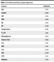
Binary logistic regression showed that a lower PR, absence of CVA, absence of HLP, shorter time to visit, higher blood pressure, and higher blood sugar were associated with a better prognosis (Table 3). Specifically, the analysis revealed that a lower PR (OR = 0.976; P = 0.030), absence of CVA (OR = 0.456; P = 0.003), and absence of HLP (OR = 0.560; P = 0.012), as well as a shorter time to visit (OR = 0.956; P = 0.002), were related to better outcomes. Additionally, higher blood pressure (OR = 1.026, P = 0.001) and higher blood glucose (OR = 1.002, P = 0.049) were associated with better outcomes.
| Feature | Odd Ratio | 95% CI | P-Value |
|---|---|---|---|
| Age | 1.001 | (1.013 - 0.989) | 0.873 |
| Gender | 0.889 | (1.198 - 0.660) | 0.441 |
| SBP | 1.026 | (1.042 - 1.011) | 0.001 |
| DBP | 0.992 | (1.018 - 0.992) | 0.542 |
| RR | 1.003 | (1.013 - 0.992) | 0.636 |
| PR | 0.976 | (0.998 - 0.954) | 0.030 |
| Temperature | 0.981 | (1.242 - 0.776) | 0.877 |
| O2 sat | 1.018 | (1.043 - 0.994) | 0.137 |
| Blood glucose | 1.002 | (1.004 - 1.000) | 0.049 |
| Time to visit | 0.956 | (0.984 - 0.929) | 0.002 |
| GCS | 1.091 | (1.210 - 0.984) | 0.099 |
| IHD | 2.147 | (3.433 - 1.342) | 0.001 |
| CVA | 0.456 | (0.766 - 0.272) | 0.003 |
| HLP | 0.560 | (0.883 - 0.335) | 0.012 |
| HTN | 1.153 | (1.865 - 0.712) | 0.563 |
| Diabetes | 0.754 | (1.131 - 0.503) | 0.172 |
Abbreviations: GCS, Glasgow Coma Scale; IHD, ischemic heart disease; CVA, cerebra-vascular accident; HTN, hypertension; HLP, hyperlipidemia; SBP, systolic blood pressure; DBP, diastolic blood pressure; PR, pulse rate; RR, respiratory rate; O2 sat, O2 saturation.
5. Discussion
This study was performed to evaluate the etiology of non-traumatic LOC and the prognosis of these patients referred to two academic EDs. A total of 1000 participants were evaluated. Sepsis was the most common cause in these patients. There are several ways in which sepsis can cause someone to lose consciousness. One common way is through septic shock, a condition in which blood pressure drops to dangerously low levels, depriving the brain of oxygen and causing the person to lose consciousness. In severe cases, septic shock can lead to coma or even death (17, 18).
A study published by Lutz et al. (19) showed that the cause of LOC in 835 non-trauma patients was cerebral hemorrhage with 224 cases (26.8%), followed by epilepsy, cerebral infarction, metabolic, and cardiac causes. They did not evaluate the patients' outcomes. Schmidt et al. conducted a study on the etiology of comatose patients and found that the most common cause is primary brain lesions (39%), followed by secondary pathologies that affected the brain (36%) (20).
Our results differed from these studies. We found that the most common cause of LOC is sepsis, which was not reported as a primary cause in the Lutz and Schmidt studies.
However, in general, cardiac, cerebral, and metabolic causes were relatively common in both studies, and the results were somewhat similar in this respect. The difference in the results of the two studies cannot be determined exactly, but the high number of sepsis patients in our study may be due to the lack of prevention, necessary care, and early treatment of infectious diseases for geriatric and disabled patients.
Kermani et al. (21) conducted another study in 2021 to investigate the causes of LOC in children at Mashhad University of Medical Sciences. They showed that infectious diseases (40.6%), seizures (23.8%), and poisonings (19.8%) were the most common causes of LOC in children. Overall, the mortality rate due to LOC was reported to be 18.8%, with infection being the most important cause. Duyu et al. designed another study on this age group (children) and showed that the most common cause of non-traumatic coma was neuroinfection (31.4%) (22).
Our study showed similar evidence but with a higher mortality rate, which could be attributed to the older age of patients and their underlying conditions. In another study conducted in 2016 by Sarin et al. (23) in India, patients over 13 years with GCS < 9 and non-traumatic coma were included. After a one-year follow-up, they showed that the most common causes of coma were cerebrovascular events, metabolic encephalopathies, and infections, respectively. Despite the similar causes of LOC in their study, it should be noted that the methodology was fundamentally different. Patients with a GCS of more than 9 were not included in Sarin's study, which may explain the differences in the findings between the two studies.
A systematic review study conducted in 2015 by Horsting et al. (3) assessed the etiology and outcomes of patients with non-traumatic coma in the ICU. They showed that the most common causes of non-traumatic coma were stroke, hypoxia, intoxication, and some metabolic causes, respectively, with poisoned patients having the best outcomes. The most significant difference between our study and theirs is the absence of sepsis. However, this study, like the Sarin study, examined only patients who were in a coma (GCS < 9), while we assessed many patients with higher GCS.
Forsberg et al. in Stockholm, the Netherlands evaluated 875 patients with a reduced level of consciousness and GCS < 11, dividing them into two groups: Those with decreased levels of consciousness due to metabolic (72% of cases) and structural causes of the brain (28% of cases) (24). They showed that young adults with non-traumatic LOC and low or normal blood pressure without focal neurological deficits might have an underlying metabolic disorder, making neuroimaging unnecessary as the first-line evaluation method. These results are similar to our findings, as the mean age of patients with diabetic ketoacidosis and those who had consumed toxic alcohols was lower than other patients. Additionally, patients with expected pathological findings on brain imaging (such as stroke and intracranial hemorrhage) were middle-aged.
In 2004, Aboutalebi and Fotouhi Ghiam studied the causes of decreased non-traumatic levels of consciousness in 392 patients. The results showed that the most common causes were metabolic (42.9%), structural (40.1%), and infectious diseases (6%) (25). However, in 11% of cases, the cause remained unclear. The mortality rates due to infectious, structural, and metabolic causes were 33.3%, 26.8%, and 22%, respectively. The most important difference between this study and ours is the high proportion of patients with sepsis in our study, which was not observed in other similar studies. Regarding the mortality rate, the number of patients who died was higher in our study.
In this study, we found that half of the patients referred to the ED with LOC complaints died within a month of follow-up. Sepsis was also the most common diagnosis in our patients. With these findings, it may be suggested that patients who present to the ED with LOC are more likely to die, and therefore, LOC can be used as a prognostic factor of death at hospitalization. Researchers can design more studies in the future to determine the cause of the high rate of sepsis in our patients.
5.1. Limitations and Strengths
Our study, like other studies, had its strengths and limitations. One of the limitations was the lack of investigation into the specific causes of sepsis in patients with septic shock. It would also have been better if we had considered mixed causes; for example, some patients had both uremia and sepsis, but we only considered their sepsis. In contrast, our study had a relatively large sample size, which could increase the accuracy of the results, and this is a strength of the study.
