1. Background
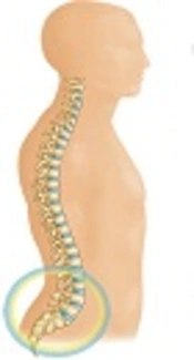
Spondylolisthesis is a common skeletal disorder, which refers to the slippage of the upper vertebra on the lower one. This might happen in anterior or posterior spondylolisthesis forms (1). Spondylolisthesis does not usually have a clear symptom; however, it is one of the important causes of back pain and disability in the low back area.
Spondylolisthesis usually occurs in the lumbosacral area because of 2 reasons including spondylolysis and degenerative diseases (2). In the spondylotic cases, a fracture usually occurs in the pars interarticularis manifested initially in childhood and early adulthood (3, 4). In the degenerative cases, however, perfect spondylolisthesis occurs in the posterior lumbar vertebra. Whereas there are diverging opinions about the causes of this disorder, the degenerative type usually does not occur at ages earlier than 50 years. It seems that the facet joints and discs degenerative changes due to aging are related to this disorder (5).
There are a number of risk factors considered to cause spondylolisthesis especially in the lumbosacral area, including age increase (6, 7), the increase of sagittal alignment angle, reduction of the diameter of para-spinal muscles (8, 9), increase of sacrum slope, and increase of the alignment of L1 to S1 (10).
L4 and L5 are the most commonly vertebrae afflicted with spondylolisthesis (9). There are various reports on the prevalence of spondylolisthesis in different communities. The prevalence among females ranges from 6% in Taiwan to 25% in the US, and among males, from 3% in Taiwan to 31% in the US (6, 10-14).
Unfortunately, there is no accurate statistics about the prevalence of spondylolisthesis in Iran. Listhesis is 3 to 4 times more frequent among females than males. In addition, the prevalence of spondylolisthesis is 3 times more in black females than the white ones (1).
The precise reason of higher prevalence of spondylolisthesis among females is unknown. However, it is very likely that this higher prevalence is due to factors such as pregnancy, ligament laxity, total oophorectomy, and post-menopause posture (15-17).
Elderly females have a more limited disc space when compared with the same age males. These degenerative changes make females more prone to degenerative spondylolisthesis (18, 19).
Numerous physiological changes and development happen during pregnancy in a female including ligament and joint laxity of this area (20) as the results of relaxin hormone (21). The precise effects of this hormone are still unknown; however, it increases the level of collagenase (22). The collagenase influences the anolous collagen in lumbar intervertebral discs, leading to increased risk of spondylolisthesis (23, 24).
In females with pregnancy experience, there is considerable laxity in the pelvis when compared with nulliparous ones. This, of course, occurs over the first pregnancy, and does not get intensified over the subsequent pregnancies (25). The ligament laxation has long-term effects on the joints, even years after delivery. On the other hand, the relaxin hormone is likely to influence the stability of facet joints; thereby resulting in rotation of vertebra on each other (26).
Another issue considered as a risk factor for spondylolisthesis is the increased lumbar lordosis during pregnancy. This theory, however, is rejected by some researchers. These researchers believe that the extension happening in the hip joint during pregnancy compensates for the increased lordosis. Therefore, the lordosis angle does not undergo considerable changes during pregnancy (27).
The most important factor contributing to the increased risk of degenerative spondylolisthesis in pregnancy, particularly in multiple pregnancies, is the laxation of anterior abdominal muscles, which is more noticeable in multipara females than nulliparous females and males (28-30).
Investigations showed that the most prevalent area for anterolisthesis is L4-L5, and for retrolisthesis is L3-L4 and L5-S1 (13).
The Meyerding classification model is used to categorize spondylolisthesis:
Grade 0, no slip; grade I, ≥ 5% and < 25%; grade II, 26% - 50%; grade III, 51% - 75 %; grade IV, 76% - 100 %; and grade V, complete slippage (30).
Based on the conducted studies, it seems that early age pregnancy, particularly below 20 years, along with multiple pregnancies can seriously increase the risk of spondylolisthesis. To date, no study investigated this issue in Iran. Therefore, the current study aimed at investigating the risk factors in spondylolisthesis in females afflicted with it.
2. Methods
A total of 113 female patients with spondylolisthesis referring to the physical medicine and rehabilitation clinic were included in the study. The inclusion criteria were female gender, and confirmed affliction with spondylolisthesis in lumbosacral X-ray or magnetic resonance imaging (MRI). The exclusion criteria comprised affliction to severe diseases such as rheumatologic diseases, and history of malignancy. The patients were examined by a specialist in physical medicine and rehabilitation. The related demographic questionnaires including age, trauma history, weight, and height were filled in and pregnancy and delivery information was also recorded. The data were analyzed with SPSS version 20.0
3. Results
In the current study, 113 females with the mean age of 53.79 (SD: 10.79) years were enrolled. The demographic information and the maternity records of the patients are demonstrated in Tables 1 and 2. The median for pregnancies was 4, and the mode was 5.
| Variables | Minimum | Maximum | Mean | Std. Deviation |
|---|---|---|---|---|
| Age | 30.00 | 82.00 | 53.79 | 10.79 |
| BMI | 20.69 | 37.11 | 28.23 | 3.98 |
| Pregnancy number | 1.00 | 11.00 | 4.17 | 2.01 |
The Demographic Information and Maternity Records of the Participants
| Variables | Number | Percentage |
|---|---|---|
| First delivery | ||
| Below 20 year | 84 | 74.3 |
| Above 20 year | 29 | 25.7 |
| Job | ||
| Employee | 24 | 21.2 |
| Housewife | 89 | 78.8 |
| History of trauma | ||
| Yes | 10 | 8.8 |
| No | 103 | 91.2 |
The Demographic Information and Maternity Records of the Participants
About 74.3% of the patients had a history of birth delivery below the age of 20 years. In addition, 89 patients (78.8%) were housewives and the rest had other jobs; 8.8% of the patients had a history of direct trauma to their cords.
The area with highest frequency of listhesis in the results was L4-L5 (49.6%), followed by L5-S1 ranked the second with a frequency of 40.7%. The third frequency belonged to 2 vertebrae including L5-S1, and L4-L5 with 1.8% of the total frequency.
In the current study, the most frequent grade of spondylolisthesis was grade I with 82.3%. However, 7.3% of the patients had vertebrae spondylolisthesis grade II and above (Table 3).
| Variables | Grade I | Grade II | Grade III | Grade IV |
|---|---|---|---|---|
| Frequency | 93 | 19 | 1 | 0 |
| Percentage | 82.3 | 16.8 | 0.9 | 0 |
Frequency of Spondylolisthesis in Patients, According to Their Grading
4. Discussion
The study by Lai-Chang showed that in elderly males and females, lower height, higher body mass index (BMI), higher hip and spine BMD (bone mass measurement), and degenerative clinical arthritis increased the risk of spondylolisthesis. In the current study, the mean age and BMI of the patients were 53.79 (± 10.79) years and 28.23 (± 3.98) kg/m2 respectively, classified as elderly and overweight patients.
The study by Paul, probing the role of pregnancy on the occurrence of spondylolisthesis, reported that females with pregnancy experiences had significantly higher affliction rates to spondylolisthesis compared with males and nulliparous females (9). In the current study, the average delivery was 4, which can be indicative of this fact.
Studies reported the most frequent occurrence of spondylolisthesis in anterolisthesis of L4-L5 (13). Similarly, in the current study, the most frequent occurrences of listhesis were in L4-L5, and then in L5-S1.
In the study by Lai-Chang, 97% of the patients were afflicted with grade I listhesis, and 2.7% had grade II (1). Likewise, in the current study, a similar trend was observed as a substantial majority of the patients (82.3%) were afflicted with grade I listhesis, and 16.8 % of them had grade II.
In the current study, 74.3% of the patients had a history of pregnancy prior to age 20 years. In other studies there were no comments on this conception.
4.1. Implications for Practice
Based on the results of the current study, a correlation was observed between the early age pregnancy, multiple pregnancies, higher BMI, aging, and the rate of spondylolisthesis.
4.2. Conclusion
Due to the limitations of the current study, it cannot be concluded that the early pregnancy can be considered as a risk factor for spondylolysthesis. It is necessary to design other forms of research such as a case-control study, but community, especially females, awareness of pregnancy health including avoiding early and multiple pregnancies can be helpful and may reduce the risk of low back pain and improve community health.
