1. Background
Pain classically defined as an unpleasant sensory and emotional experience which is usually associated with tissue damage (1). Two clinical types of pain including acute and chronic are reported, the former one is a protective mechanism which alerts the individual to certain condition that is immediately harmful to the body; whereas, the chronic pain is persistent or intermittent. Acute pain rarely needs medical attention; when it does, nonsteroidal anti-inflammatory drugs (NSAIDs), powerful opioid analgesics, or local anesthetics can adequately control the pain. Chronic pain differs from acute pain not only due to its onset and duration, but more importantly in its mechanisms. Chronic pain may not have identifiable ongoing injury or inflammation, and often responds poorly to NSAIDs and opioids (2). Neuropathic pain (NP) known as a form of chronic pain results from any kind of damage to the central or peripheral nervous system (3, 4). Patients with NP often have spontaneous pain, allodynia, and hyperalgesia. NP may have delayed onset after initial nerve injury; therefore, pain may be present in the absence of apparent lesion or injury, making proper diagnosis and early treatment difficult (5, 6). The chronic constriction injury (CCI) model, developed by Bennett and Xie, is a well-known model of mononeuropathy which produces signs of NP (7). The CCI shows inflammatory characterization related to the condition/disease and is reported that the inflammatory component of NP in the case of CCI is present mainly in the first phase of the disease (8). Mechanical test of paw withdrawal latencies and observations of guarding behavior to certain mechanical stimuli and thermal stimulation including the tail flick test (9), the hind limb withdrawal plantar test (10), and the hotplate test (11, 12) have been extensively used in assessing pain behavior in animals (13, 14). Certain types of therapy including prescription of analgesic drugs, electrical stimulation, and ultrasound and laser therapy have been developed during the recent years to decrease the regenerative process and return the function (15). LLLT is a special type of laser therapy in which the irradiation used is red or near infrared beams with a wavelength of 600 - 1100 nm and an output power of 1 - 500 mW. This type of radiation is a continuous wave or pulsed light which consists of a constant beam of relatively low energy density (0.04–50 J/cm2) (16, 17). Since 1970s LLLT has been used in several clinical and experimental research studies on peripheral nerve injuries. Irradiation parameters and properties of LLLT, such as dose, intensity, time and application methods are notably varied among different clinical reports. The clinical effects of LLLT including cell apoptosis, improving cell proliferation, migration, cell adhesion, enhancing the cells’ mitotic activity, increasing blood flow, and local microcirculation are reported in many studies (18). Reis et al. reported effectiveness of laser at 660 nm for recovery of sciatic nerve in rat model following neurotmesis (19). Belchior et al. reported clinical and functional recovery of injured sciatic nerve by using GaAlAs laser at a wavelength of 660 nm, density of 4 J/cm2 for 21 days consecutively. It is also reported that the use of low level laser (660 nm) could significantly promote neural regeneration (20). Barbosa et al. used laser at 660 nm and 830 nm for recovery of sciatic nerve regeneration following crushing injuries, and reported that 660 nm provided early functional nerve recovery in comparison to 830 nm (21). Hsieh et al. demonstrated that 660 nm GaAlAs laser at a dose of 9 J/cm2 significantly reduced neuropathic allodynia in rats with CCI and significantly promoted functional recovery (22), Bertolini et al. also reported the same results (23).
2. Objectives
The controversy on energy densities and wavelengths of LLLT for peripheral neuropathies and the lack of specific pain evaluation among different studies led us to design the present research.
3. Materials and Methods
3.1. Animals
Twenty adult male Wistar rats (250 - 320 g) were used in this study. The animals were divided into three groups (n = 10) as follows:
•CCI group: animals that were subjected to surgical procedure, without undergoing irradiation.
•CCI + Laser 660nm therapy group (LLLT 660 nm): animals that were subjected to surgical procedure and also received laser irradiation with energy density of 4 J/cm2 and intensity of 0.354 (W/cm2)
•Control group: self-control
Animals received food and water ad libitum, and all procedures were performed according to the Ethic Committee for Animal Research of IUMS. All the animals were subjected to the functional evaluation before the operation (self-control). To induce neuropathic pain, the sciatic nerve injury model described by Bennett and Xie was used which is explained elsewhere (7).
3.2. Laser Therapy
A couple of CW diode lasers emitter with following specification were used in this study. A laser with the wavelength of 660 nm, the power of 100 mW (Heltschl; model: ME-TL10000-SK), the energy density of 4 J/cm2, and the power density of 0.354 W/cm2. Calibration was performed before use. Three points of the surgical incision were irradiated transcutaneously with no direct skin contact as follows: two points on two ends of surgical incision, and another at the midpoint of them. The laser therapy was started on the first day after the operation and was continued for two weeks daily in same time between 10 - 12 AM.
3.3. Functional Analysis
3.3.1. Thermal Withdrawal Threshold
By using a Plantar Test apparatus (UgoBasile, Italy) thermal hyperalgesia, the latency to withdrawal of the hind paws from a focused beam of radiant heat applied to the plantar surface, was studied (10). The animals were placed in an acrylic box with glass floor and the plantar surface of their hind paw was exposed to a beam of infrared radiant heat. The paw withdrawal latencies were recorded at infrared intensity of 50, and three trials for the right hind paws were performed and for each reading, the apparatus was set at a cut-off time of 25s. Each trial was separated by an interval time of 5 minutes.
3.3.2. Mechanical Withdrawal Threshold
Mechanical paw withdrawal thresholds were assessed with the Randall–Selitto method by using an Analgesy-meter apparatus (UgoBasile, Italy (24)). This instrument exerts a force that increases at a constant rate. The force was applied to the hind paw of the rat, which was placed on a small plinth under a cone shaped pusher with a rounded tip (1.5 mm in diameter).The rat was held upright with the head and limb to be tested free, but most of the rest of the body cradled in the hands of the experimenter. The paw was then put under the pusher until the rat withdrew the hind paw was occurred. Each hind paw was tested twice, with a 10 min interval between the measurements, and mechanical paw withdrawal thresholds were calculated as the average of two consecutive measurements.
3.4. Statistical Analysis
Statistical analysis was performed using SPSS 19.0. The results were presented as means ± SD, P Value less than 0.05 was considered to be significant.
4. Results
Plantar Test and Randall-Selitto method were used for thermal and mechanical withdrawal thresholds respectively.
4.1. Plantar Test
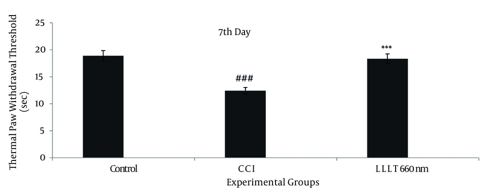
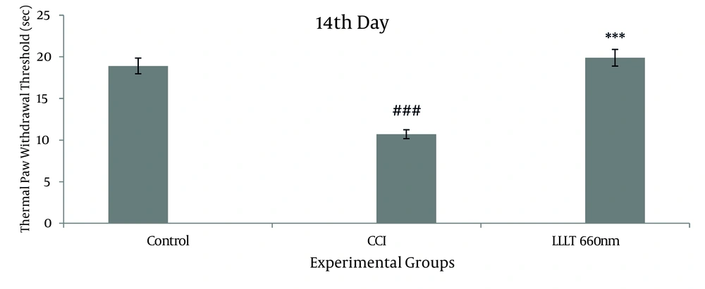
The thermal withdrawal threshold of the control group was, on average, 18.91 ± 4.08 seconds of the data collected prior to the injury. For the CCI group it was 12.42 ± 4.82 seconds and 10.70 ± 5.02 seconds on the 7 th and 14 th days after the operation respectively. For the LLLT 660 nm group, the mean value was 18.34 ± 4.29 seconds and 19.88 ± 3.13 seconds on the 7 th day and 14 th days after the operation respectively. There was a significant difference between LLLT 660 nm and CCI groups on the 7 th , and 14 th postoperative days (P < 0.001). There was no significant difference between the 7 th , and 14 th postoperative days of the LLLT 660 nm group and the control group (Figure 1 and 2)
Mean values of the thermal withdrawal threshold obtained from the groups during the study period (before the operation (control), 7 days after the operation). Asterisks represent significant differences from CCI group (*** P < 0.001) and represent significant differences from Control group (### P < 0.001)
Mean values of the Thermal Withdrawal Threshold obtained from the groups during the study period (before the operation (control), 14 days after the operation). Asterisks represent significant differences from CCI group (*** P < 0.001) and represent significant differences from Control group (### P < 0.001)
4.2. Randall-Selitto Method
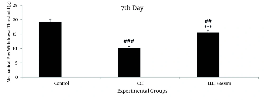
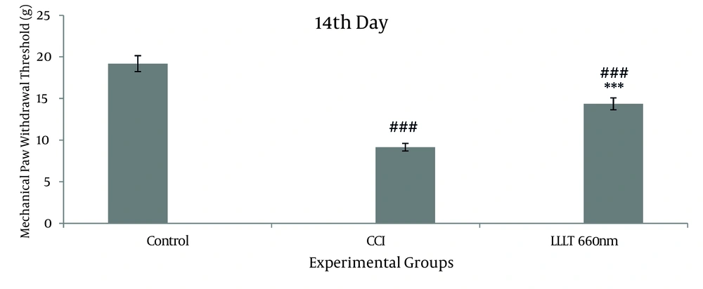
The mean of mechanical withdrawal threshold of the control group was 19.18 ± 4.66g before the operation. For the CCI group were 10.17 ± 4.18 g and 9.15 ± 4.20 g on the 7 th and 14 th days after the operation respectively. For the LLLT 660 nm group, the mean values were 15.54 ± 4.50g and 14.36 ± 5.43g on the 7 th and 14 th days after the operation correspondingly. Statistical analysis indicated that the difference between control group and the CCI group on the 7th, and 14 th postoperative days, was significant (P < 0.001); also the difference between LLLT 660 nm group and the CCI group on the 7 th , and 14 th postoperative days, was significant (P < 0.001); and there was a significant difference between the 7 th , and 14 th postoperative days of the LLLT 660nm group and the control group (P < 0.01, P < 0.001, respectively) (Figure 3 and 4).
Mean values of the Mechanical Withdrawal Threshold obtained from the groups during the study period (before the operation (control), the 7 days after the operation). Asterisks represent significant differences from CCI group (*** P < 0.001), and represent significant differences from Control group (###P < 0.001; ##P < 0.01).
Mean values of the Mechanical Withdrawal Threshold obtained from the groups during the study period (before the operation (control), 14 days after the operation). Asterisks represent significant differences from CCI group (*** P < 0.001), and represent significant differences from Control group (### P < 0.001).
5. Discussion
Our data significantly confirmed the therapeutic effects of LLLT on pain reduction in the CCI model; the wavelength of 660 nm was effective. In this section, we discuss the mechanisms of LLLT effectiveness on pain reduction. It is known that CCI induces an inflammatory condition which activates inflammatory cascade marked by the increase of proinflammatory cytokines such as IL-1β, IL-6, and TNF-α which play important role in the etiology and continuation of neuropathic pain (25-28). In addition, prostaglandin E2 (PGE2) or prostaglandin I2 (PGI2) administration induces hyperalgesia and enhances the sensitivity of primary afferents to either mechanical or chemical stimulation (29-31). During the preinflammatory phase, the production of reactive oxygen species (ROS) increases, which in turn activates NF-κB (32, 33). The activated NF-κB increases the expression of the iNOS and subsequent synthesis of NO (34, 35). NO and its reactive nitrogen intermediates may destruct cells and tissues and play significant role in the pathology of certain inflammatory conditions (36). Regarding the LLLT effectiveness on pain reduction various mechanisms were postulated. Rabelo et al. reported the therapeutic effects of LLLT on wound healing of diabetic rat throughout reducing the inflammatory progress and reduction of inflammatory cell density (37). Same evidences are also reported for reducing of rat paw edema by red LLLT with wavelengths of 632.8nm (He-Ne) and 650nm (38, 39). It is also shown that 660 nm and 684 nm from red diode lasers are effective in reducing edema (40). In another study on post traumatic muscular tissue repair, it was shown that LLLT reduced the inflammatory response, collagenesis, expression of iNOS, and the activation of NF-κB (41, 42). It is reported that the expression of the proinflammatory gene such as IL-1β is suppressed by LLLT in human keratinocytes (43, 44). Aimbire et al. reported that LLLT (650nm) reduced expression of TNF-α, after acute immune complex lung injury, in rats (45). It is also shown that LLLT is able to inhibit production of PGE2 and decrease the mRNA levels of cyclooxygenase-2 (46). The role of ROS as a natural cytotoxic production of the normal metabolism of oxygen is reported. ROS has important roles in cell signaling, regulating nucleic acid synthesis, protein synthesis, enzyme activation, and cell cycle progression (47, 48). There are also reports on the effects of different wave lengths of LLLT. Wu et al. used transcranial LLLT with 36 J/cm2 of a 665nm, 810nm and 980 nm laser four hours after traumatic brain injury. They reported the effectiveness of LLLT in improving the motor performance during the succeeding four weeks (49), and thus concluded that the absorption spectrum of the different chromophores located in the mitochondria and the cell membrane is important for LLLT therapeutic effects (49-51). Regarding photochemical effects of LLLT, the role of cytochrome c oxidase (complex IV mitochondrion) was also discussed (49). Cytochrome c oxidase is the photoreceptor in the red region of the spectrum which is responsible for activating the synthesis of ATP and consequently, better cell metabolism (52). The ability of the cell to have a greater energy source during the repair process might be the reason for the result in the group treated with laser 660 nm. LLLT transmits energy at low levels and therefore does not release heat, sound, or vibrations. Other experiments using LLLT have shown that the immediate increase in heat of the target tissue is negligible (53). In addition to above mechanisms, it is also reported that wavelength of 660nm such as He-Ne laser leads to photo reactivation of cellular superoxide dismutase (54). Although some mechanisms for the effectiveness of LLLT are known or claimed, unknown ones remain to be studied. The significance of this study is to provide new ways in laser therapy for clinical trials to reduce certain types of pain in patients with peripheral nerve injuries.


