1. Background
Fibroblasts, as wide spread cells of complex organisms contribute to wound healing of injured tissues with their ability to synthesize collagen resulting in wound edges closure (1-4). Wound healing is an intricate process composed of a set of physiological events that recruits various cell types to fulfill their function in order to close the wound. Fibroblasts play a pivotal role in the proliferation phase of this procedure (4). In adults, remodeling of a wound in damaged organs generally lasts about 2 - 3 weeks and complete normal wound healing process usually needs more time, sometimes years. Despite the fact that deposition of extracellular matrix in the fetal wound healing process is similar to the adult one, the temporal and spatial distribution of fetal extracellular matrix is different from its adult counterpart. Compared to adults, the production and precipitation of extracellular matrix by fetal fibroblasts is much more coordinated with stronger synthetic and secretory phenotype (5, 6). Fetal skin repair before the third trimester of pregnancy shows minimal inflammation due to the production of particular cytokines, growth factors, faster extracellular matrix deposition, and turnover, which could probably be responsible for scarless fetal wound healing (6-8). Regarding the crucial role of fibroblast in collagen deposition as the key element of skin extracellular matrix, it has been suggested that fetal fibroblasts are the central pillar of wound repair process (9). Some other studies have revealed that different wound healing properties of fetal skin is much more related to the fetal tissue instead of the uterus environment of the fetus, which have raised the intriguing possibility of their application to adult wound healing since 1970s (10, 11). It is not surprising that fetal fibroblasts with low immunogenic property and different wound healing process could be considered as a suitable alternative to neonatal foreskin cell based products in regenerative medicine and cell therapy (12). Cell therapy, as an almost newly introduced method in the medical field, is a challenging issue, which should assure the safety of the final product and the absence of viral or bacterial contamination with minimal immunogenic response (13, 14). Good manufacturing practice (GMP), as a quality assurance system, ensures that the quality of final product, focusing on all steps of manufacturing is in accordance with current specifications. Therefore, based on the current GMP (cGMP) principles, it is a matter of great importance to monitor raw materials’ quality and the conformity of all production procedures including cell culture, collection, and cryopreservation with validated standard operating procedures (SOPs) (15-17). In this respect, it has been highly recommended to perform cell production in a professionally designed GMP-compliant clean room facility using xeno-free media. Present relevant guidelines focus on progression, production, quality control, and non-clinical and clinical advances in cellular therapy (18). Therefore, when cell preparation complies with GMP regulations, the safety and quality of the final product will be increased (19). To achieve this aim, safe and pure reagents must be used during the entire cell production processes and research-grade reagents must be replaced by valid GMP-grade materials, to extend possible (20). Cell bank establishment is considered as one of the fundamental steps in cell therapy and clinical applications of desired cells (21). In order for GMP-grade cell manufacturing, we designed and established a clean room facility, affiliated to the Cell Therapy and Regenerative Medicine Research Center, to produce the most suitable clinical-grade cells for cell therapy. Based-on our previous studies to establish GMP-grade cell banking, in the current study we demonstrated the GMP-compatible and clinical-grade fetal fibroblast cell banking to be used for clinical applications.
2. Methods
Informed consent was obtained from the donor and the fetus was transported to the GMP facility via a tissue container with ice. Fetal skin was harvested from the back of the fetus with a gestational age of less than 12 weeks, which was legally and therapeutically aborted. Following skin harvesting, specimens were immediately immersed in transport medium (DMEM PAA, Austria) / Penicillin-Streptomycin and transferred to the clean room facility in less than 6 hours. Harvested tissue was transformed into the culture plate. All steps of isolation and culturing were performed under sterile condition under laminar airflow hood, according to cGMP guidelines in clean room. During the cell culture procedures, bacteriological tests were performed in order to identify the probable contamination of the samples.
2.1. Tissue Processing and Cell Culture
Skin sample was gently washed in phosphate-buffered saline (PBS; CliniMACS®, MiltenyiBiotec, Germany). The dermal side of the skin was scraped in order to remove its subcutaneous part and then it was sliced into small strips. After that, it was transferred to a 37°C water bath and was incubated with 0.5% (dispase; MP Biomedicals, USA/PBS) for four hours. After incubation, epidermis was removed as a sheet from the skin and dermal sample was washed and gently shaken in PBS. Approximately 10 pieces of sample were placed in a 35-mm tissue culture dish and a cover glass was placed over the tissue pieces. The space between sample pieces under the cover glass was filled with 4°C (DMEM-low glucose; PAA, Austria) supplemented with 10% fetal bovine serum (FBS; Pharma grade, Australian origin and gamma irradiated, PAA, Austria) and also 2 mL of 4°C mentioned medium was added gently to the culture dish. Dishes were placed into the incubator at 37°C, 5% CO2, and 97% humidity. Medium renewal was done every 72 hours. When the cells reach approximately 85% - 90% confluency, the cover glass was removed and expanded cells were washed twice with 4°C PBS and were enzymatically harvested with TrypLE™ Express (life technologies, USA). Cells were subcultured after evaluating the cell count, purity, and viability using 0.4% Trypan blue solution (Invitrogen, USA) and hemocytometer. Moreover, chromosomal stability of cultivated fibroblasts was assessed at the10th subculture. In order for long term storage of cultured cells, enzymatically detached fibroblasts were cryopreserved using freezing cold media composed of a final concentration of 10% dimethylsulfoxide (DMSO; WAK - Chemie Medical GmbH, Germany), 50% FBS, and 40% DMEM-low glucose. Cryo vials were placed in a pre-cooled Mr. Frosty freezing container and were frozen overnight at -80°C. Then, crypreserved cells transfer to liquid nitrogen at -196˚C. All banked cryovials were labelled carefully and their data was documented correctly.
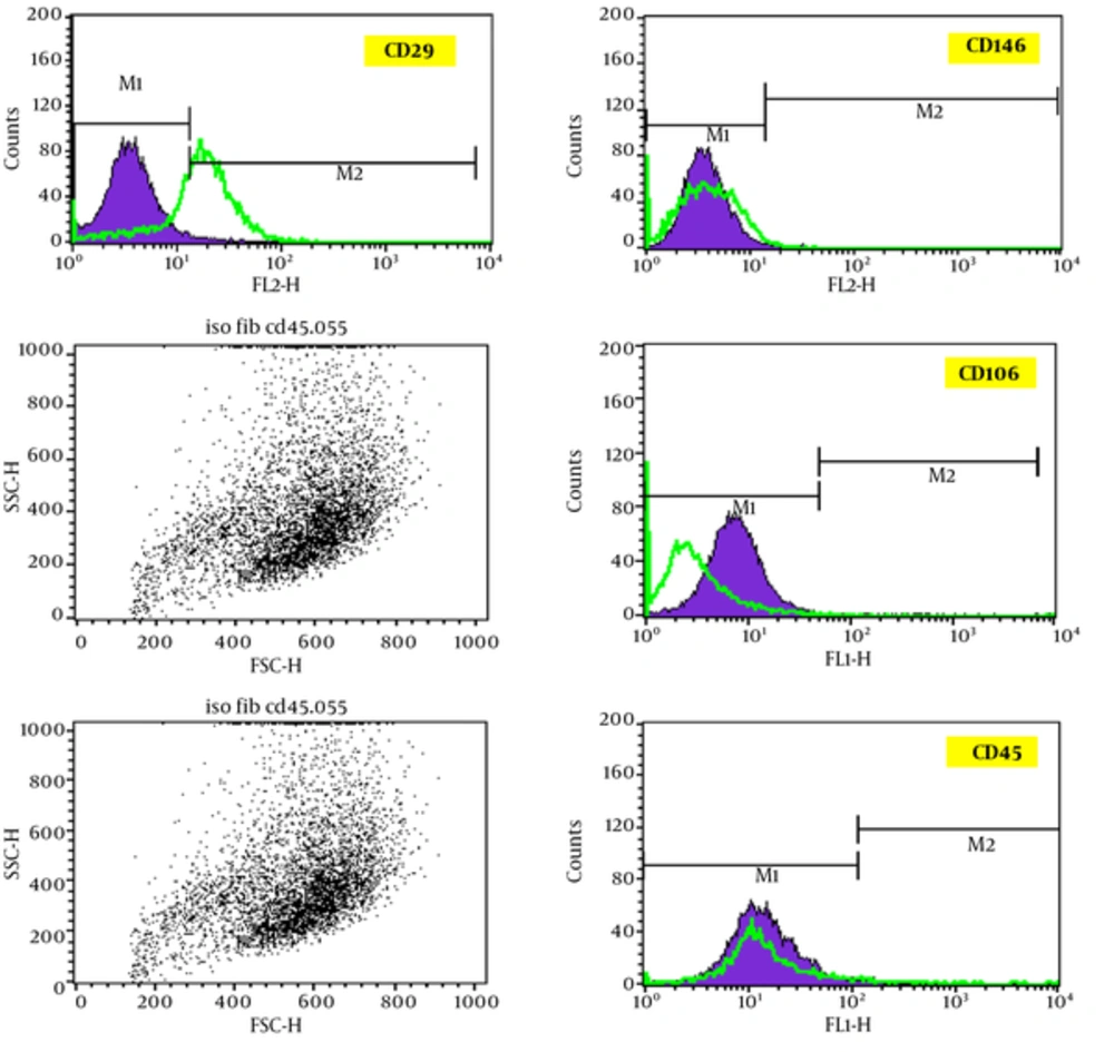
Approximately 5 × 10⁵ subcultured fibroblasts were re-suspended in FACS-buffer (3% BSA in PBS) in order to analyze the specific cell surface markers using flow cytometry. Following that, cell suspension was incubated with desired Fluorescein Isothiocyanate (FITC) - conjugated antibodies, CD146, CD106, CD45, and CD29 for 60 minutes at 4°C and fibroblasts were analyzed on a fluorescence-activated cell sorter (FACS; Partec, Denmark).
3. Results
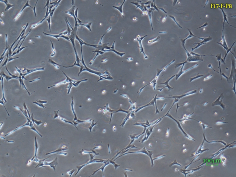
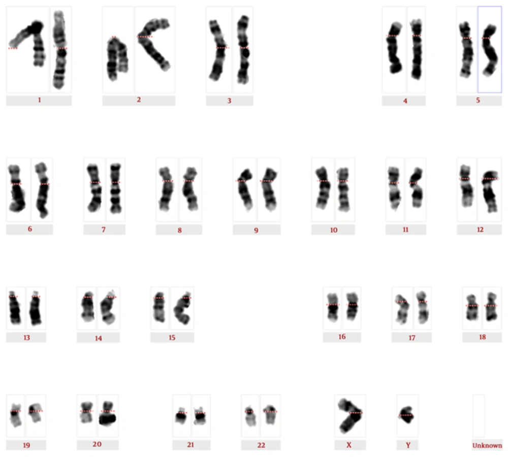
Isolated fetal dermal fibroblasts were harvested and stored at early passages. The viability of fibroblasts was ≥ 98% indicating successful establishment of fibroblast banking. Spindle-shaped morphological features of dermal fibroblasts are shown in Figure 1. Karyotyping of fibroblasts at the 10th subculture shows a normal diploid male pattern (Figure 2).
Screening CD markers have demonstrated the presence of CD106 and CD29 and the absence of CD146 and CD45 molecules on the fibroblasts surface (Figure 3). Therefore, according to flow cytometric data analysis, the lack of CD146 expression, which is positive in mesenchymal cells, has revealed the high purity of isolated and cultured fibroblastic population.
4. Discussion
Fibroblasts, as the key player of connective tissue, have a crucial role in the structural integrity of the tissue by secreting extracellular proteins such as proteinases leading to regulate the structure of the whole tissue. Dermal fibroblasts existing in the skin are considered as an important component of wound healing process and could be isolated and purified from skin specimens (22). These cells are similar to mesenchymal stem cells (MSCs) phenotypically. Therefore, the discrimination between these two types of cells is crucially important. However, there are no specific markers to discriminate between MSCs and fibroblasts. As CD146 is expressed only in MSCs, it could help scientists identify MSCS. On the other hand, CD106 is positive in both cells while they are expressed in MSCs around 10-fold higher than in fibroblasts. Additionally, CD146 are express just in MSCs (23). The most commonly used dermal derivatives appropriate for clinical applications are developed from foreskin tissues obtained from young donors. On the other hand, using fetal dermal fibroblast based products demonstrated that complete healing process of burns and wounds does not need further surgery or grafting methods. Thus, fetal skin is introduced as a prompt and effective treatment solution for wound healing regardless of the etiology of the wound (24). This aroused great interest in using fetal skin cells instead of isolated cells from conventional neonatal or young foreskin. Some studies have revealed that the use of fetal dermal cell derivatives for pediatric burn patients have led to a rapid and complete wound closure with the lack of retraction and a little hypertrophy of new skin in all subjects involved in the study (25, 26). Scientists have shown that using fetal skin cells on an incurable ulcer (has remained unhealed for 14 years) led to its efficient and also scarless healing. Surprisingly, Hirt-Burri et al., reported promising results indicating that the etiology of the wound is not a determinant of wound healing efficacy of fetal skin cells (21). The fact that fetal skin wound healing does not leave scars is due to more rapid re-epithelialization, no inflammation, and normal tissue recovery architecture (1). Since Shiraha et al., reported that skin fibroblast’s growth and migratory properties decrease with age (27, 28). Elucidation of unique fetal fibroblasts properties involved in wound healing process turned the spotlight on their potential therapeutic application for acute and chronic wound repair and regeneration. Obviously, some major obstacles to clinical trials of fetal skin derivatives could be problems with taking legal steps to contract the fetus donation, harvesting and culturing desired cells, as well as clinical-grade fetal cell banking (12). Although fetal cell banking has been used for medical purposes for many years, the GMP-grade and xeno-free cell banking is a new perspective on this field leading to supply much safer cell based products in case of compatibility with human health. Prathalingam et al., reported the production of GMP-grade human fibroblast line, Ncl1Fed1A, as a validated resource of feeder cell line for human embryonic stem cells (hESCs) and iPSCs in clinical community (29). Regarding mesenchymal stem cell harvesting, it has been demonstrated that common enzymatic cell isolation procedure may led to protein degradation while explant culture method, as a less invasive alternative, resulted in reduced probability of cellular damage and increased cellular viability (30). Enzymatic digestion with consequent mechanical damage during centrifugation times seems as a laborious procedure that may lead to reduced cell viability (31). It has been indicated that the characteristics of isolated cells may be altered by the use of enzymatic digestion method as a repercussion of different environment of the cells (32). In explant culture method, the processing time for the tissue is faster than conventional enzymatic method. Moreover, it has been shown that the less use of enzymes in cell isolation procedure, the more elimination of xeno- or bacterial-derived agents. This makes the final products safer and more suitable for further clinical applications (31). Due to this fact, in this study explant culture method was performed to reduce the potential destroying effect caused by enzymatic method. The majority of human embryonic stem cell lines used in clinical trials are not cultivated and isolated according to GMP standards all over the world. In order for clinical application of manufactured cells, their establishment needs strict clean room conditions, exclusive use of GMP compatible materials, and careful documentation of all required data (33, 34). Thus, all steps of cell manufacturing process must be implemented in accordance with necessities of GMP, if possible. When there is no alternative to research-grade reagents, the animal origin could be used if the manufacturer officially confirms their compliance with GMP regulations. We previously reported the clinical-grade cultivation and banking of human Schwann cells, human fetal liver-derived mesenchymal stem cells, and human adipose tissue-derived mesenchymal stem cells (19, 35, 36). In the current study, in order for clinical-grade fibroblasts establishment, according to the Stringent Ethical guidelines, informed consent was obtained from the donor. In the next step, all procedures of cell cultivation and banking were performed under sterile condition in clean rooms using GMP-compatible raw materials as far as possible. Moreover, to assure that the final product will meet safety standards, the karyotype and sterility of GMP-grade fetal fibroblasts were investigated. To avoid further transmission of xenogenic, infectious reagents, and transmissible spongiform encephalopathy (TSE) existing in fetal bovine serum (FBS), according to European medicines agency (EMEA) recommendations the use of FBS must be prohibited (37, 38). This special kind of FBS is compatible with the current guidelines of biopharmaceutical products. Based on these safety measures, we have chosen it as an appropriate alternative to conventional FBS. TrypLE Select (life technologies, USA) was another substitute for animal origin enzymes, as a xeno-free and very gentle detachment enzyme (39). In summary, in this investigation we establish GMP-compliant human fetal skin fibroblast banking as a potent candidate to further clinical applications for acute and chronic wounds healing. To this end, all production procedures were performed in clean rooms in accordance with GMP guidelines and standards. The use of GMP certified raw materials ensure the high quality and efficacy of final product for therapeutic purposes. Harvested fetal fibroblasts were labeled and stored in liquid nitrogen at -196˚C.
4.1. Conclusion
Preparing cell based products for clinical applications needs optima quality and safety adhering to GMP guidelines, which are represented by both European medicines agency and the food and drug administration. To achieve this goal, all parameters involved in the manufacturing process must be strictly controlled to assure the compatibility of the final product with current GMP regulations. The manufactured cells, according to the highest level of safety, are considered as the most appropriate treatment option for cellular therapy. Based on the current evidence, clinical-grade fetal fibroblasts throw new light on efficient and prompt healing of acute and chronic wounds of any etiology. While it is true that this valuable source of fibroblast is possibly an ideal candidate for scarless healing of wounds, further studies are required to clarify the exact mechanism and potential risk treatments of this new therapy.


