
1. Background
The human immunodeficiency virus (HIV) epidemic has been threatening to healthcare systems worldwide for over three decades. Although the highest number of infected patients are reported from Africa, India, and Eastern Asia, more than a million patients are infected with HIV in North America and Europe (1). In 2015 alone, 2.1 million people have become newly infected with HIV (2). In our region, the Middle East and North Africa, a 4% rise in HIV incidence has been reported from 2010 to 2015 (3).
Systemic complications due to direct or indirect effects of HIV infection are observed in almost half of the patients living with HIV during the course of the disease; while neurologic symptoms both manifest as acute retroviral syndrome and as complications of the disease (4, 5). Most prevalent of the neurologic manifestations include opportunistic central nervous system (CNS) infections, malignancies, cerebral vascular disease, dementia/neurocognitive impairment, and peripheral neuropathy (6-8). In the pre-antiretroviral era, frequency of neurologic complications was reported around 30% to 60% and was associated with low baseline CD4+ T-cell counts (5, 9-11). Introduction of antiretroviral therapy (ART) has largely decreased the incidence of opportunistic infections and CNS neoplasia in the recent two decades; however, virus replication persists in the cerebrospinal fluid (CSF) and neuronal tissues due to variable drug penetration as well as development of drug resistance (7, 12-14); neurocognitive impairment is also a bothersome complication for patients and clinicians, despite appropriate ART prescription (15, 16). On the other hand, ART has changed the face of the disease into a chronic illness and people living with HIV (PLWH) are now prone to cerebrovascular diseases similar to non-infected patients of similar age groups; although with higher morbidity and mortality rates (17).
Although many previous studies have addressed the presence of neurologic manifestations in the course of HIV infection; an update on the type of neurologic involvement, presenting signs and symptoms, radiologic findings, and response to treatment is essential.
2. Objectives
In the present study, we evaluated the frequency of different types of neurologic presentations of HIV as well as their response to ART in a group of PLWH at a central referral clinic. We hypothesized that the primary clinical picture and radiologic findings predict mortality in our patient population.
3. Methods
3.1. Study Design
In a case series, we enrolled all patients (n = 42) with neurologic presentations and concurrent HIV infection admitted to the Department of Infectious Diseases of Imam Khomeini Hospital affiliated with Tehran University of Medical Sciences, an academic tertiary hospital, during the years 2015 and 2016. To investigate the etiology of neurologic presentations, we performed and documented para-clinical tests including CSF analysis (for opportunist infections) and imaging. An expert radiologist reported the result of MRI brain imaging, according to number, shape, size, location, and enhancement of lesions. Response to treatment that was initiated based on the mentioned findings was documented for each patient. If patients had no response to the chosen treatment, biopsy (stereotaxis) would be the next step.
3.2. Patients
We recruited all patients 14 years or older diagnosed with HIV who had been presented with neurologic signs/symptoms at the time of admission for 12 months.
Neurologic signs/symptoms at the time of admission included: altered level of consciousness, seizures, fever, headache, cognitive impairment, behavioral impairment, swallowing dysfunction, ataxia, sphincter dysfunction, visual disturbance, paresthesia, cerebellar dysfunction, meningeal irritation, upper motor neuron involvement, and lower motor neuron involvement. HIV diagnosis was based on the results of the enzyme linked immunoassay and Western Blot tests performed in the current hospitalization or previous assessments.
3.3. Checklists
“Neurologic presentations” at the time of admission were documented, as explained above. “Diagnostic pattern” of different neurologic diseases was documented as follows:
• Meningitis [acute bacterial, aseptic, cryptococcal, tuberculosis, syphilitic, lymphomatoid].
• Focal neurologic lesions [primary CNS lymphoma, brain toxoplasmosis, Varicella zoster virus (VZV) encephalitis, progressive multifocal leukoencephalopathy (PML), Cryptococcoma, Tuberculoma, Ischemia/hemorrhagic stroke].
• Nonfocal neurologic lesions [mild neurocognitive disorder (MND), Toxoplasma encephalitis, CMV encephalitis, Aspergillus encephalitis, Herpes encephalitis].
The following “radiologic findings” were also documented in the checklist for each patient:
• Size, location, and number of lesions, enhancement, symmetry, evidence of Ischemia/hemorrhage, mass effect, and hydrocephalus.
Based on the above-mentioned findings, besides laboratory test results, one of these final “etiologies” would be defined for each patient:
• Toxoplasmosis, tuberculosis, bacterial/viral/fungal infection, Cytomegalovirus (CMV), Herpes simplex virus (HSV), HIV-associated neurologic deficit (HAND), non-adherence to anticonvulsant agents, non-neurologic causes.
“Response to treatment” would be defined as improvement observed in clinical, laboratory and imaging examinations. “Mortality” would be considered as death during the course of admission and in follow-up evaluation.
3.4. Ethical Considerations
Since the study protocol included documentation of findings as provided during patient admission and based on routine care, no informed consent was necessary to be filed by patients. A biopsy was only performed in patients based on the physician requirement according to diagnostic and therapeutic procedures. These patients had biopsy indication regardless of our study. Confidentiality of data was assured with the anonymity of checklists; all other procedures were in complete concordance with the 1975 Declaration of Helsinki.
3.5. Analysis of Data
All data acquired through checklists were entered into the Statistical Package for Social Sciences Software (SPSS version 16.0). To check for normal distribution, Kolmogorov-Smirnov and Shapiro-Wilk tests were performed. Mean (standard deviation) and median (inter-quartile range) were utilized to describe quantitative variables; frequency and percentage were used for qualitative variables. In comparison studies between survival and non-survival groups, we used Mann-Whitney U test for quantitative variables and chi-square for qualitative variables and Fisher’s exact test if needed. P values lower than 0.05 were considered statistically significant; if, 0.05 < P < 0.1 a marginally significant value would be reported.
4. Results
4.1. General and Clinical Characteristics
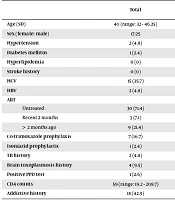
In total, we recruited 42 patients presenting with neurologic symptoms/signs and diagnosed with HIV. The mean age of the patients was 39.74 (SD = 10.88); 25 (59.5%) were men, 19 (45.2%) were newly diagnosed. Thirteen patients (31%) died within four weeks of treatment when they were hospitalized; from them, six were newly diagnosed. As shown in Table 1, other demographic and clinical characteristics of the patients were evaluated in relation to their survival status. The CD4 level in dead patients was significantly lower than alive individuals (P = 0.002); also, patients using drugs had significantly higher mortality rates (P = 0.004). In addition, the lowest CD4 level was found in patients with Toxoplasmosis (12) and HAND (16); the highest level of CD4 (280 cells/mm3) was found in a patient with herpes encephalitis. Duration of treatment with ART was also significantly different in survivors vs. non-survivors. A total of 92.3% of deceased patients had no history of ART treatment, and only one patient received this medication within two months. While, in surviving subjects, 62.1% were not receiving ART.
Table 1.
Demographic and Clinical Characteristics of HIV Infected Patients in Relation to their Survival Statusa
| Total | Survival Status (N = 42) | P Value | ||
|---|---|---|---|---|
| Live (N = 29) | Dead (N = 13) | |||
| Age (SD) | 40 (range: 32 - 46.25) | 38 (range: 32.5 - 43) | 45 (range: 32 - 47.5) | 0.19 |
| Sex (female: male) | 17:25 | 10:19 | 7:6 | 0.24 |
| Hypertension | 2 (4.8) | 1 (3.4) | 1 (7.7) | 0.53 |
| Diabetes mellitus | 1 (2.4) | 1 (3.4) | 0 | 0.99 |
| Hyperlipidemia | 0 (0) | 0 (0) | 0 (0) | NA |
| Stroke history | 0 (0) | 0 (0) | 0 (0) | NA |
| HCV | 15 (35.7) | 11 (37.9) | 4 (30.8) | 0.74 |
| HBV | 2 (4.8) | 1 (3.4) | 1 (7.7) | 0.53 |
| ART | ||||
| Untreated | 30 (71.4) | 18 (62.1) | 12 (92.3) | |
| Recent 2 months | 3 (7.1) | 2 (6.9) | 1 (7.7) | 0.04* |
| > 2 months ago | 9 (21.4) | 9 (31) | 0 (0) | |
| Co-trimoxazole prophylaxis | 7 (16.7) | 6 (20.7) | 1 (7.7) | 0.41 |
| Isoniazid prophylactic | 1 (2.4) | 1 (3.4) | 0 (0) | 0.99 |
| TB history | 2 (4.8) | 2 (6.9) | 0 (0) | 0.99 |
| Brain toxoplasmosis history | 4 (9.5) | 4 (13.8) | 0 (0) | 0.29 |
| Positive PPD test | 1 (2.6) | 1 (3.8) | 0 (0) | 0.99 |
| CD4 counts | 59 (range: 19.2 - 208.7) | 112 (range: 29 - 280) | 18 (range: 9.5 - 65) | 0.002* |
| Addictive history | 18 (42.9) | 9 (31) | 9 (69.2) | 0.04* |
aValues are expressed as No. (%), unless it was mentioned.
4.2. Baseline Neurologic Presentations
In Table 2, there was a significant association between level of consciousness and survival rate (P = 0.007); in addition, there was a marginally significant relation between survival and presence of seizures, cognitive impairment, signs of meningeal irritation, and upper motor neuron involvement in non-survivors (P < 0.01). A total of 72.4% of the survivors had a completely normal level of consciousness at the beginning of hospital stay, while the percentage in the dead was 24.1%. One patient was admitted in the hospital in a coma, which ultimately died. There was a significant correlation between the level of consciousness and the survival status of the patients (P = 0.007). The majority of the deceased (76.9%) had a decreased level of consciousness or was in a coma, while in the survivor group, this ratio was 27.7 %.
Table 2.
Baseline Neurologic Presentations of HIV Infected Patients in Relation to Their Survival Statusa
| Total | Survival Status (N = 42) | P Value | ||
|---|---|---|---|---|
| Live (N = 29) | Dead (N = 13) | |||
| Level of consciousness | 0.007* | |||
| Stupor/lethargy | 17 (40.5) | 8 (27.6) | 9 (69.2) | |
| Coma | 1 (2.4) | 0 (0) | 1 (7.7) | |
| Alert | 24 (57.1) | 21 (72.4) | 3 (24.1) | |
| Fever | 17 (40.5) | 11 (37.9) | 6 (46.2) | 0.61 |
| Headache | 16 (42.1) | 10 (35.7) | 6 (60.0) | 0.18 |
| Seizure | 9 (22) | 4 (13.8) | 5 (41.7) | 0.09 |
| Cognitive impairment | 5 (13.5) | 2 (6.9) | 3 (37.5) | 0.06 |
| Behavioral disorder | 2 (5.3) | 2 (6.9) | 0 (0) | 0.99 |
| Swallowing disorders | 2 (5.7) | 2 (7.4) | 0 (0) | 0.99 |
| Ataxia | 6 (17.6) | 5 (17.9) | 1 (16.7) | 0.99 |
| Language disorders | 5 (13.5) | 3 (10.3) | 2 (25) | 0.29 |
| Sphincter dysfunction | 3 (7.9) | 1 (3.4) | 2 (22.2) | 0.13 |
| Visual impairment | 4 (10.8) | 3 (10.3) | 1 (12.5) | 0.99 |
| Burning and tingling of extremities | 4 (12.1) | 3 (11.5) | 1 (14.3) | 0.99 |
| Cerebellar disorders | 3 (9.4) | 2 (7.7) | 1 (16.7) | 0.47 |
| Signs of meningeal irritation | 2 (4.8) | 0 (0) | 2 (15.4) | 0.09 |
| Upper motor neuron disorders | 19 (45.2) | 9 (31) | 10 (76.9) | 0.006* |
| Lower motor neuron disorders | 2 (5.9) | 2 (7.1) | 0 (0) | 0.99 |
aValues are expressed as No. (%).
4.3. Laboratory and Tissue Biopsy Findings
Median CSF white blood cells counts were significantly higher in non-survivors (Table 3). However, we failed to show any significant relation between other factors and survival rate (P > 0.1). (A) The cases diagnosed with CSF PCR results were two patients with fungal etiology, four cases (21%) JC, one (5%) CMV, and one HSV (5%). Due to the low number of smear/culture positive samples, we were unable to analyze their relation with survival rate. Tissue biopsy was performed for three patients only; one diagnosed with Nocardiosis, one with Toxoplasmosis, and one with Cladosporium. All patients who were clinically diagnosed with Toxoplasmosis had positive serology. (B) Out of the 28 patients with focal cerebral lesions, 22 had positive IgG toxo. All eight known brain toxoplasmosis (positive test or diagnostic based on appropriate therapeutic response) had positive IgG toxo.
Table 3.
Laboratory Findings of HIV Infected Patients with Neurologic Presentations in Relation to their Survivala
| Status | Total | Survival Status (N = 42) | P Value | |
|---|---|---|---|---|
| Live (N = 29) | Dead (N = 13) | |||
| CSF glucose level | 47.5 (range: 37.2 - 59.5) | 47.5 (range: 31.7 - 56.7) | 48.5 (range: 39 - 64) | 0.82 |
| CSF protein level | 67.15 (range: 38 - 84.5) | 68 (range: 84.7 - 46.9) | 62 (range: 14.7 - 81.5) | 0.44 |
| CSF WBC level | 17.5 (range: 3.5 - 61.0) | 8.5 (range: 1 - 123.7) | 40 (range: 6.25 - 48.75) | 0.44 |
| CSF RBC level | 11.5 (range: 1.7 - 95) | 11.5 (range: 1 - 142.5) | 15 (range: 4 - 65.5) | 0.97 |
| CSF differential | 0.99 | |||
| PMN | 2 (18.2) | 1 (16.7) | 1 (20) | |
| Lymph | 9 (81.8) | 5 (83.3) | 4 (80) | |
| CSF smear | 0.99 | |||
| Fungi | 2 (9.5) | 1 (7.7) | 1 (12.5) | |
| Negative | 19 (90.5) | 12 (92.3) | 7 (87.5) | |
| CSF PCR | NA | |||
| Fungi | 2 (10) | 1 (8.3) | 1 (14.3) | |
| JC | 4 (21) | 2 (16.7) | 2 (28.6) | |
| Negative | 11 (57) | 7 (58.3) | 3 (42.9) | |
| CMV | 1 (5) | 1 (8.3) | 1 (14.3) | |
| HSV | 1 (5) | 1 (8.3) | 0 (0) | |
| CSF culture | 0.99 | |||
| Fungi | 2 (10) | 1 (7.7) | 1 (14.3) | |
| Negative | 18 (90) | 12 (92.3) | 6 (85.7) | |
| Brain biopsy | NA | |||
| Nocardia | 1 (33.3) | 0 (0) | 1 (50) | |
| Toxoplasma | 1 (33.3) | 1 (100) | 0 (0) | |
| Fungi | 1 (33.3) | 0 (0) | 1 (50) | |
aValues are expressed as No. (%), unless it was mentioned.
4.4. Radiologic Findings
Imaging was assessed in association with the survival rate in Table 4. The most common diagnosis was “focal brain lesion” (28 individuals, 71.7%); most reported etiologies included brain toxoplasmosis (8 individuals, 20.5%) and tuberculosis (4 individuals, 10.1%). Most involvement areas in toxoplasmosis were basal ganglia and cortical regions (62.5%), while all the tuberculosis lesions were in the cortical region. The proportion of compression effect of basal ganglia was significantly higher in non-survivors (P < 0.05).
Table 4.
Findings of Brain Imaging Studies in HIV Infected Patients in Relation to their Survival Statusa
| Total | Survival Status (N = 42) | P Value | ||
|---|---|---|---|---|
| Live (N = 29) | Dead (N = 13) | |||
| Evidence of edema | 19 (46.3) | 12 (42.9) | 7 (53.8) | 0.51 |
| Supratentorial involvement | ||||
| Basal ganglion | 12 (28.6) | 5 (17.2) | 7 (53.8) | 0.30 |
| Pre-ventricular | 11 (26.2) | 7 (24.1) | 45 (30.8) | 0.65 |
| Cortical | 24 (57.1) | 15 (51.7) | 9 (69.2) | 0.29 |
| Ventriculitis | 1 (2.4) | 0 (0) | 1 (7.7) | 0.31 |
| Basal ganglia individually | 2 (5) | 0 (0) | 2 (15.4) | NA |
| Periventricular individually | 8 (20.5) | 7 (26.9) | 1 (7.7) | NA |
| Cortical individually | 13 (32.5) | 10 (37.5) | 3 (23.1) | NA |
| No involvement of supratentorial | 6 (15) | 5 (18.5) | 1 (7.7) | NA |
| Periventricular + cortical + basal | 2 (5) | 0 (0) | 2 (15.4) | NA |
| Cortical + basal | 7 (17.5) | 5 (18.5) | 2 (15.4) | NA |
| Ventriculitis + basal + cortical | 1 (2.5) | 0 (0) | 1 (7.7) | NA |
| Periventricular + cortical | 1 (2.5) | 0 (0) | 1 (7.7) | NA |
| Involvement of infratentorium | 14 (35.9) | 7 (26.9) | 7 (53.8) | 0.16 |
| Parallel lesions | 3 (7.9) | 3 (12) | 0 (0) | 0.54 |
| Evidence of ischemia | 8 (20.5) | 4 (15.4) | 4 (30.8) | 0.44 |
| Evidence of hemorrhage | 7 (17.9) | 3 (11.5) | 4 (30.8) | 0.19 |
| Compression effect | 9 (23.1) | 3 (11.5) | 6 (46.2) | 0.04 |
| Lesion Enhancement | 22 (56.4) | 15(36.5) | 7 (17.9) | 0.54 |
| Hydrocephalus | 2 (5.1) | 2 (7.7) | 0 (0) | 0.55 |
| The largest diameter of the lesion | 12.5 (range: 5 - 22.7) | 10.5 (range: 4.25 - 18.5) | 8 (range: 3.25 - 22.25) | 0.13 |
| Number of lesions | 10 (range: 3.5 - 20.5) | 18.5 (range: 12.5 - 27.5) | 12 (range: 5 - 31.5) | 0.8 |
aValues are expressed as No. (%), unless it was mentioned.
The most prevalent causes of death in patients with focal brain lesions were reported as “unknown” (5 individuals, 15.3%), PML (2 individuals, 15.3%), and stroke (2 individuals, 15.3%).
4.5. Response to Treatment
It should be noted that the most definite and empirical therapy was anti-toxoplasmosis (17 subjects, 38.1%). This treatment was used as monotherapy (23.8%) or anti-bacterial/anti-TB (14.3%). The duration of clinical response in patients with fully or partially recovered disease was estimated to be about six days. The lesions were completely removed in their imaging studies after 15 to 60 days.
Overall, 23 of 29 live patients (79%) had totally recovered with the target treatment, three individuals (10%) had partially recovered, and three (10%) showed no significant recovery. Eight out of 11 (72.7%) patients were followed-up by radiologic studies; one of them was treated with anti-toxoplasmosis therapy, and another was treated with corticosteroid and ART (diagnosed with IRIS PML), and patients was treated with Itraconazole (diagnosed with Cladosporium). Patients with Tuberculosis and Cladosporium had a significant recovery in their radiological findings after 60 - 90 days. The final diagnosis of patients infected with HIV based on neurologic classification is presented in Table 5.
Table 5.
Final Diagnosis of Patients Infected with HIV Based on Neurologic Classificationa
| Total (N = 39) | Survival Status (N = 42) | ||
|---|---|---|---|
| Live (N = 29) | Dead (N = 13) | ||
| Meningitis | |||
| Fungi | 2 (50) | 1 (33.3) | 1 (100) |
| TB | 1 (25) | 1 (33.3) | 0 (0) |
| Reactive mastoidits | 1 (25) | 1 (33.3) | 0 (0) |
| Non-focal | |||
| HAND | 3 (75) | 3 (75) | 0 (0) |
| Encephalitis | 1 (25) | 1 (25) | 0 (0) |
| Focal | |||
| Toxoplasmosis | 8 (28.6) | 8 (50) | 0 (0) |
| PML | 3 (10.7) | 1 (6.4) | 2 (16.7) |
| TB | 3 (10.7) | 3 (18.8) | 0 (0) |
| Fungi | 1 (3.6) | 1 (6.3) | 0 (0) |
| Stroke | 3 (10.7) | 1 (6.3) | 2 (16.7) |
| Bacterial emboli | 1 (3.6) | 1 (6.3) | 0 (0) |
| Iris PML | 1 (3.6) | 1 (6.3) | 0 (0) |
| Toxoplasmosis/stroke | 1 (3.6) | 0 (0) | 1 (3.8) |
| Nocardiosis | 1 (3.6) | 0 (0) | 1 (8.3) |
| Unknown fungal | 5 (17.9) | 0 (0) | 5 (41.7) |
| CMV | 1 (3.6) | 0 (0) | 1 (8.3) |
| Non-neurologic | |||
| Sepsis | 2 (66.7) | 2 (66.7) | 0 (0) |
| Not taking anticonvulsant medicines | 1 (33.3) | 1 (33.3) | 0 (0) |
aValues are expressed as No. (%).
5. Discussion
In the present study, we evaluated 42 patients diagnosed with HIV and concomitant neurologic presentations, as well as their response to treatment (if diagnosed). The mean age of the patients was 40 years, and 19 patients (45.2%) were diagnosed with HIV in the current admission, neurologic complications being their first manifestation of the disease.
Up to our knowledge, this is the first prospective study of all patients with HIV and neurologic presentations in an academic tertiary hospital in our region. A thorough examination of clinical, laboratory, and radiologic findings of patients’ neurologic conditions, besides their association with survival status, is the main strength of the current study.
In a similar study conducted in Ethiopia from 2002 to 2009, the mean age of the patients was 35 years with 33.7% being newly diagnosed. As of our findings, CD4+ T-cell counts were significantly lower in non-survivors; and serum levels of CD4+ T-cells was a determining factor in the prognosis of patients living with HIV (6). A 2006 study in the US also confirmed that the decline in CD4+ T-cell counts had been associated with neurologic complications and prognosis of this disease (9). This factor has been confirmed as a prognostic one in other studies as well (7, 18).
A systematic review at Johns Hopkins, in 2005, introduces a list of the factors involved in the neurologic complications of HIV including older age, female gender, and presence of other systemic diseases (5). Although we found no association between demographic features or systemic diseases and survival in the presence of neurologic presentations, injection drug use was significantly higher among non-survivors. In this study, there was a significant association between the levels of consciousness and patients’ survival rates; the majority of non-survivors were in stupor or coma state when admitted. Other common neurologic presentations among non-survivors compared to survivors included seizure (41.7% vs. 13.8%), cognitive impairment (37.5% vs. 6.9%), signs of meningeal irritation (15.4% vs. 0%), and upper motor neuron signs (76.9 vs. 31%). In a study by Kaplan et al. (19), in California, decreased the level of consciousness was known as a predictor of mortality. The Ethiopian study also introduced fluctuating levels of consciousness and seizures as prognostic factors of mortality (6). Although a seizure has been reported as the main presentation of neurologic involvement in HIV and associated with high mortality rates (14) since we did not evaluate seizure disorders we cannot make sure about non-presence of epileptic activities before loss of consciousness in our patient population.
The most common diagnosis in our study was “focal brain lesion” and the most common of etiology was “cerebral toxoplasmosis” (8 individuals, 20.5%), followed by TB (4 patients, 10.1%) and then fungal and bacterial diseases (3 cases, 7.6%). In the Ethiopian study, the incidence of cerebral toxoplasmosis was 35.2%, TB meningitis was 22.5%, Cryptococcal meningitis was 22.2%, and bacterial meningitis was 6.9%. Neuro-imaging was performed in 38%, which 56.8% of them had a mass lesion.
Neurologic presentations of HIV are concomitant with high mortality rates. Not receiving ART, low baseline CD4+ T-cell counts, and history of drug use were associated with higher mortality among our patients; although the sample size is not large enough to generalize the findings, all three factors highlight the role of early diagnosis and compliance to treatment in the reduction of mortality associated with HIV infection. Infections remain the common cause of neurologic disease in this patient population.
Designing a study with longer follow up periods and performing general laboratory assessments could be helpful in reaching more generalizable findings.