Abstract
Introduction:
SARS-CoV-2 infection is spreading worldwide, and due to multi-organ involvement, it could mimic other well-known diseases.Case Presentation:
Herein, we describe the case of a pediatric patient with acute promyelocytic leukemia (APL), who developed severe respiratory illness with diffuse pulmonary involvement after consuming all-transretinoic acid during the COVID-19 pandemic.Conclusions:
All-transretinoic acid syndrome is a very similar condition to COVID-19 both in clinical presentations and radiologic findings; thus, the treatment of such patients may be challenging.Keywords
SARS-CoV-2 COVID-19 ATRA syndrome differentiation Syndrome Acute Promyelocytic Leukemia
1. Introduction
Acute promyelocytic leukemia (APL) is characterized by the morphology of cancer cells with a reciprocal chromosomal translocation (15;17) (q22;q12-q21) and coagulopathy. The recommended treatment includes anthracycline-based chemotherapy (i.e., daunorubicin and idarubicin), cytarabine, and all-transretinoic acid (ATRA) (1). ATRA syndrome is a multisystem disorder characterized by shortness of breath, fever, weight gain, hypotension, and high permeability of pulmonary microcirculation. This condition is commonly seen in patients with APL, after the administration of ATRA at a rate of 2% - 27% (2).
SARS-CoV-2 pneumonia could mimic ATRA syndrome by its similar clinical presentations and imaging findings. SARS-CoV-2 infection usually manifests with mild upper respiratory tract signs and symptoms and may lead to lower respiratory tract involvement and multi-organ failure. The mortality of this disease ranges between 0.2% and 15%, depending on age, underlying diseases, comorbidities, secondary bacterial infection, and timely diagnosis and treatment of the disease (3).
The duration of onset of symptoms is generally between 2 days to two weeks., and real-time polymerase chain reaction (RT-PCR) is the point-of-care diagnostic testing for this condition (4). Primary diagnosis could usually be made based on clinical signs and symptoms, epidemiologic links with the index case, and high-resolution computed tomography (HRCT) scan. Here, we present the case of an APL patient during the COVID-19 pandemic, who primarily presented with an acute respiratory illness suspected to SARS-CoV-2 pneumonia.
2. Case Presentation
A 7-year-old boy referred to our center with a chief complaint of easy bruising for one month. On admission, white blood cells (WBC) count was 3,200 cell/mm3 (i.e., 20% neutrophils, 18% lymphocytes, 2% eosinophils, and 60% promyelocytes), hematocrit was 36.2%, and platelet was 20,000 cell/mm3. On peripheral blood smear, numerous fragmented red blood cells were found.
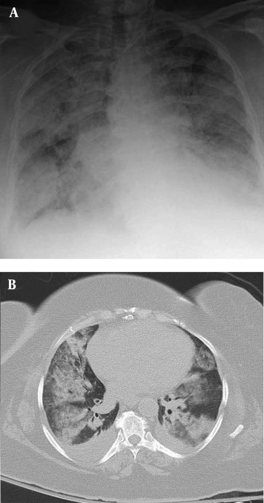
Bone marrow (BM) aspiration/biopsy showed hypercellularity with a marked increase in promyelocytes (80%).The diagnosis of acute promyelocytic leukemia was confirmed by bone marrow cytogenetics, which was positive for (t[15;17][q22;q12]] translocation, and the patient was treated with ATRA 45 mg/m2 on day one and intravenous idarubicin 10 mg/m2 on days 7, 9, and 11. After three days, he developed fever, respiratory distress, low systolic blood pressure, and extremity edema. A chest X-ray was performed, which revealed bilateral pulmonary infiltration (Figure 1A), and a chest CT scan demonstrated mixed ground-glass opacity (GGO), consolidation with peripheral distribution, and pericardial effusion (Figure 1B). Blood urea nitrogen (BUN) and creatinine increased, and the patient became oliguric.
A, Chest X-ray showed diffused ground-glass opacity (GGO) and consolidation in both lungs, with a “white lung” appearance; B, multifocal GGO in both lungs with patchy consolidation, multi-segmental pulmonary distribution, air bronchogram, and bilateral pleural effusion.
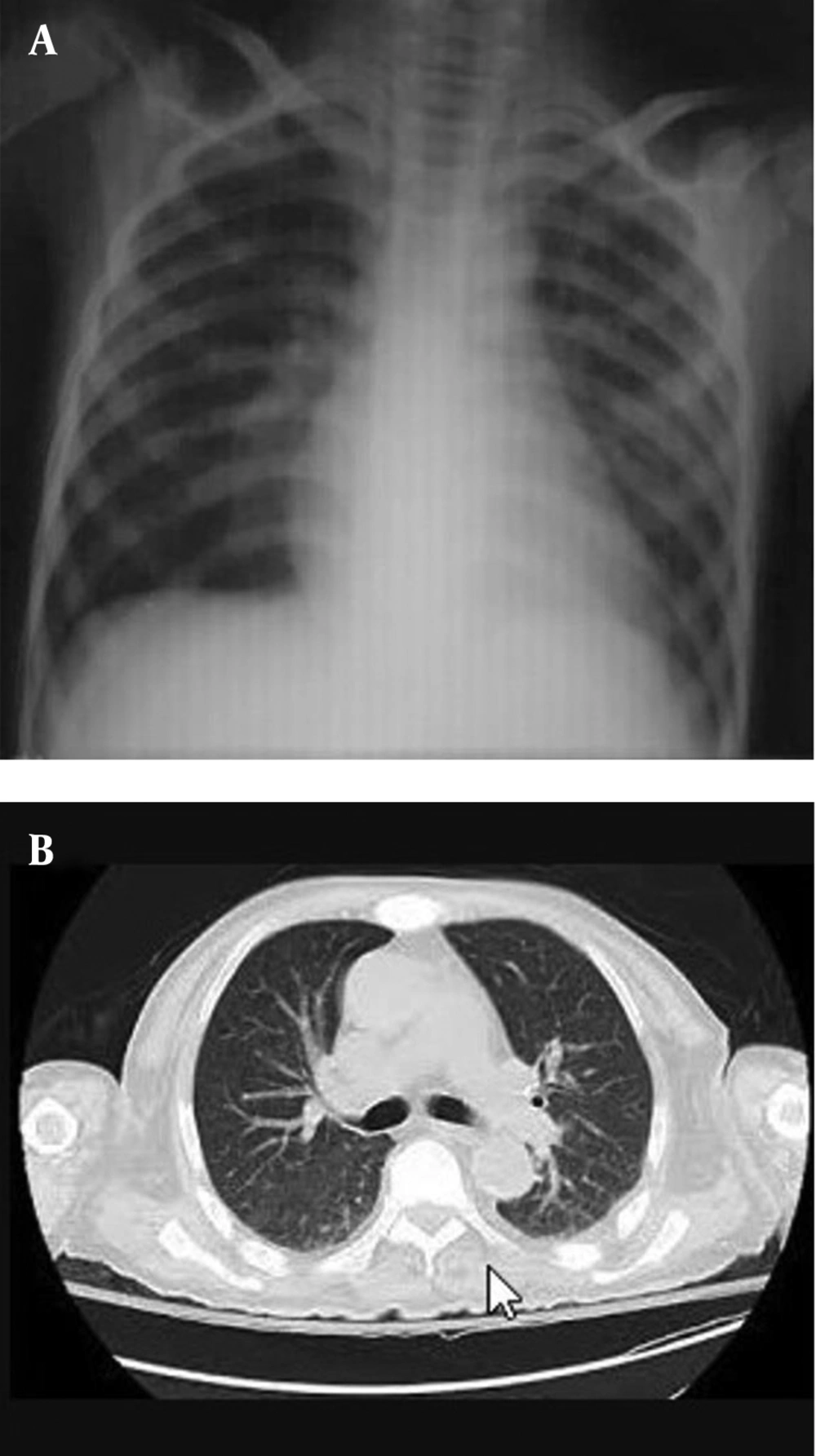
SARS-CoV-2 RT-PCR was repeatedly negative for the patient and his family members. After ruling out COVID-19 pneumonia, the diagnosis of ATRA syndrome was suspected, and ATRA was discontinued, and parenteral dexamethasone 2 mg every 8 hours was started accordingly. The patient’s symptoms improved dramatically after the discontinuation of ATRA and starting dexamethasone. He was subsequently given cytarabine and idarubicin at a subtherapeutic dose. Radiologic findings significantly improved after the treatment modification and corticosteroid therapy (Figure 2).
A and B, Four days after starting dexamethasone, chest c-ray and CT showed disappearance of the lung infiltrations
3. Discussion
ATRA or differentiation syndrome is characterized by fever, hypoxemia, lung opacities, pleural effusion, and pericardial effusion in a patient treated with ATRA (5-7). Differentiation syndrome is a life-threatening condition usually seen in patients with APL. The reported incidence of ATRA syndrome varies between 2% and 27% in different clinical trials on patients receiving ATRA (8).
ATRA prevents releasing inflammatory cytokines such as interleukin (IL) 6 and IL-8, and tumor necrosis factor-alpha (TNF-α) from leukemic cells. The pathogenesis of ATRA syndrome is not completely clear, but it seems some vasoactive inflammatory cytokines lead to capillary leakage (9). The maturation of promyelocytes and tissue infiltration of these matured cells is the next declared etiology (10). The final mechanism of injury is endothelial damage, followed by edema, bleeding, fibrinous exudates, leukocyte infiltration, and finally, respiratory failure (8).
The pathogenesis of the SARS-CoV-2 infection is almost similar to ATRA syndrome with cytokine storm, diffuse alveolar damage, and acute respiratory distress syndrome (ARDS) (11, 12). Tubular epithelial cells of the kidney, the central nervous system, and various types of immune cells may also be impaired. It seems that both abnormal immune responses and cell injury may be critical factors in SARS-CoV-2 infection (13, 14).
The diagnosis of ATRA syndrome is usually made after excluding other differential diagnoses such as viral and bacterial infections. The mortality rate of this condition varies between 8% and 28%, and dexamethasone is the recommended treatment (15, 16).
In our patient, bacterial and fungal blood cultures were negative. COVID-19 PCR was also negative in the patient and his family. The patient was treated with corticosteroids and responded quickly by reducing the symptoms and radiological disturbances. This rapid response to glucocorticoids is consistent with the diagnosis of ATRA syndrome, while steroids may lead to worsening of the SARS-CoV-2 infection. Routine use of systemic corticosteroids for COVID-19 pneumonia is discouraged in the updated guidance released by the World Health Organization (WHO) on May 18, 2020 (17), because of the possibility of prolonged viral replication, as seen in middle east respiratory syndrome (MERS) pneumonia, unless there is another reason for use, for example, exacerbation of chronic obstructive pulmonary disease or septic shock (18).
As with previous illnesses, such as severe acute respiratory distress syndrome (SARS) and MERS, corticosteroids are not commonly recommended as they may exacerbate COVID-19-related lung damage (19). Based on the results of a systematic review and meta-analysis of the impact of corticosteroid therapy on various outcomes of patients with SARS-CoV-2, SARS-CoV, and MERS-CoV infection, no significant effect on reducing the risk of death, reducing hospitalization days, ICU admission rate, and use of mechanical ventilation could be found (20).
Preventive steroid therapy during induction therapy with ATRA is not routinely recommended; however, despite the lack of supportive evidence in randomized trials, based on some case series, preventive strategy with corticosteroids is proposed in those with leukocyte count > 5 - 10 × 109/L (21).
Chest radiographs of ATRA syndrome also mimic COVID-19 pneumonia. GGO (60%), consolidation, nodular opacities, and pleural effusion can be seen that may be similar to COVID-19 pneumonia or other lung infections (22, 23). GGO, with or without consolidations, is the most common CT finding in patients with COVID-19 pneumonia (24-26). Chest CT abnormalities are usually bilateral, with lower lobes preference and peripheral distribution. Other less common findings include pleural effusion, hilar lymphadenopathy, crazy paving pattern, cavitation, interlobular septal thickening, and linear opacities (26, 27).
It has been shown that chest CT scan is more sensitive than RT-PCR in the early stages of COVID-19 pneumonia, and CT scan usually shows abnormal findings up to seven days before positive results of RT-PCR assay (27). Therefore, it is essential to rule out other differential diagnoses because symptoms, lab data, and imaging findings may overlap with other diseases. Pneumonia, pulmonary thromboembolism, and heart failure are the most important differential diagnoses of ATRA syndrome (28).
3.1. Conclusions
COVID-19 infection is a multisystem disease with clinical and imaging similarities to ATRA syndrome. A high index of suspicion is necessary when treating patients receiving ATRA during the COVID-19 pandemic. While corticosteroids are the treatment of choice for patients complicating with ATRA syndrome, they may be harmful in patients with COVID-19.
References
-
1.
Thomas X. Acute Promyelocytic Leukemia: A History over 60 Years—From the Most Malignant to the most Curable Form of Acute Leukemia. Oncology and Therapy. 2019:1-33. [PubMed ID: 29977766]. https://doi.org/10.1007/s40487-018-0091-5.
-
2.
Metage C, Hazarika B, Sarma J, Karwa R. Retinoic acid syndrome in a elderly male with psoriasis-A case report. Respiratory medicine case reports. 2018;24:81-3. [PubMed ID: 32169119]. https://doi.org/10.1016/j.rmcr.2018.04.007.
-
3.
Guo Y, Cao Q, Hong Z, Tan Y, Chen S, Jin H, et al. The origin, transmission and clinical therapies on coronavirus disease 2019 (COVID-19) outbreak–an update on the status. Military Medical Research. 2020;7(1):1-10. https://doi.org/10.1186/s40779-020-00240-0.
-
4.
Patel A, Jernigan DB. Initial public health response and interim clinical guidance for the 2019 novel coronavirus outbreak—United States, December 31, 2019–February 4, 2020. Morbidity and Mortality Weekly Report. 2020;69(5):140.
-
5.
Reddy R, Yaghmour B. Severe Acute Respiratory Distress Syndrome Secondary to All-trans Retinoic Acid Lung Toxicity. Chest. 2016;150(4):494A. https://doi.org/10.1016/j.chest.2016.08.508.
-
6.
Ariza-Prota M, Pando-Sandoval A, García-Clemente M. Lung injury caused by all-trans-retinoic acid in the treatment of acute promyelocytic leukemia. Archivos de Bronconeumolog a (English Edition). 2016;8(52):441-2. [PubMed ID: 26915524]. https://doi.org/10.1016/j.arbres.2016.01.007.
-
7.
Baris HE, Yalindag-Ozturk N, Girgin FI, Koc A. A Child Patient with Acute Promyelocytic Leukemia Presenting with White Lung: Differentiation Syndrome in the Differential Diagnosis: Case Report. Türkiye Klinikleri Journal of Case Reports. 2016;24(2):114-7. https://doi.org/10.5336/caserep.2015-48895.
-
8.
Patatanian E, Thompson DF. Retinoic acid syndrome: a review. Journal of clinical pharmacy and therapeutics. 2008;33(4):331-8. [PubMed ID: 18613850]. https://doi.org/10.1111/j.1365-2710.2008.00935.x.
-
9.
Luesink M, Jansen JH. Advances in understanding the pulmonary infiltration in acute promyelocytic leukaemia. British journal of haematology. 2010;151(3):209-20. [PubMed ID: 20735400]. https://doi.org/10.1111/j.1365-2141.2010.08325.x.
-
10.
Lin C, Huang M, Chang IY, Lin W, Sheu Y. Retinoic acid syndrome induced by arsenic trioxide in treating recurrent all-trans retinoic acid resistant acute promyelocytic leukemia. Leukemia & lymphoma. 2000;38(1-2):195-8. [PubMed ID: 10811463]. https://doi.org/10.1016/j.biopha.2020.110195.
-
11.
Zhang Y, Geng X, Tan Y, Li Q, Xu C, Xu J, et al. New understanding of the damage of SARS-CoV-2 infection outside the respiratory system. Biomedicine & Pharmacotherapy. 2020:110195. [PubMed ID: 32361161].
-
12.
Huang C, Wang Y, Li X, Ren L, Zhao J, Hu Y, et al. Clinical features of patients infected with 2019 novel coronavirus in Wuhan, China. The Lancet. 2020;395(10223):497-506. https://doi.org/10.1016/S0140-6736(20)30183-5.
-
13.
Liu J, Zheng X, Tong Q, Li W, Wang B, Sutter K, et al. Overlapping and discrete aspects of the pathology and pathogenesis of the emerging human pathogenic coronaviruses SARS‐CoV, MERS‐CoV, and 2019‐nCoV. Journal of medical virology. 2020. https://doi.org/10.1002/jmv.25709.
-
14.
Li H, Liu S, Yu X, Tang S, Tang C. Coronavirus disease 2019 (COVID-19): current status and future perspective. International Journal of Antimicrobial Agents. 2020:105951. https://doi.org/10.1016/j.ijantimicag.2020.105951.
-
15.
De Botton S, Dombret H, Sanz M, Miguel JS, Caillot D, Zittoun R, et al. Incidence, clinical features, and outcome of all trans-retinoic acid syndrome in 413 cases of newly diagnosed acute promyelocytic leukemia. Blood, The Journal of the American Society of Hematology. 1998;92(8):2712-8. https://doi.org/10.1182/blood.V92.8.2712.420k03_2712_2718.
-
16.
Montesinos P, Bergua JM, Vellenga E, Rayón C, Parody R, de la Serna J, et al. Differentiation syndrome in patients with acute promyelocytic leukemia treated with all-trans retinoic acid and anthracycline chemotherapy: characteristics, outcome, and prognostic factors. Blood, The Journal of the American Society of Hematology. 2009;113(4):775-83.
-
17.
World Health Organization. Clinical management of COVID-19 interim guidance. WHO; 2020, [updated 2020 May 31]. Available from: https://www.who.int/publications-detail/clinical-management-of-covid-19.
-
18.
Russell B, Moss C, Rigg A, Van Hemelrijck M. COVID-19 and treatment with NSAIDs and corticosteroids: should we be limiting their use in the clinical setting? ecancermedicalscience. 2020;14. https://doi.org/10.3332/ecancer.2020.1023.
-
19.
Russell CD, Millar JE, Baillie JK. Clinical evidence does not support corticosteroid treatment for 2019-nCoV lung injury. The Lancet. 2020;395(10223):473-5. https://doi.org/10.1016/S0140-6736(20)30317-2.
-
20.
Li H, Chen C, Hu F, Wang J, Zhao Q, Gale RP, et al. Impact of corticosteroid therapy on outcomes of persons with SARS-CoV-2, SARS-CoV, or MERS-CoV infection: a systematic review and meta-analysis. Leukemia. 2020:1-9. [PubMed ID: 32372026]. https://doi.org/10.1038/s41375-020-0848-3.
-
21.
Sanz MA, Montesinos P. How we prevent and treat differentiation syndrome in patients with acute promyelocytic leukemia. Blood, The Journal of the American Society of Hematology. 2014;123(18):2777-82. [PubMed ID: 24627526]. https://doi.org/10.1182/blood-2013-10-512640.
-
22.
Frankel SR, Eardley A, Lauwers G, Weiss M, Warrell RP. The retinoic acid syndrome in acute promyelocytic leukemia. Annals of internal medicine. 1992;117(4):292-6. [PubMed ID: 1637024]. https://doi.org/10.7326/0003-4819-117-4-292.
-
23.
Vahdat L, Maslak P, Miller WJ, Eardley A, Heller GASD, Scheinberg DA, et al. Early mortality and the retinoic acid syndrome in acute promyelocytic leukemia: impact of leukocytosis, low-dose chemotherapy, PMN/RAR-alpha isoform, and CD13 expression in patients treated with all-trans retinoic acid. Blood. 1994;84(11):3843-9. [PubMed ID: 7949141]. https://doi.org/10.1182/blood.V84.11.3843.bloodjournal84113843.
-
24.
Shi H, Han X, Jiang N, Cao Y, Alwalid O, Gu J, et al. Radiological findings from 81 patients with COVID-19 pneumonia in Wuhan, China: a descriptive study. The Lancet Infectious Diseases. 2020. https://doi.org/10.1016/S1473-3099(20)30086-4.
-
25.
Zhao W, Zhong Z, Xie X, Yu Q, Liu J. Relation between chest CT findings and clinical conditions of coronavirus disease (COVID-19) pneumonia: a multicenter study. American Journal of Roentgenology. 2020:1-6. [PubMed ID: 32125873].
-
26.
Salehi S, Abedi A, Balakrishnan S, Gholamrezanezhad A. Coronavirus disease 2019 (COVID-19) imaging reporting and data system (COVID-RADS) and common lexicon: a proposal based on the imaging data of 37 studies. European Radiology. 2020:1. [PubMed ID: 32346790].
-
27.
Ai T, Yang Z, Hou H, Zhan C, Chen C, Lv W, et al. Correlation of chest CT and RT-PCR testing in coronavirus disease 2019 (COVID-19) in China: a report of 1014 cases. Radiology. 2020:200642. [PubMed ID: 32101510]. https://doi.org/10.1148/radiol.2020200642.
-
28.
Montesinos P, Sanz MA. The differentiation syndrome in patients with acute promyelocytic leukemia: experience of the pethema group and review of the literature. Mediterranean journal of hematology and infectious diseases. 2011;3(1). [PubMed ID: 22220256]. https://doi.org/10.4084/mjhid.2011.059.