1. Introduction
Since the worldwide pandemic of novel coronavirus 19 disease (COVID-19), different clinical presentations have been reported in children with various disease courses. At first, the main presentations were respiratory, and it seemed that this virus could not affect children under one year old. However, different case reports showed that the symptoms and clinical courses in the pediatric population could range from respiratory involvement to gastrointestinal symptoms and even Kawasaki disease that has been reported lately (1, 2). Here, we report a 2-months old boy with dehydration and electrolytes imbalance referring to our hospital with lung involvement in his CT-scan.
2. Case Presentation
On 3rd of March 2020, a 2-months old boy referred to our hospital with severe dehydration and fever from 4 days ago. He had received cefixime suspension and oral ondansetron and oral acetaminophen for fever in the outpatient clinic with no obvious improvements in the clinical course. His mother had respiratory symptoms such as cough and mild chest pain, and there was a positive history of contact with a person who had returned from one of the epidemic areas in Iran. His first physical examination revealed severe dehydration. He had a 38°C fever, and his heart rate was 180 beats/min, respiratory rate was 40/min, and blood pressure of 80/50 mmHg. His first O2 saturation detected by finger pulse oximetry was 92%.
He was admitted to the pediatric ward in Northeast of Iran (North Khorasan), and we started intravenous (IV) fluid resuscitation with 20 cc/kg normal saline and then IV dextrose 5% and half saline with 40 meq/L KCl 15% (calculating based upon maintenance and dehydration). Blood tests were carried out, and the initial therapy was based on the diagnosis of possible sepsis with IV ampicillin and cefotaxime. He had no respiratory distress, but his O2 sat was constantly lower than 90%, so we started free O2 flow 5 - 6 lit/min. His first Chest X-Ray was normal.
His laboratory test results were as follows:
A complete sepsis workup was done for him. The laboratory results revealed: White blood cell (WBC) = 25000 (with 50% Neutrophil), a positive C-reactive protein (CRP) and erythrocyte sedimentation rate (ESR) was 25 in one hour. Urine analysis and culture were performed that were negative. The lumbar puncture was not remarkable. His blood sodium level was 168 mEq/L, and potassium was 3.2 mEq/L; so the IV fluid changed to dextrose 5% and normal saline with 40 meq/L KCl 15%. The first arterial blood gas (ABG) showed metabolic acidosis. Nebulization with a 5% saline solution and Budezonide was administrated. Serum sodium and potassium levels were measured every 6 hours, and O2 therapy was continued. One day after the admission, he had moderate dehydration, and breast milk was started. Serial sodium levels were continuously high, and changes in volume and amount of IV fluid’s sodium had no effect on it. After 24 hours, he had poor response hypernatremia, moderate dehydration, and O2 dependency.
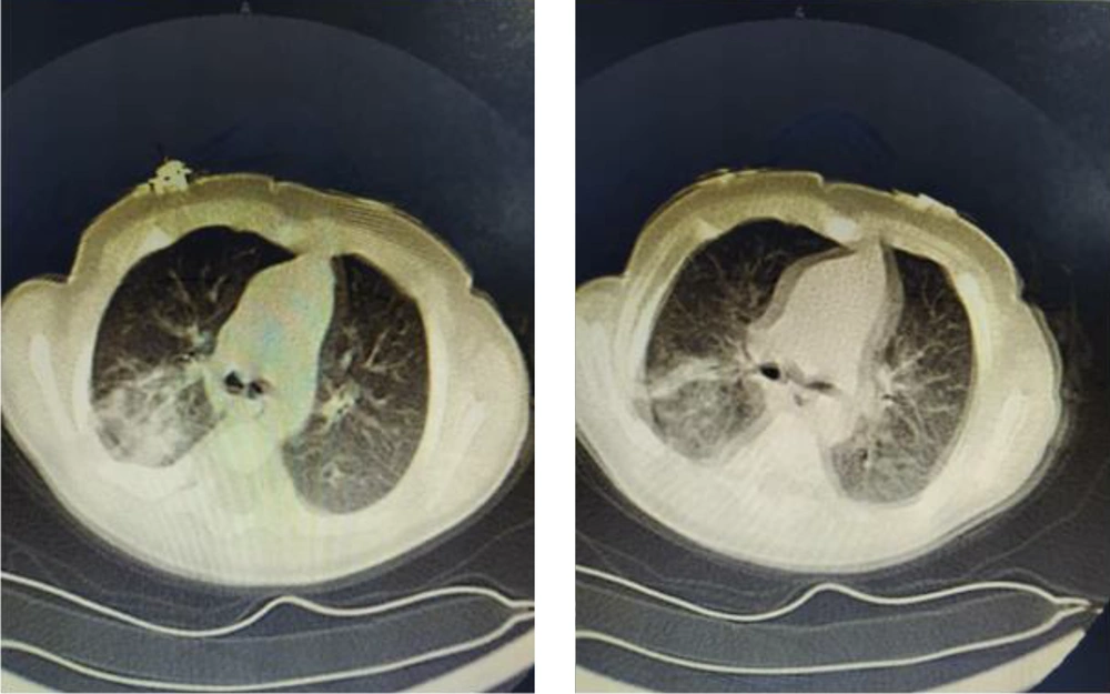
Considering the outbreak of the novel coronavirus in our country, we took an oropharyngeal swap sample and a chest CT-scan. A blood test was done for lactate dehydrogenase (LDH) and creatine phosphokinase (CPK), which were high (1320 and 371, respectively). His test result was negative for coronavirus, but his chest CT-scan showed consolidation in the upper lobe and inferior lobe’s posterior segments of the right lung and ground-glass opacity (GGO) in the left lung’s inferior lobe lateral base and posterior base (Figure 1). Thereafter, we started oral azithromycin and hydroxychloroquine and admitted him to the isolated COVID-19 ward. Intravenous ampicillin was discontinued, but we continued cefotaxime along with fluid therapy.
On day 3 of the admission, his situation improved, and the hemodynamic status had been stabled. The fever was resolved, and he had mild dehydration. Serial sodium and potassium check were discontinued after two consequent normal tests. His O2 saturation was above 95%, and there was no need for O2 therapy anymore. Unfortunately, his father discharged him despite the specialist opinion. He was stable in further telephone follow-ups.
3. Discussion
Pediatric case reports in COVID-19 are limited, and it seems that the disease has a milder course compared to the adult patients, but the range of the clinical features are wide (3). Most of the cases are reported in family clusters, and there is a strong correlation between the disease in family members and the child. The incidence of COVID-19 has been reported by 18.1% in children under 1 year old (2). We here presented a 2-months old boy with fever and gastrointestinal symptoms and a suspected history of contact, as his mother showed symptoms suspected to COVID-19.
Fever is the most common sign in children like our patient, but vomiting and diarrhea are very rare (3-5). Our case presented with fever and severe dehydration as well.
The oropharyngeal swap test results were negative in our case. As it has been reported to be less positive in pediatrics, and it should be kept in mind among the differential diagnosis with all aspects of the patient (2). Although the positive PCR test is considered the gold standard test for the diagnosis of COVID-19, false-negative patients are the source of the infection (3) and repeating the test increases the accuracy, which was not done in our patient due to the shortage of diagnosis facilities in the area (Northeast of Iran).
Other studies in pediatrics showed normal laboratory tests (4, 5), but our patient had leukocytosis, a positive CRP and high levels of LDH and CPK that can be seen in any viral infection. So, further specific diagnostic methods were needed.
In this case, the base of our clinical diagnosis was the CT scan’s remarkable pattern, despite the normal chest X-ray (CXR). As reported in other studies, the radiologic changes in the lungs can be seen in the first CXR, but a normal graph would not rule out the diagnosis of COVID-19. Chest CT-scan can detect the pathologic lesions better than a plain X-ray, and ground-glass opacity (32.7% in one study) and segmental consolidation in bilateral lungs have been reported in pediatric patients (2, 4) that was seen in our patient as well.
Our patient had hypernatremia due to severe dehydration, and the response to the initial fluid resuscitation was not quite satisfactory. However, after starting oral azithromycin and hydroxychloroquine (based on the management protocol), he got better and, finally, discharged in a good health situation.

