1. Introduction
The first cases of coronavirus disease 2019 (COVID-19) were identified in Wuhan, China, in December 2019, and then it immediately spread to other parts of the world. The pathogenesis of this disease is a new type of coronavirus called severe acute respiratory syndrome coronavirus 2 (SARS-CoV-2) (1). Cough, fever, dyspnea, diarrhea, myalgia, and fatigue can be cited as the most important signs and symptoms of the disease (2). Conjunctivitis has also been reported as one of the manifestations of the disease, and some studies have suggested the transmission of the disease through teardrops in adults, but only a few studies have evaluated this probability in children (3-5). In this study, we report a three-year-old child with a confirmed diagnosis of COVID-19 developing conjunctivitis in Iran.
2. Case Presentation
Institutional Review Board approval was obtained for this study, and we followed the declaration of Helsinki in all procedures. Written informed consent was obtained from the patient’s parents.
The patient was a three-year-old male child who was referred to Namazee Hospital (Shiraz) due to fever, dry cough, tachypnea, and respiratory distress. He was admitted with the impression of a COVID-19 infection. The patient was well until five days before admission that developed with fever and dry cough. With developing tachypnea, the patient was referred to Namazee Hospital and admitted to the Pediatric Invasive Care Unit (PICU). He was intubated due to severe respiratory distress. The parents mentioned no significant medical or family history for their child.
On the physical examination, the child was in respiratory distress. The temperature and the respiratory rate were 38°C and 50, respectively. Bilateral rales were detected. Endotracheal swab specimens were taken to assess the coronavirus by the reverse-transcription polymerase chain reaction (RT-PCR) assay, which proved to be positive after two days. Also, an endotracheal sample was sent for culture, which was also revealed to be positive for methicillin-resistant Staphylococcus aureus (MRSA).
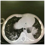
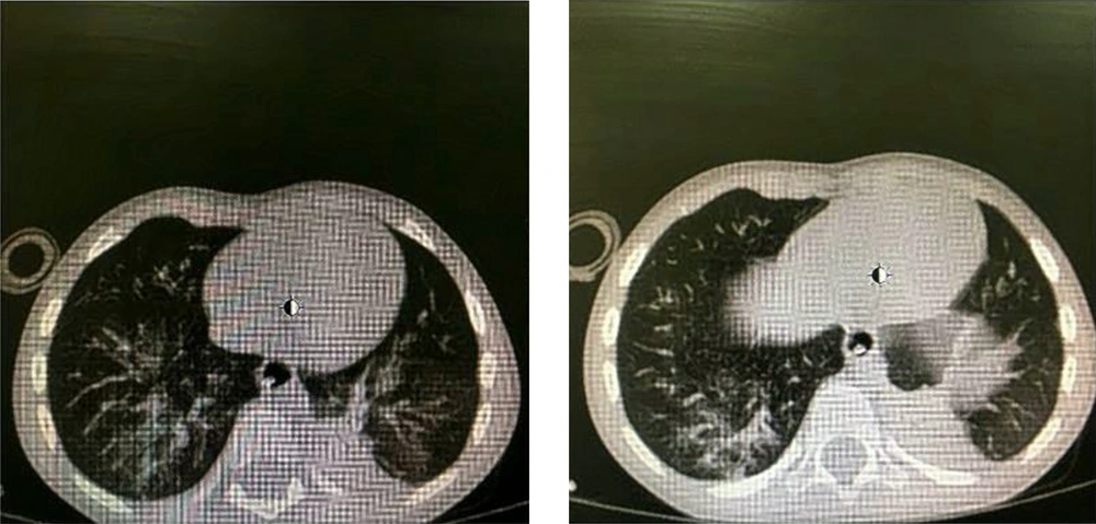
Blood tests were done, and supportive and therapeutic care along with antibiotic therapy began for him. He received meropenem (500 mg/q12h) and clindamycin (15 mg/kg/q6h). Moreover, a portable chest X-ray and chest computed tomography (CT) scan were requested for him, which proved lung involvement (Figure 1). After four days of admission, the patient’s condition improved, and he was extubated.
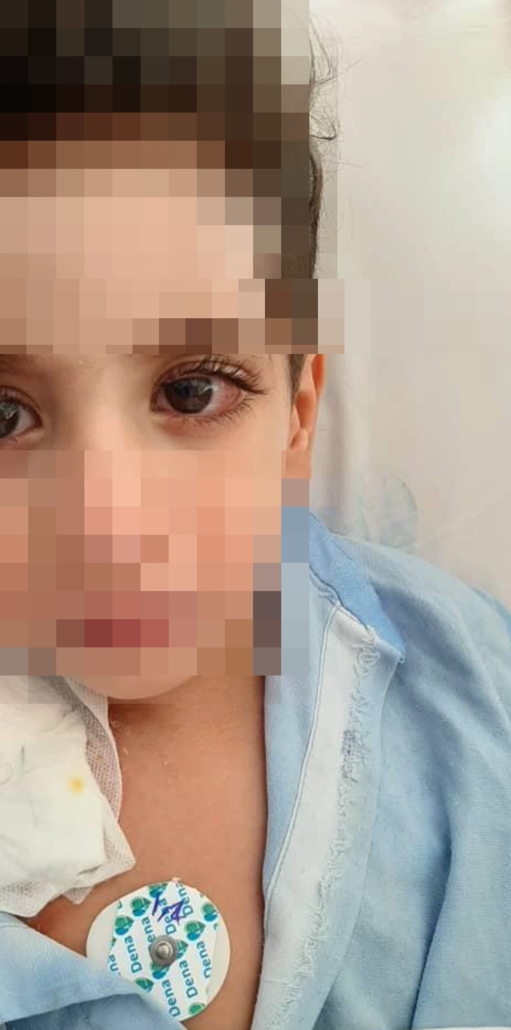
On the sixth day of admission, the patient developed unilateral red-eye and foreign body sensation in the left eye. Hence, we consulted an ophthalmologist. Slit-lamp examination identified conjunctival injection, which was not purulent, and no lesions were detected on the corneal area. Also, the exam did not detect any anterior chamber inflammation. Left eye conjunctivitis in the patient can be seen in Figure 2.
A conjunctival swab was done for collecting tears and conjunctival secretions from the lower eyelid fornix without topical anesthesia and was sent for assessing the presence of SARS-CoV-2. After two days, it was demonstrated to be positive. The results of the patient’s blood test were: WBC = 9.8 (neutrophil: 82.1%, lymphocyte: 15.7%); Hb = 10.6; platelet = 117,000; ESR = 19; CRP = 90; BUN = 11; creatine = 0.5; AST = 36; ALT = 14; CPK = 1769; LDH = 635. The symptoms, especially conjunctivitis, alleviated after 11 days of admission, so the patient was discharged from the hospital with clindamycin (15 mg/kg/q6h; PO) and OPD follow-up.
3. Discussion
We presented a case with ocular complications of COVID-19, which appeared after 11 days of illness onset. Chen et al. (6) presented a 30-year-old adult case with confirmed COVID-19 that developed conjunctivitis after 13 days of disease onset. Xia et al. (5) evaluated tear and conjunctival secretion specimens for SARS-CoV-2 RNA in 30 adult patients. The specimens of tears and conjunctival secretions from only one patient with conjunctivitis showed positive RT‐PCR results (5).
There are only limited pediatric cases with a definite diagnosis of COVID-19 for which the viral RNA was detected in their conjunctival specimens. Valente et al. (4) carried out a study on 27 children. Four of them showed ocular manifestations on admission, of whom just one was proven to have a positive RT‐PCR result for COVID-19 in conjunctival specimens. Moreover, two other patients had positive findings for SARS-CoV-2 in their conjunctival specimens, without having any clinical signs of conjunctivitis (4).
Our findings show the presence of COVID-19 in tears; so, it can be a source for transmission, like in adults (7). Additionally, it should be taken into account that ocular complications may not appear in the early stages of infection.


