Etiological Agents of Pediatric Diarrhea in Ardebil, Northwestern Iran

authors:

avatar Leila Asadi 1 , avatar Tala Pourlak 2 , avatar Behrooz Ahmadi 3 , avatar Mina Aghamali 4 , avatar Mohammad Asgharzadeh 4 , avatar Mohammad Aghazadeh 5 , avatar Elham Zeinalzadeh 1 , avatar Hossein Samadi Kafil ORCID 6 , *

Iranian Center of Excellence in Health Management, Tabriz University of Medical Sciences, Tabriz, Iran
Connective Tissues Disease Research Center, Tabriz University of Medical Sciences, Tabriz, Iran
Department of Parasitology, Tabriz University of Medical Sciences, Tabriz, Iran
Biotechnology Research Center, Tabriz University of Medical Sciences, Tabriz, Iran
Hematology and Oncology Research Center, Tabriz University of Medical Sciences, Tabriz, Iran
Drug Applied Research Center, Tabriz University of Medical Sciences, Tabriz, Iran

how to cite: Asadi L, Pourlak T, Ahmadi B, Aghamali M, Asgharzadeh M, et al. Etiological Agents of Pediatric Diarrhea in Ardebil, Northwestern Iran. Arch Pediatr Infect Dis. 2018;6(1):e11771. https://doi.org/10.5812/pedinfect.11771.

Abstract

Background:

Gastroenteritis is known as the leading cause of morbidity and mortality in children worldwide. Diarrhea is the second leading cause of infant death in the developing countries. Identification of gastroenteritis-causing pathogens helps to control fecal-oral transmission and prevents further outbreaks.

Objectives:

The current study aimed at evaluating the prevalence of bacterial, viral, and parasitic pathogens associated with children’s diarrhea in Ardebil, Iran.

Methods:

A total of 400 stool specimens were collected from children within the age range of 2 months to 12 years from December 2015 to June 2016 in Bu-Ali hospital in Ardebil, Iran. The samples were tested for bacterial, viral, and parasitic agents.

Results:

The results showed that parasites had the largest proportion of pathogens in the collected samples, followed by bacteria and viruses. The most commonly detected pathogens were: rotavirus, Escherichia coli, and Entamoeba histolytica, respectively. Protozoan parasites showed higher prevalence compared with helminthic infections.

Conclusions:

Rotavirus, E. coli and E. histolytica were the most prevalent agents of diarrhea in children’s aged 2 month to 12 years in this study. Although detection of few micro organisms were considered in our study, the results indicate importance of hygiene improvement in this area. More specified molecular epidemiology studies with evaluating the most common etiologies will be helpful for a better understanding of the frequency of common infections and their transmission in this area.

1. Background

Gastroenteritis is known as the leading cause of morbidity and mortality in children worldwide and diarrhea ranks second in the fatality rate of infants in the developing countries (1). Although diarrhea is prevalent in all ages, children are affected more due to rapid dehydration (2, 3). Diarrheal diseases cause 1 in 10 deaths in children under 5 years old (4). Poor sanitation, lack of safe drinking water, improper preparation of food and inadequate hygiene practices are the main risk factors of diarrhea (2). In recent years, numerous bacterial, viral, and parasitic agents are considered the main cause of gastroenteritis. Viral agents are the cause of the high rates of gastroenteritis, followed by bacterial and parasitic agents (1, 5). The human rotavirus is the most common etiologic agent of pediatric infections, which annually causes 125 million cases of diarrhea and more than 600 000 childhood deaths worldwide (6, 7). Bacteria account for 20% - 40% of diarrhea cases (8). Campylobacter jejuni, diarrheagenic Escherichia coli, non-typhoidal Salmonella spp. and Shigella spp. are frequently associated with diarrhea (9-11). Non-typhoidal Salmonella spp. annually causes 93,800,000 cases of gastroenteritis and 155,000 deaths globally (12, 13). Protozoan diarrhea is also distributed around the globe. Giardia lamblia, Entamoeba histolytica, and Cryptosporidium parvum are the mostly reported protozoan pathogens that cause gastroenteritis (4, 5). Studies showed that approximately 100,000 deaths followed by gastroenteritis are related to protozoan species (12). Asymptomatic amebiasis is one of the most common complications of E. histolytica, but it is life threatening if parasite invades the intestinal mucosa. In this case, amebiasis may lead to dysentery, liver abscess, and even death. Amebiasis is regarded as the third cause of mortality after malaria and schistosomiasis (14). Identification of gastroenteritis-causing pathogens helps to establish surveillance systems and guides priorities to control the transmission of infections and prevents outbreaks (15).

2. Objectives

The current study aimed at elucidating the prevalence of bacterial, viral, and parasitic pathogens associated with children’s diarrhea in Ardebil, Iran.

3. Methods

The current retrospective study was conducted from December 2015 to June 2016 in Bu-Ali Hospital in Ardebil, Northwestern Iran. During the period, all stool specimens were collected from patients referred to Bu-Ali hospital as the study samples. Children within the age range of 2 months to 12 years with the symptoms of diarrhea, fever, vomiting, abdominal pain, and (in some cases) dehydration were selected. Children with bloody diarrhea were excluded from the study. The samples were tested for bacterial, viral, and parasitic agents. The viral samples were examined for rotavirus and adenovirus using the immunochromatographic strip method (Immunospark, Spain) (16, 17). Bacterial samples were plated onto eosin methylene blue (EMB) agar and Salmonella-Shigella agar (SS agar) media using standard microbiological techniques (18). After 24 hours of incubation at 37C°, bacterial cultures were identified by biochemical tests (9). Parasitic samples were examined by an immunofluorescence assay, microscopy, and trichrome staining. The enzyme-linked immunosorbent assay (ELISA) was used to differentiate Entamoeba histolytica from non-pathogenic Entamoebadispar.

4. Results

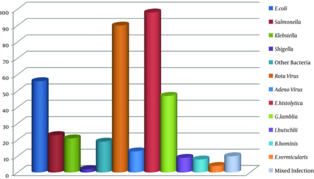
The study was conducted on 400 stool specimens of children within the age range of 2 months to 6 years. Of all samples, there were 100 (25%) viral, 121 (30.25%) bacterial, and 166 (41.5%) parasitic agents. Mixed infection was detected in 10 (2.5%) (2.5% confidence interval (CI): 1 - 4.5) cases (Figure 1). Out of 100 viral samples, the rotavirus constituted the largest proportion with 90 (22.5%) (21.75% CI: 17 - 26) cases, and the adenovirus made up the remaining 10 (3.25%) (3.25% CI: 1.9 - 5.4) cases. Of all bacterial pathogens, the most common one was Escherichia coli with 56 (14%) (14% CI: 10.94 - 17.74) cases, followed by non-typhoidal Salmonella spp., Klebsiella spp. and other bacteria with 23 (5.75%) (5.75% CI: 3 - 8), 21 (5.25%) (5.25% CI: 3.4 - 7.8) and 19 (4.75%) (3.75% CI: 2.29 - 6.09) cases, respectively. Parasitic pathogens were identified as Entamoeba histolytica with 98 (24.5%) (24.5% CI: 20.5 - 28.9), Giardia lamblia with 47 (11.75%) (11.75% CI: 8.9 - 15.2), Iodamoeba butschlii with 9 (2.25%) (2.25% CI: 1.19 - 4.2), Blastocystis hominis with 8 (2%) (2% CI: 0.01 - 3.9), and Enterobius vermicularis with 4 (1%) (1% CI: 0.03 - 2.0) cases, respectively. The results showed that parasitic agents had the largest proportion, followed by bacterial and viral agents in the study samples. Thus, the most prevalent viral, bacterial, and parasitic pathogens were rotavirus, E. coli, and E.histolytica.

Number of Pathogens Isolated from 400 Stool Specimens in the Study

5. Discussion

Gastrointestinal infections remain a major health problem despite the advances in infection control measures. Several studies are conducted to evaluate the prevalence of gastrointestinal pathogens in Iran. The current study found that 41.5% of samples were positive for parasites, 30.25% for bacteria, and 25.75% for viruses. Although bacterial and viral agents are traditionally considered the main causes of diarrhea, the current study indicated that protozoan pathogens were very important causes of diarrhea in this region. The current study found E. histolytica as the most prevalent parasitic agent. This result was in contrast with those reported by Sayyari et al. (19). The latter study reported G. lamblia as the major parasite in Iran, whereas the current study found it as the second most important causative agent. On the other hand, the current study result was compatible with the reports from Southwestern Iran (20). Pestehchian et al. and some other studies demonstrated a high prevalence of E. histolytica in the Southwest Iran (20, 21). In regards to the fact that intestinal protozoan infections are associated with lack of hygiene (22), sanitary measures should be improved in order to control infection and prevent fecal-oral transmission. The most effective preventive sanitary measures include: hand washing, proper food preparation, consumption of healthy water, and safe human waste disposal. Due to high costs of drugs and side effects followed by drug therapy, scientist suggest vaccination program as a favorable alternative (23). Since humans are the only known reservoir for E. histolytica, an effective vaccine can eradicate global E. histolytica infection (24, 25). In agreement with Zebardast et al., and some other studies, the current study results showed a higher prevalence of protozoan parasites compared with the helminthic agents (26). Similar to E. histolytica, E. coli infections are predominately due to lack of sanitary condition. In the current study, most viral infections were associated with the rotavirus, which was in accordance with several other studies from Iran (27-29). Previously conducted studies from other cities of Iran showed the large proportion of the rotavirus infection among children (27). Esteghamati et al., reported that, generally, more than half of all hospitalizations for severe diarrhea in Iran were associated with the rotavirus (30). A study in Tehran revealed a prevalence of 19% for rotavirus infections, while another study from Ahvaz showed the prevalence of 35% for rotavirus infection (31, 32). In another study, the rate of rotavirus positivity (42%) in Shiraz was higher than that of the current study results and the ones reported from other cities (33). It is considered that variety in the incidence of infection is associated with geographical conditions. Data from many other studies revealed the global seasonal occurrence of rotavirus infections (34, 35). Based on the preceding reports, rotavirus diarrhea frequently occurs during the winter months (34). The current study results supported the findings of the previously published studies and confirmed the abundance of rotavirus infections in cooler months (36). However, a report from Saudi Arabia showed the high incidence of rotavirus infection during dry and hot season (28). In addition, the seasonal pattern is not reported from countries with tropical climates such as Malaysia. With regard to the importance of the rotavirus, as the main cause of diarrhea in children (37), the rotavirus vaccination is a promising alternative to prevent the infection. The recommended vaccines by the world health organization (WHO) provide good protection and show a remarkable decrease in the seasonal prevalence of severe rotavirus infections. Rotavirus spread mainly occurs through fecal-oral route and contaminated hands (35); therefore, personal hygiene has a great impact on virus transmission. In the current study, similar to previous reports from Iran as well as other countries, the adenovirus had a lower rate compared with rotavirus (33). Also, there was no seasonal pattern for the adenovirus infection. Understanding the etiology of diarrhea advances in rapid and accurate diagnosis of microorganisms, and rational management certainly helps to prevent the outbreaks.

4.1. Conclusion

In conclusion, rotavirus, E. coli and E. histolytica were the most prevalent agents of diarrhea in children’s aged 2 month to 12 years in this study. Although detection of few micro organisms were considered in our study, the results indicate importance of hygiene improvement in this area. More specified molecular epidemiology studies with evaluating the most common etiologies will be helpful for a better understanding of the frequency of common infections and their transmission in this area.

Acknowledgements

References

  • 1.

    Wilhelmi I, Roman E, Sanchez-Fauquier A. Viruses causing gastroenteritis. Clin Microbiol Infect. 2003;9(4):247-62. [PubMed ID: 12667234]. https://doi.org/10.1046/j.1469-0691.2003.00560.x.

  • 2.

    A. L-Kubaisy W, A. L-Talib H, Al-khateeb A, Shanshal MM. Intestinal parasitic diarrhea among children in Baghdad--Iraq. Trop Biomed. 2014;31(3):499-506. [PubMed ID: 25382477].

  • 3.

    Nascimento DSF, Oenning Martins AL, Schuelter-Trevisol F. Incidence of acute diarrhea among children aged 0 - 1 year in southern Brazil, 2012. Arch Pediatr Infect Dis. 2015;3(4). https://doi.org/10.5812/pedinfect.28054.

  • 4.

    Ahmed T, Khanum H, Uddin MS, Barua P, Arju T, Kabir M. Entamoeba histolytica, giardia lamblia and cryptosporidium spp. infection in children in an urban slum area of Bangladesh. Biores Commun. 2016;2(1):175-81.

  • 5.

    Huh JW, Moon SG, Lim YH. A survey of intestinal protozoan infections among gastroenteritis patients during a 3-year period (2004-2006) in Gyeonggi-do (province), South Korea. Korean J Parasitol. 2009;47(3):303-5. [PubMed ID: 19724707]. https://doi.org/10.3347/kjp.2009.47.3.303.

  • 6.

    Azaran A, Makvandi M, Samarbafzadeh A, Neisi N, Hoseinzadeh M, Rasti M, et al. Study on Rotavirus Infection and Its Genotyping in Children Below 5 Years in South West Iran. Iran J Pediatr. 2016;26(2). e2080. [PubMed ID: 27307959]. https://doi.org/10.5812/ijp.2080.

  • 7.

    Sherchand JB, Tandukar S, Sherchan JB, Rayamajhi A, Gurung B, Shrestha L, et al. Hospital-based study in children with rotavirus gastroenteritis and other enteropathogens. J Nepal Health Res Counc. 2012;10(21):130-5. [PubMed ID: 23034375].

  • 8.

    Nic Fhogartaigh C, Dance DAB. Bacterial gastroenteritis. Medicine. 2013;41(12):693-9. https://doi.org/10.1016/j.mpmed.2013.09.010.

  • 9.

    Bialvaei AZ, Kafil HS, Asgharzadeh M, Aghazadeh M, Yousefi M. CTX-M extended-spectrum beta-lactamase-producing Klebsiella spp, Salmonella spp, Shigella spp and Escherichia coli isolates in Iranian hospitals. Braz J Microbiol. 2016;47(3):706-11. [PubMed ID: 27268117]. https://doi.org/10.1016/j.bjm.2016.04.020.

  • 10.

    Talebreza A, Memariani M, Memariani H, Shirazi MH, Eghbali Shamsabad P, Bakhtiari M. Prevalence and antibiotic susceptibility of shigella species isolated from pediatric patients in Tehran. Arch Pediatr Infect Dis. 2015;4(1). https://doi.org/10.5812/pedinfect.32395.

  • 11.

    Mazaheri M, Haji Rezaei M, Aalinezhad M, Sharif MR, Akhavan T. Clinical and laboratory characteristics of pediatric campylobacter spp. Acute gastroenteritis. Arch Pediatr Infect Dis. 2016;4(4). https://doi.org/10.5812/pedinfect.35730.

  • 12.

    Zhang SX, Zhou YM, Xu W, Tian LG, Chen JX, Chen SH, et al. Impact of co-infections with enteric pathogens on children suffering from acute diarrhea in southwest China. Infect Dis Poverty. 2016;5(1). https://doi.org/10.1186/s40249-016-0157-2.

  • 13.

    Bialvaei AZ, Pourlak T, Aghamali M, Asgharzadeh M, Gholizadeh P, Kafil HS. The Prevalence of CTX-M-15 Extended-spectrum beta-Lactamases Among Salmonella spp. and Shigella spp. Isolated from three Iranian Hospitals. Eur J Microbiol Immunol (Bp). 2017;7(2):133-7. [PubMed ID: 28690880]. https://doi.org/10.1556/1886.2017.00004.

  • 14.

    Hegazi MA, Patel TA, El-Deek BS. Prevalence and characters of Entamoeba histolytica infection in Saudi infants and children admitted with diarrhea at 2 main hospitals at South Jeddah: a re-emerging serious infection with unusual presentation. Braz J Infect Dis. 2013;17(1):32-40. [PubMed ID: 23287546]. https://doi.org/10.1016/j.bjid.2012.08.021.

  • 15.

    Jin HI, Lee YM, Choi YJ, Jeong SJ. Recent viral pathogen in acute gastroenteritis: a retrospective study at a tertiary hospital for 1 year. Korean J Pediatr. 2016;59(3):120-5. [PubMed ID: 27186218]. https://doi.org/10.3345/kjp.2016.59.3.120.

  • 16.

    Asadi Faezi N, Zahedi Bialvaei A, Ebrahimzadeh Leylabadlo H, Soleimani H, Yousefi M, Samadi Kafil H. Viral infections in patients with acute respiratory infection in Northwest of Iran. Mol Gen Microbiol Virol. 2017;31(3):163-7. https://doi.org/10.3103/s0891416816030046.

  • 17.

    Asgharzadeh M, Shahbabian K, Samadi Kafil H, Rafi A. Use of DNA Fingerprinting in Identifying the Source Case of Tuberculosis in East Azarbaijan Province of Iran. J Med Sci. 2007;7(3):418-21. https://doi.org/10.3923/jms.2007.418.421.

  • 18.

    Kafil HS, Mobarez AM. Spread of Enterococcal Surface Protein in Antibiotic Resistant Entero-coccus faecium and Enterococcus faecalis isolates from Urinary Tract Infections. Open Microbiol J. 2015;9:14-7. [PubMed ID: 26161154]. https://doi.org/10.2174/1874285801509010014.

  • 19.

    Sayyari AA, Imanzadeh F, Bagheri Yazdi SA, Karami H, Yaghoobi M. Prevalence of intestinal parasitic infections in the Islamic Republic of Iran. East Mediterr Health J. 2005;11(3):377-83. [PubMed ID: 16602457].

  • 20.

    Pestehchian N, Nazary M, Haghighi A, Salehi M, Yosefi H. Frequency of Entamoeba histolytica and Entamoeba dispar prevalence among patients with gastrointestinal complaints in Chelgerd city, southwest of Iran(*). J Res Med Sci. 2011;16(11):1436-40. [PubMed ID: 22973344].

  • 21.

    Hooshyar H, Rezaian M, Kazemi B, Jeddi-Tehrani M, Solaymani-Mohammadi S. The distribution of Entamoeba histolytica and Entamoeba dispar in northern, central, and southern Iran. Parasitol Res. 2004;94(2):96-100. [PubMed ID: 15293045]. https://doi.org/10.1007/s00436-004-1175-8.

  • 22.

    Calegar DA, Nunes BC, Monteiro KJ, Santos JP, Toma HK, Gomes TF, et al. Frequency and molecular characterisation of Entamoeba histolytica, Entamoeba dispar, Entamoeba moshkovskii, and Entamoeba hartmanni in the context of water scarcity in northeastern Brazil. Mem Inst Oswaldo Cruz. 2016;111(2):114-9. [PubMed ID: 26841049]. https://doi.org/10.1590/0074-02760150383.

  • 23.

    Quach J, St-Pierre J, Chadee K. The future for vaccine development against Entamoeba histolytica. Hum Vaccin Immunother. 2014;10(6):1514-21. [PubMed ID: 24504133]. https://doi.org/10.4161/hv.27796.

  • 24.

    Roncolato EC, Teixeira JE, Barbosa JE, Zambelli Ramalho LN, Huston CD. Immunization with the Entamoeba histolytica surface metalloprotease EhMSP-1 protects hamsters from amebic liver abscess. Infect Immun. 2015;83(2):713-20. [PubMed ID: 25452550]. https://doi.org/10.1128/IAI.02490-14.

  • 25.

    Gaucher D, Chadee K. Prospect for an Entamoeba histolytica Gal-lectin-based vaccine. Parasite Immunol. 2003;25(2):55-8. [PubMed ID: 12791100].

  • 26.

    Zebardast N, Gharavi MJ, Abadi A, Tabaei SJS, Yeganeh F, Khazan H, et al. Frequency of intestinal parasites in patients with gastrointestinal disorders, in different parts of Iran during 2012-2013. Int J Enteric Pathog. 2015;3(1).

  • 27.

    Moradi-Lakeh M, Shakerian S, Yaghoubi M, Esteghamati A, Shokraneh F, Baradaran HR, et al. Rotavirus Infection in Children with Acute Gastroenteritis in Iran: A Systematic Review and Meta-analysis. Int J Prev Med. 2014;5(10):1213-23. [PubMed ID: 25400878].

  • 28.

    Kazemi A, Tabatabaie F, Agha-Ghazvini MR, Kelishadi R. The role of rotavirus in acute pediatric diarrhea in Isfahan, Iran. Pak J Med Sci. 2006;22(3):282-5.

  • 29.

    Memariani M, Najar Peerayeh S, Shokouhi Mostafavi SK, Zahraei Salehi T. Detection of class 1 and 2 integrons among enteropathogenic Escherichia coli isolates. Arch Pediatr Infect Dis. 2014;3(2). https://doi.org/10.5812/pedinfect.16372.

  • 30.

    Eesteghamati A, Gouya M, Keshtkar A, Najafi L, Zali MR, Sanaei M, et al. Sentinel hospital-based surveillance of rotavirus diarrhea in Iran. J Infect Dis. 2009;200 Suppl 1:S244-7. [PubMed ID: 19821714]. https://doi.org/10.1086/605050.

  • 31.

    Modaress S, Rahbarimanesh AA, Edalat R, Sohrabi A, Modarres S, Gomari H, et al. Human rotavirus genotypes detection among hospitalized children, a study in Tehran, Iran. Arch Iran Med. 2011;14(1):39-45. [PubMed ID: 21194260].

  • 32.

    Ziaei Kajbaf T, Shamsizadeh A, Kalvandi G, Macvandi M. Relative frequency of rotavirus and adenovirus among children aged 1-60 months hospitalized with acute diarrhoea in South-Western, Iran. Jundishapur J Microbiol. 2012;6(1):47-50. https://doi.org/10.5812/jjm.4072.

  • 33.

    Motamedifar M, Amini E, Talezadeh Shirazi P. Frequency of rotavirus and adenovirus gastroenteritis among children in shiraz, iran. Iran Red Crescent Med J. 2013;15(8):729-33. [PubMed ID: 24578843]. https://doi.org/10.5812/ircmj.4415.

  • 34.

    Patel MM, Pitzer VE, Alonso WJ, Vera D, Lopman B, Tate J, et al. Global seasonality of rotavirus disease. Pediatr Infect Dis J. 2013;32(4):e134-47. [PubMed ID: 23190782]. https://doi.org/10.1097/INF.0b013e31827d3b68.

  • 35.

    Parashar UD, Nelson EA, Kang G. Diagnosis, management, and prevention of rotavirus gastroenteritis in children. BMJ. 2013;347:f7204. [PubMed ID: 24379214]. https://doi.org/10.1136/bmj.f7204.

  • 36.

    Ghorashi Z, Behbahan AG, Oskouei SA. Rotavirus enteric infection in children of northwest Iran. Pediatr Infect Dis J. 2011;30(7):616-8. [PubMed ID: 21217537]. https://doi.org/10.1097/INF.0b013e31820a45cb.

  • 37.

    Bwogi J, Malamba S, Kigozi B, Namuwulya P, Tushabe P, Kiguli S, et al. The epidemiology of rotavirus disease in under-five-year-old children hospitalized with acute diarrhea in central Uganda, 2012-2013. Arch Virol. 2016;161(4):999-1003. [PubMed ID: 26724820]. https://doi.org/10.1007/s00705-015-2742-2.