1. Background
COVID-19 in pediatric patients is often a milder disease (1-3), with fever and cough as the most common symptoms. Gastrointestinal symptoms are also frequently observed in children (4, 5), along with neuropsychiatric manifestations in some hospitalized cases (6). The primary diagnostic tool for children with COVID-19 is polymerase chain reaction (PCR) (7, 8). Chest imaging findings in pediatric COVID-19 cases are usually mild or even normal, with lower lobes often affected by patchy ground-glass opacities (9). Although computed tomography (CT) scans are commonly used in children and can detect COVID-19 pneumonia even before clinical symptoms arise, they involve radiation exposure (10).
Lung ultrasound (LUS) is a potentially useful imaging modality in children with COVID-19, as it avoids radiation exposure (11). Lung ultrasound can assess the severity of COVID-19 pneumonia and may replace chest CT for the initial evaluation of pulmonary involvement in many patients with confirmed COVID-19 pneumonia (12). Studies indicate that LUS and chest CT have similar accuracy in diagnosing COVID-19 pneumonia in suspected cases. Lung ultrasound can also rule out COVID-19 pneumonia, aiding in diagnosis (13).
2. Objectives
However, few studies compare LUS, chest CT, and CXR in pediatric COVID-19 cases. Thus, the aim of our study was to assess the correlation between LUS and chest CT, as well as CXR, in diagnosing COVID-19 pneumonia in children.
3. Methods
This single-center cross-sectional study was conducted at Mofid University-affiliated Hospital in Tehran, Iran. The inclusion criteria included patients under 18 years of age with a definite or probable COVID-19 diagnosis based on PCR testing, chest CT, clinical symptoms, and signs, admitted to the COVID-19 ward and ICU at Mofid Pediatric Hospital between December 2021 and August 2022. Definite COVID-19 cases were defined as those with symptoms and signs suggestive of COVID-19, along with an abnormal chest CT scan and a positive PCR test. Probable cases were defined as children with compatible symptoms, an abnormal chest CT scan suggestive of COVID-19, and a negative PCR test (14).
3.1. Imaging Modalities
Nearly all patients in the study underwent LUS. Ultrasound was performed on admission, up to 8 days later (average 3 days after admission), while CT scans and CXRs were conducted at the time of admission. A Samsung H60 ultrasound machine with a linear probe was used. A 12-zone protocol divided each hemithorax into anterior, lateral, and posterior regions, each further split into upper and lower sections. Each zone received a score from 0 to 3, with a total LUS score ranging from 0 - 36, calculated by summing the scores for each zone (15).
Thirty-five patients had chest X-rays (CXRs), and 29 patients had CT scans. Computed tomography scans were performed using a 16-slice Siemens CT Scanner, and images were classified according to the Radiological Society of North America (RSNA) classification system for children into four categories: Typical appearance, indeterminate appearance, atypical appearance, and negative for pneumonia (16). For CT severity scoring, all five lung lobes were assessed for involvement. Scores were assigned as follows: Zero% = 0, 1 - 25% = 1, 26 - 50% = 2, 51 - 75% = 3, and 76 - 100% = 4. A total CT severity score ranging from 0 - 20 was calculated by summing the scores for each lobe (17).
The study received ethical approval from the Research Institute for Children’s Health of Shahid Beheshti University of Medical Sciences Ethics Committee (ethical code IR.SBMU.RICH.REC.1400.070). Verbal consent was obtained from all participants or, for those under 16, from a parent and/or legal guardian.
3.2. Statistical Analysis
Data were presented as mean ± standard deviation (SD) or percentages. Pearson correlation was used to assess the correlations between imaging modalities, with statistical significance set at P < 0.05. Statistical analysis was performed using SPSS (version 24).
4. Results
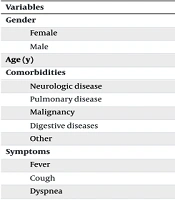
Sixty patients (39 males, 21 females; mean age 4.9 ± 4.0 years) were included in this study. The most common symptoms were fever (86%), cough (66%), and dyspnea (35%). Eighteen patients had comorbidities, two of which were related to lung diseases. Fifty patients (83%) tested positive for COVID-19 via PCR. The demographic and clinical characteristics of the patients are presented in Table 1.
| Variables | Values |
|---|---|
| Gender | |
| Female | 21 (35) |
| Male | 39 (65) |
| Age (y) | 4.92 ± 4.01 |
| Comorbidities | 18 (30) |
| Neurologic disease | 4 (22.2) |
| Pulmonary disease | 2 (11.1) |
| Malignancy | 6 (33.3) |
| Digestive diseases | 2 (11.1) |
| Other | 4 (22.2) |
| Symptoms | |
| Fever | 52 (86.7) |
| Cough | 40 (66.7) |
| Dyspnea | 21 (35.0) |
| Laboratory results | |
| WBC | 8.85 ± 5.19 |
| RBC | 4.76 ± 5.01 |
| Lymphocyte percentage | 34.54 ± 16.65 |
| Neutrophil percentage | 58.91 ± 18.25 |
| Hemoglobin | 11.68 ± 1.86 |
| Platelets | 342.81 ± 171.36 |
| C-reactive protein | 22.52 ± 14.21 |
| ESR | 30.66 ± 19.96 |
| AST | 61.53 ± 144.95 |
| LDH | 621.21 ± 231.25 |
| SARS-CoV-2 (RT-PCR) test | 59 (98.3) |
| Positive | 50 (84.7) |
| Negative | 9 (15.3) |
Abbreviations: WBC, white blood cell count; RBC, red blood cell count; ESR, erythrocyte sedimentation rate; AST, aspartate aminotransferase; LDH, lactate dehydrogenase; SARS-CoV-2, severe acute respiratory syndrome coronavirus 2; RT-PCR, reverse transcription-polymerase chain reaction.
a Values are expressed as No. (%) or mean ± SD.
Nearly all patients (98%) underwent a LUS examination. Pleural effusion was detected in two patients (3.4%). The mean LUS score was 6.28 ± 4.49. Chest X-rays were performed on 35 patients (58%); 19 (54%) had normal findings, and 9 (25%) exhibited increased bronchovascular markings (Table 2).
| Variables | Values |
|---|---|
| CT findings | 29 (48.3) |
| Lesion characteristics | 28 |
| Consolidation | 13 (46.4) |
| Ground-glass opacity | 3 (10.7) |
| Mixed pattern | 12 (42.9) |
| Nodular | 5 (17.9) |
| Round or well-defined opacity | 2 (7.1) |
| Halo sign | 1 (3.6) |
| Type of ground-glass opacity | 9 |
| Pure ground-glass opacity | 7 (77.8) |
| Crazy paving | 2 (22.2) |
| Type of consolidation | 18 |
| Non lobular | 14 (77.8) |
| Lobular | 4 (22.2) |
| Pleural effusion | 3 (10.3) |
| Mild | 2 (66.7) |
| Severe | 1 (33.3) |
| Lymphadenopathy | 4 (13.8) |
| COVID-19 imaging classification | 28 |
| Typical appearance | 12 (42.9) |
| Indeterminate appearance | 15 (53.6) |
| Atypical appearance | 1 (3.6) |
| Negative for pneumonia | 0 |
| Average of CT severity score | 5.82 ± 3.82 |
| LUS findings | 59 (98.3) |
| Pleural effusion | 2 (3.4) |
| Average of LUS score | 6.28 ± 4.49 |
| CXR findings | 35 (58.3) |
| Normal | 19 (54.3) |
| Increased bronchovesicular marking | 9 (25.7) |
| Consolidation | 4 (11.4) |
| Ground-glass opacity | 2 (5.7) |
| Pleural effusion | 1 (2.9) |
Abbreviations: LUS, lung ultrasound; CT, computed tomography; CXR, chest X-ray.
a Values are expressed as No. (%) or mean ± SD.
Approximately half of the patients (48%) had a chest CT scan. The most common lesion characteristics were consolidation (46%) and a mixed pattern (42%). Pleural effusion was noted in three patients (10.3%): Two mild cases and one severe case. Twelve patients (42.9%) displayed a typical appearance on CT, while 15 (53.6%) had an indeterminate appearance. The mean CT severity score was 5.82 ± 3.82. Findings from the imaging modalities are summarized in Table 2.
For the 29 patients who underwent both LUS and chest CT, a significant correlation was observed between the LUS score and the CT severity score (correlation coefficient = 0.467, P = 0.011). Lesion distribution on LUS was similar to that on chest CT.
Table 3 presents results for the definitive and probable diagnosis of COVID-19 based on PCR positivity alongside abnormal findings on LUS, chest CT, and CXR.
No significant correlation was found between LUS scores and CXR findings (P = 0.392). Of the 19 patients with normal CXR findings, 16 had a LUS score ≤ 4, while the LUS scores of the remaining three were 8, 14, and 14.
| Abnormal Imaging | Definite (PCR Positive); N = 50 | Probable (PCR Negative); N = 10 |
|---|---|---|
| LUS (38) | 31 (62) | 8 (80) |
| Chest CT (29) | 19 (38) | 10 (100) |
| CXR (16) | 15 (30) | 1 (10) |
Abbreviations: PCR, polymerase chain reaction; LUS, lung ultrasound; CT, computed tomography; CXR, chest X-ray.
a Values are expressed as No. (%).
5. Discussion
In our study, 12 patients (42.9%) showed typical changes for COVID-19, 15 patients (53.6%) had indeterminate chest CT findings, and 1 patient (3.6%) had atypical chest CT findings. Lopes et al. in Brazil observed similar findings among 45 adult COVID-19 patients, where 29 (64.4%) CT images were classified as typical, 8 (17.8%) as indeterminate, and another 8 (17.8%) as negative. We found a significant correlation between LUS scores and CT severity scores in pediatric COVID-19 patients (correlation coefficient = 0.467, P = 0.011). This association aligns with the findings of Lopes et al., who also observed a correlation between LUS scores and lung lesion extent on CT (P < .0001) (18).
Similarly, studies by Nouvenne et al. and Tung-Chen et al., conducted in Italy and Spain respectively, also reported a significant correlation between LUS and CT scores (P < 0.001) (19, 20). In China, Deng et al. demonstrated a strong correlation between LUS scores and CT scores in critically ill COVID-19 pneumonia patients (r = 0.891, P < 0.01) (21). Similar results have been documented in studies of pregnant populations, with a high correlation between LUS and CT scores (r = 0.793, P < 0.01) as reported by Deng et al. (22), and a significant positive correlation (rICC = 0.946; P < 0.001) found by Biancolin et al. (23).
In terms of lesion distribution, we observed comparable lung involvement on both LUS and chest CT. The study by Giorno et al. in Brazil also reported a similar topographical distribution of lesions on LUS and chest CT in pediatric COVID-19 patients (11). Our results further showed no significant correlation between LUS scores and CXR findings (P = 0.392), consistent with the findings of Volpicelli et al. in Italy (24). In their descriptive study, LUS and CXR results disagreed in 60 (43.2%) adult patients suspected of COVID-19, underscoring the limitations of CXR in identifying COVID-19 lesions compared to LUS.
In our study, sixteen out of the nineteen patients with normal CXR findings had a LUS score of ≤ 4, while the remaining three with normal CXRs had LUS scores of 8, 14, and 14 (these patients had ALL and neurological diseases). This suggests that LUS was more sensitive than CXR. A similar observation was made in a pediatric COVID-19 study by Giorno et al. in Brazil, where eight patients with LUS abnormalities had normal CXRs (11). In line with our findings, Ture et al. in Turkey also found that LUS was more sensitive than CXR in early-stage and mild COVID-19 pneumonia in pediatric patients suspected of COVID‐19; in their study, four patients had normal CXRs but displayed LUS findings (25). Consistently, Mateos Gonzalez et al. reported that 20 adult COVID-19 patients with normal CXRs (55%) had pulmonary infiltrates detected by LUS. Their study in Spain found that LUS was more sensitive than CXR (81% vs. 63%) in detecting COVID-19 pneumonia (26). Similarly, in a study by Gibbons et al., LUS showed higher sensitivity than CXR in diagnosing COVID-19 pneumonia in adults (97.6% vs. 69.9%) (27).
5.1. Limitations
Our study had several limitations. First, the sample size was relatively small. Second, LUS, chest CT, and CXR were not performed simultaneously, with ultrasound conducted an average of three days after admission, which affects the assessment of correlations among the different imaging modalities. In our study, 83% of patients had a positive PCR test. While a positive PCR test is the gold standard for diagnosing COVID-19, during the early pandemic, patients with abnormal chest CT findings, a contact history, and clinical symptoms were also treated as COVID-19 cases.
5.2. Conclusions
Lung ultrasound demonstrated greater sensitivity than CXR and outperformed it in detecting COVID-19 pneumonia. Lung ultrasound showed a significant correlation with CT findings, making it a promising, safer alternative for diagnosing COVID-19 pneumonia in children, although it may not fully replace chest CT in all cases.
