1. Background
The human gastrointestinal system hosts a diverse microbiota, including various viral species. Numerous viral agents have been identified in the stool samples of individuals suffering from gastroenteritis, such as rotavirus, astrovirus, calicivirus, hepatitis E virus, and various strains of coronaviruses like SARS-CoV-2 (1). COVID-19 is commonly known as a respiratory illness; however, digestive symptoms are also acknowledged as part of the illness (2). SARS-CoV-2 primarily spreads through respiratory droplets, but some studies report the presence of infectious viral particles in stool samples.
Reverse transcription polymerase chain reaction (RT-PCR) has been utilized to identify viral genetic material in stool samples for various types of viruses, including pathogenic viruses and small round structured viruses (SRSV) (3). The extraction of high-quality total RNA from stool samples poses significant challenges due to the inherent complexity of the sample matrix. Stool is a heterogeneous mixture containing a diverse range of microorganisms, host cells, dietary components, and inhibitory substances such as bile salts, complex polysaccharides, and humic acids (4). These inhibitors can interfere with RNA isolation and downstream applications such as RT-PCR and RNA sequencing, making the process highly demanding. To address this issue, various methods have been developed that involve capturing viral material in stool samples using antigens or specific oligonucleotides attached to magnetic beads. This allows for the concentration of viral RNA separated from other components in the stool sample. Alternative methods have integrated phenol/chloroform extractions with additional extraction processes involving freon and cetyl hexadecyltrimethylammonium bromide (CTAB) in conjunction with polyethylene glycol (PEG) precipitations to isolate viral material from inhibiting substances (5).
Recent advances in nanotechnology have provided innovative solutions to improve biomolecule extraction. Among these, mesoporous silica nanoparticles (MSNs) have garnered attention for their unique physicochemical properties, including high surface area, tunable pore size, and selective adsorption capabilities. These properties make MSNs particularly suitable for capturing nucleic acids, even in complex biological matrices. In the context of stool samples, MSNs offer the potential to efficiently bind and isolate RNA while minimizing contamination from inhibitors (6, 7).
The MSN has a high surface area and pore volume, allowing for efficient RNA binding and minimizing contaminants in the extracted RNA. Thus, the use of MSNs leads to higher RNA yield and purity compared to traditional methods (8).
2. Objectives
In this study, we aim to evaluate the performance of MSNs for extracting total RNA from stool samples. Specifically, we compare the RNA yield, purity, and integrity achieved with MSNs to those obtained using a commercial RNA extraction kit. Additionally, we assess the usability of the extracted RNA in downstream applications such as RT-PCR. By addressing the limitations of existing methods, this work seeks to establish MSNs as a robust alternative for RNA extraction from challenging sample types, with broad implications for molecular diagnostics, microbiome research, and beyond.
3. Methods
3.1. Study Population
In this descriptive study with a margin of error of 0.5 and a 95% confidence level, based on our total population size with the online sample size calculator Raosoft Inc, 94 samples were needed for examination and study in order to have only 1 error in 100 tests with a probability of 95% (9-11).
3.2. Preparation of Stool Samples
One hundred clinical stool samples were collected from patients hospitalized at Mofid Children's Hospital with suspected COVID-19 and gastrointestinal symptoms. All samples were stored at -80°C without any preservatives. They were then resuspended in sterile DNase RNase-free water in a 1:5 ratio (w:v) prior to RNA extraction. As a homogenizer for these samples, we added 200 mg of stool to 1 mL of sterile DNase RNase-free water. After that, the microtube was vortexed well so that the stool and DNase RNase-free water were thoroughly mixed and homogenized. In the next step, the microtube was centrifuged at 5000 rpm for 5 minutes, and then 300 microliters of the clarified supernatant were used for the extraction process.
3.3. Study Design
The homemade lysis buffer (LB) solution was used in this study for lysing virus and human cells. It contains guanidinium thiocyanate, Tris-Cl, EDTA, and cetyl trimethyl ammonium bromide. In the next step, two extraction methods were prepared for collecting RNA: (1) Silica membrane column and (2) Fe-doped mesoporous silica nanoparticle column (Fe-MSN). The efficiency of extracted RNA was compared with an automated magnetic bead-based commercial extraction kit (Tianlong kit T180H, China) as a standard method by evaluating the cycle threshold (Ct) in multiplex real-time PCR for the envelope (E) gene, spike protein (S) gene of the SARS-CoV-2 virus, and RNase P gene. Additionally, the amount of extracted total RNA was determined by a Qubit fluorometer (Invitrogen, USA).
3.4. RNA Extraction with Silica Membrane
Viral RNA was extracted from stool samples using a silica membrane-based protocol. Briefly, 300 µL of clarified supernatant from pre-processed stool was mixed with 600 µL of LB, 60 µL of proteinase K, and 20 µL of RNA carrier. The mixture was incubated at 57°C for 10 minutes. Subsequently, the lysate was transferred onto a silica membrane spin column and centrifuged at 10,000 rpm for 1 minute. The column was then washed twice with absolute ethanol and centrifuged at 14,000 rpm for 1 minute to remove residual ethanol and dry the membrane. For RNA elution, 50 µL of sterile, DNase/RNase-free water was added to the membrane, followed by centrifugation at 10,000 rpm for 1 minute. The eluted RNA was collected in a DNase/RNase-free microtube and stored at -80°C until further use in real-time PCR assays.
3.5. RNA Extraction with Fe-doped Mesoporous Silica Nanoparticle Column
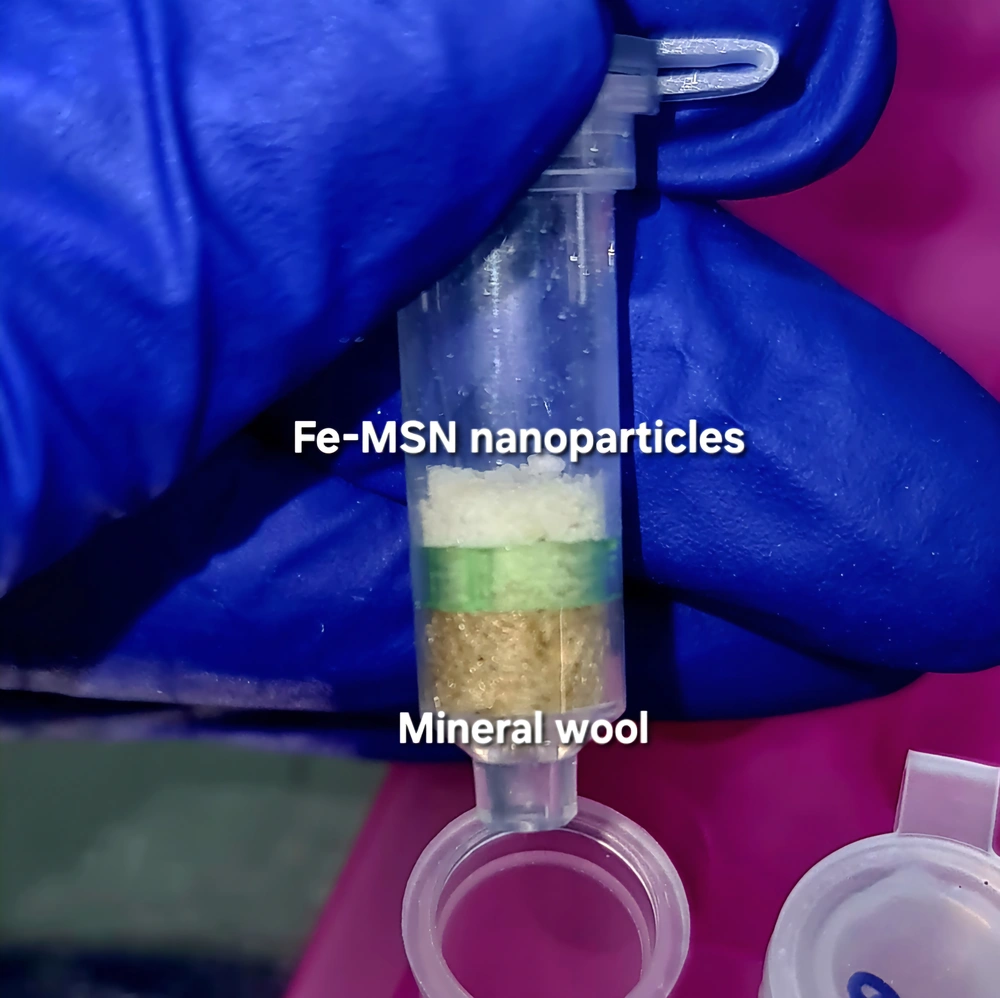
In this study, an in-house extraction column was designed using mineral wool as a physical barrier and filled with Fe-MSN nanoparticles (Figure 1). The Fe-MSNs were obtained from the Nanotechnology Research Group at the Islamic Azad University, North Tehran Branch. These nanoparticles were employed as the active component of a custom-designed RNA extraction column, in which mineral wool was used as a physical support matrix. To characterize the structural and physicochemical properties of Fe-MSNs, Fourier-transform infrared spectroscopy (FTIR), X-ray diffraction (XRD), scanning electron microscopy (SEM), and nitrogen adsorption-desorption analysis (BET) were performed (Appendix 1 in Supplementary File).
Viral RNA was extracted from stool samples using Fe-MSN following a modified protocol. Briefly, 300 µL of clarified stool supernatant was mixed with 600 µL of LB, 60 µL of proteinase K, and 20 µL of RNA carrier. The mixture was incubated at 57°C for 10 minutes to facilitate viral lysis and protein digestion. Following incubation, the entire volume was transferred into an Fe-MSN-based extraction column. The mixture was allowed to pass slowly through a bed of Fe-MSN particles and mineral wool under gravity into a collection tube. Subsequent washing steps followed the general procedure of silica membrane-based RNA extraction, except that no centrifugation was required during the binding and washing steps. Finally, RNA was eluted by adding elution buffer to the column, followed by centrifugation at 5000 rpm for 30 seconds. The eluate containing purified RNA was collected in a DNase- and RNase-free microtube and stored for downstream real-time PCR analysis.
3.6. RNA Extraction with Magnetic Bead-based Commercial Extraction Kit
In this step, viral RNA was extracted using a commercial nucleic acid extraction kit (Tianlong Nucleic Acid Extraction Kit, China), following the manufacturer’s instructions. The extraction process was performed on the Tianlong Libex automated extraction system. Stool samples were loaded into the designated sample wells of the extraction cartridge, along with the appropriate reagents provided in the kit. The automated protocol included lysis, binding of nucleic acids to magnetic beads, sequential washing steps, and elution. At the end of the run, purified RNA was collected in DNase/RNase-free elution tubes and stored at -80°C until use in downstream applications, including real-time PCR.
3.7. Deletion of DNA from Extraction Solution
The DNase I kit from Sinnagen Company, Iran, was used for the removal of DNA contamination from extractions, following the kit protocol.
3.8. Qubit
The concentration of the extracted RNA through all three methods was measured using the Qubit fluorometer (Invitrogen, USA).
3.9. Multiplex Real-time Polymerase Chain Reaction
At this stage, the E gene, S protein of the SARS-CoV-2 virus, and RNase P gene as an internal control were identified. Additionally, the Ct value of the RNase P gene was evaluated to measure the quality of the extracts with a commercial real-time PCR kit (COVITECH, Iran) according to the company's protocols and sheet information. The real-time PCR instrument used was the Corbett real-time PCR Rotor-Gene 6000.
3.10. Statistical Analysis
In this study, a paired t-test was used to compare the mean Ct between the commercial total RNA extraction kit and the two designed methods pairwise.
4. Results
Quantification of the 100-extracted total RNA using the Qubit fluorometer revealed that the average RNA concentration obtained using the Fe-MSN-based nanoparticle extraction kit was 9.8 µg/mL, compared to 8.3 µg/mL with the silica column-based kit, and 7.6 µg/mL using the commercial kit with an automated extraction device.
Comparative analysis of real-time PCR results from 100 stool samples demonstrated that the Fe-MSN nanoparticle-based kit outperforms the commercial extraction kit in terms of RNA yield and purity. Additionally, the Ct of the genes examined in this study was lower when using nanoparticles than with the silicon column method. Specifically, the average Ct of the internal control gene in the samples extracted by the kit method designed based on Fe-MSN nanoparticles was 24.8 ± 2.3. The average Ct of the internal control gene in the samples extracted by the kit method based on the silicon column was 26.04 ± 2.42. The extraction method with the commercial kit and the automatic device yielded an average Ct of 29.27 ± 2.48, indicating a lower average for the extraction method designed based on Fe-MSN nanoparticles. Similarly, for the Ct of the S gene and E gene of the SARS-CoV-2 virus, we observed a lower average for extractions based on the Fe-MSN method, as shown in Table 1.
| Methods | Ct Average | ||
|---|---|---|---|
| IC Gene | S Gene | E Gene | |
| Magnetic bead-based | 29.27 ± 2.48 | 28.54 ± 4.32 | 30.23 ± 4.73 |
| Silica membrane column | 26.04 ± 2.42 | 25.63 ± 3.18 | 27.39 ± 3.81 |
| Fe-MSN | 24.8 ± 2.3 | 23.8 ± 2.5 | 25.27 ± 4.32 |
Abbreviations: Ct, cycle threshold; S gene, spike protein gene; E gene, envelope gene; Fe-MSN, Fe-doped mesoporous silica nanoparticle column.
The lower Ct values observed with the Fe-MSN indicate a higher RNA yield and purity, likely due to the increased surface area and pore volume of the nanoparticles, facilitating better RNA adsorption and reducing inhibitors.
5. Discussion
The global outbreak of SARS-CoV-2 has highlighted the critical need for more efficient and reliable methods of viral detection, particularly in the context of RNA extraction and purification. This necessity extends beyond respiratory viruses to include RNA viruses affecting the gastrointestinal tract, which are often present in stool samples and pose unique diagnostic challenges. This unforeseen crisis highlighted the need for technological advances to better diagnose and combat such infectious diseases (12)). The first step in conducting molecular tests is the extraction of the genetic material from the microorganism. Dealing with a clinical sample that contains a variety of microorganisms, protein substances, and potential contaminants like stool samples can make the work very challenging. Furthermore, the genetic material of RNA viruses is more vulnerable and unstable than DNA, making the extraction process even more difficult. Therefore, finding a method that can efficiently extract the total RNA with minimal damage can be very helpful in these conditions (13).
This research highlights two important factors: Introducing a new total RNA extraction kit with nano silicon columns and achieving a higher amount of RNA extraction compared to previous methods. RNA extraction is a crucial step in the molecular identification of infectious diseases. A suitable and accurate RNA extraction method can prevent false-negative results and provide the accurate Ct values needed by physicians.
In a study by Sahu et al. (9, 12), the silica column method yielded a higher amount of RNA compared to the magnetic bead-based method, which is consistent with our findings. Moreover, the magnetic bead method is not only more expensive but also requires specialized extraction equipment, making it less cost-effective than the other two methods. The limitations of this technique compared to silica column-based kits were also highlighted in the study by Sahu et al. (12).
This study showed varying results in the Ct value of the real-time PCR on stool samples depending on the method of RNA extraction utilized. The differences in Ct value in real-time PCR are due to variations in the quality of the extracted RNA using different methods. The results of real-time PCR in the current study showed that the RNA extracted with the magnetic bead method had the highest Ct in comparison with the column-based methods. This can be attributed to the low quality of the extracted RNA with magnetic beads and extraction machines.
The average Ct value of the S, E, and Human RNase P genes was reduced by utilizing the Fe-MSN nanoparticles-based kit. This indicates that we were able to extract a higher quantity of undamaged RNA with this method.
Our findings align with previous studies indicating that MSNs enhance RNA extraction efficiency. However, unlike prior methods, our Fe-MSN approach eliminates the need for automated equipment, reducing overall costs and increasing accessibility (12, 14). The strength of this study is that we were able to extract more and better RNA by using Fe-MSN nanoparticles instead of silicon membranes, which improved the efficiency of column-based extraction kits (14).
The MSN nanoparticles offer several advantages for use in biological processes, such as drug delivery and RNA absorbance (8, 15, 16). Despite a lack of prior research, using MSN for total RNA extraction in a Nano column, our study successfully demonstrated its efficacy in this capacity. In this study, we designed it for this purpose and achieved the best results in comparison to other examined methods for RNA extraction from stool (8).
5.1. Conclusions
This study aimed to evaluate and identify the most reliable and accurate method for total RNA extraction for various purposes. The results showed that Fe-MSN nanoparticles are a superior alternative for total RNA extraction from stool samples, while being cost-effective, especially in developing countries, and increasing yield and purity.

