1. Background
Hydatid disease (HD), also known as hydatidosis, is still a global problem in highly endemic areas such as Iran (1, 2). The hydatid cyst is the larval stage of Echinococcus granulosus, which is hosted by dogs and other canines (3, 4). Humans are intermediate hosts by eating the tapeworm eggs (5). According to recent estimations, echinococcosis is responsible for about 1 - 220/10000 infections in the common areas (2, 6, 7).
HD is commonly asymptomatic, and often the cysts are detected incidentally (8). In other cases, the symptoms vary largely and are non-specific, depending on the location, size, rupture, and infection of the cysts (9). The liver and lung are the most commonly involved organs in humans; and in children, the lung is more frequently infected (10-14). An elevated eosinophil count in a patient from an endemic region accompanied by non-specific symptoms such as abdominal pain, abdominal mass, and cough, is suggestive of HD (15). The diagnosis is usually made via imaging modalities (i.e. ultrasonography and chest X-ray) and confirmed by pathology (16). Surgery is the chief therapeutic method for the management of HD, although currently percutaneous aspiration-injection-reaspiration drainage is regarded as a first choice in many cases (9, 14).
The present study was performed to evaluate the epidemiological aspects, clinical presentations, paraclinical findings, and management of HD in Iranian children.
2. Objectives
We sought to review the clinical manifestations, laboratory aspects, imaging findings, and management of HD.
3. Patients and Methods
In this retrospective study, we investigated the medical records of 161 patients (1 - 15 years old) with a definite diagnosis of HD admitted to eight major referral hospitals of Iran during a 13-year period (2001 - 2013). Data were collected on sex, age, clinical manifestations, anatomical location of the cysts, size and number of the cysts, laboratory tests, and therapeutic management. Statistical analysis was carried out using statistical package for the social sciences (SPSS) version 18.
4. Results
The study population consisted of 99 (61.5%) boys and 62 (38.5%) girls (range = 1 - 15 years old) from eight teaching referral hospitals around Iran (Table 1).
| University-Hospital | Values |
|---|---|
| Isfahan-Emam Hosein | 45 (28) |
| Shanhid Beheshti-Mofid | 32 (19.9) |
| Zahedan-Ali Ebne Abitaleb | 24 (14.9) |
| Kordestan-Besat | 14 (8.7) |
| Kermanshah-Emam Reza | 13 (8.1) |
| Iran-Ali Asghar | 12 (7.5) |
| Hamedan-Besat | 12 (7.5) |
| Arak-Amir Kabir | 9 (5.6) |
| Total | 161 (100) |
a Values are presented as No. (%).
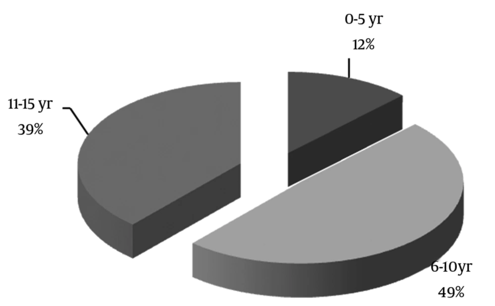
Echinococcosis was diagnosed in all our patients via pathology. The mean age of the study population was 9.25 ± 3.37 years, and most of the affected patients (49.1%) were in the age range of 6 to 10 years old (Figure 1).
The most frequent anatomical location of the hydatid cysts was the lung (in 108 (67.1%) patients), followed by the liver (in 71 (44.1%) patients). Additionally, the cysts were detected in the kidney (2.5%), spleen (2.5%), brain (1.9%), colon (1.2%), heart (0.6%) and eye (0.6%). Multiple organ involvement was found in 32 (19.8%) patients, with a single-to-multiple ratio of about 4: 1. There was no significant statistical difference between age (P = 0.551) or gender (P = 0.243) and the anatomical location of the cysts.
Our patients had totally 335 cysts: 181 (54%) with right-sided and 138 (41%) with left-sided cysts. Regarding size, 130 (39%) cysts were smaller than 5 cm in diameter, 157 (47%) cysts were between 5 and 10 cm in diameter, and 47 (14%) cysts were more than 10 cm in diameter (Table 2).
| Location | No. of Patients | No. of Cysts |
|---|---|---|
| Liver | 71 (44.1) | 137(40.5) |
| Right lobe | 54 (57.4) | 78 (56.9) |
| Left lobe | 40 (42.6) | 59 (43.1) |
| Lung | 108 (67.1) | 182 (54.3) |
| Right upper lobe | 15 (11.1) | 21 (11.5) |
| Right lower lobe | 52 (38.2) | 66 (36.3) |
| Right intermediate lobe | 11 (8.1) | 16 (8.8) |
| Left upper lobe | 19 (13.9) | 26 (14.3) |
| Left lower lobe | 39 (28.7) | 53 (29.1) |
| Other sites | 15 (9.3) | 16 (4.8) |
| Spleen | 4 (2.5) | 5 (31.25) |
| Kidney | 4 (2.5) | 4 (25) |
| Brain | 3 (1.9) | 3 (18.75) |
| Colon | 2 (1.2) | 2 (12.5) |
| Heart | 1 (0.6) | 1 (6.25) |
| Eye | 1 (0.6) | 1 (6.25) |
| Total | 161 (100) | 335 (100) |
a The values are presented as No. (%).
Fever was the most common complaint (35.4%). Cough was the most common symptom associated with the lung hydatid cyst (29.8%) and an abdominal mass was more common in the liver hydatidosis (9.3%) (Table 3).
| Chief Complaint | Values |
|---|---|
| Fever | 57 (35.4) |
| Abdominal pain | 51 (31.7) |
| Cough | 48 (29.8) |
| Chest pain | 34 (21.1) |
| Dyspnea | 34 (21.1) |
| Nausea/vomiting | 21 (13) |
| Weight loss | 20 (12.4) |
| Anorexia | 20 (12.4) |
| Abdominal mass | 15 (9.3) |
| Pulmonary effusion | 14 (8.7) |
| Hepatomegaly | 12 (7.5) |
| Hemoptysis | 11 (6.8) |
| Lung abscess | 11 (6.8) |
| Respiratory distress | 8 (5) |
| Allergic reaction | 6 (3.7) |
| Cholestasis | 3 (1.9) |
| Urinary tract infection | 3 (1.9) |
| Biliary colic | 2 (1.2) |
| Liver abscess | 2 (1.2) |
| Splenomegaly | 2 (1.2) |
| Seizure | 2 (1.2) |
| Incidental findings | 24 (14.9) |
a The values are presented as No. (%).
Laboratory data showed eosinophilia (≥ 500/micL) in 66 children, leukocytosis (WBC ≥ 15000/micL) in 47 patients, erythrocyte sedimentation rate (ESR) ≥ 30 and positive C-reactive protein (CRP) in 30 patients, and anemia (Hb ≤ 11 g/dL) in 28 patients. We assessed the abdominal sonography reports in 87 patients, which revealed the hydatid liver cyst in 83 (95.4%) cases. There were 79 chest X-ray reports, 70 (88.6%) of which suggested the lung hydatid cyst. Computed tomography (CT) scan confirmed the diagnosis in all the patients suspected of having the cyst (Table 4).
| Radiologic Findings | Values |
|---|---|
| Chest X-ray, n = 79 | |
| Normal | 9 (11.4) |
| Abnormal | 70 (88.6) |
| Pure cyst | 35 (44.3) |
| Ruptured | 21 (26.6) |
| Mass | 7 (8.9) |
| Abscess formation | 5 (6.3) |
| Calcification | 2 (2.5) |
| Sonography, n = 87 | |
| Normal | 4 (4) |
| Abnormal | 83 (96) |
| Pure cyst | 55 (52.4) |
| Daughter cyst | 14 (13.3) |
| Ruptured | 7 (6.7) |
| Calcification | 3 (2.9) |
| Coarse echo | 2 (1.9) |
| Mass | 1 (1) |
| Abscess formation | 1 (1) |
| Computed tomography scan, n = 87 | |
| Normal | 0 (0) |
| Abnormal | 87 (100) |
| Pure cyst | 45 (51.7) |
| Ruptured | 18 (20.7) |
| Abscess formation | 9 (10.3) |
| Daughter cyst | 7 (8) |
| Mass | 5 (5.7) |
| Calcification | 3 (3.4) |
a The values are presented as No. (%).
Overall, 143 (89%) patients underwent surgery, 12 (7%) patients were treated with percutaneous aspiration-injection-reaspiration drainage, and 6 (4%) patients were prescribed medication. Four out of these 6 patients were transferred to other hospitals for surgery.
5. Discussion
HD is still a health hazard in endemic countries (14). In the present study on 161 children in an age range of 1 - 15 years old, the demographical characteristics and infected sites, clinical presentations, paraclinical data, and treatment strategy were assessed. The majority of the study population were male (61.5% vs. 49.1%), and the mean age was 9.25 ± 3.37 years. Based on our findings, the lung was the most commonly involved organ (67%), followed by the liver (44%), and the combined organ involvement was seen in 19.8% of the cases. Generally, the right side was the dominant site of infection (54%). Laboratory data showed elevated eosinophil counts in 41% and ESR/CRP in 18.6% of the patients. Chest X-ray and abdominal sonography constituted the principal imaging modalities with respective accuracy rates of 88% and 96%.
The mean age of our patients was 9.25 ± 3.37 years, and 49.1% of them were in the age range between 6 and 10 years old. Males and females comprised 61.5% and 38.5% of the study population, correspondingly (male-to-female ratio = 1.6:1). Vlad et al. (8) studied 82 cases at a mean age of 10.8 years, the majority of them being between 10 and 14 years old. The female-to-male ratio was 1.6:1 (8). Djuricic et al. (18) reported the mean age of 10.1 ± 3.8 years in their study, which included 149 patients under 18 years of age. The authors found no significant difference in terms of the gender of their patients. In a large Iranian study, Mirshemirani et al. (1) reported the mean age of 11.8 ± 4.6 years in 100 patients. In our study, chiming in with the other studies, pre-adolescent years (between 8 and 12 years old) accounted for the most frequent age at clinical presentation. In Iran, various studies have demonstrated that boys are more likely to be infected by Eosinophilic gastroenteritis than girls. It should also be borne in mind that in Iran, boys tend to spend more time outdoors than girls, which can explain the higher incidence of HD in the male cases in our study.
Chaouachi et al. (17) studied 1195 cases of the hydatid cyst in children aged between 2 and 15 years at the Children’s Hospital in Tunis and reported that the most commonly involved organ was the lung (in 643 cases), followed by the liver (in 486 cases). Their result is similar to that in our study insofar as the lung was the most commonly involved organ (49.7%) in our patients, which is concordant with the results of some other similar studies on children (3, 4, 11). Nevertheless, there are some other studies that show a larger number of hydatid cysts in the liver than in the lung among children (8, 18, 19). Various genotypes of Echinococcus granulosus isolated form different animal sources can be responsible for the heterogeneity of the results and may contribute to a clinically or histologically heterogeneous disease (20, 21). Combined liver and lung involvement was detected in 15.5% of our cases, with the percentage of other combinations of the infected sites being 4.7%, which is in line with some other surveys performed in children (6% - 16%) (18, 22, 23). It is vitally important that the combined organ involvement not be missed because the maltreatment of the hydatid cyst can result in recurrence in patients.
The frequency of the uncommon anatomical locations of the hydatid cysts in the present study was 9.3%; the spleen and kidney comprised the majority of such locations. These results are slightly higher than those in the literature on the prevalence of hydatid cysts in the spleen (0.9% - 8%) and kidney (18, 23-33).
Single organ involvement was 80.2% in our study, and the single-to-multiple-organ ratio was 4:1, which is similar to those reported by Oudni-M’Rad et al. (4.3:1 in children) (23) and Talaiezadeh and Maraghi (5.7:1 in children) (34). The highest ratio was reported by Al-Shibani et al. (38:1) in cases under 20 years old (35).
Only a few studies have reported such laboratory data as eosinophil or WBC counts and ESR values (36-38). In a review article by Moro and Schantz (39), eosinophilia was found in fewer than 25% of the cases with HD. Our results showed eosinophilia (≥ 500/micL) in 41% and ESR ≥ 30 or positive CRP in 18.6% of the study population. We used a value range that was different from the one employed by Vlad et al. (8) as they found higher figures in their survey (eosinophilia ≥ 5% in 59% and ESR ≥ 10 in 68.7% of the pediatric patients). We think that the higher rate of this laboratory marker can be related to the higher risk of complications such as rupture and, accordingly, recommend that sufficient attention be paid to these results in hydatidosis patients.
Among our pediatric patients, the lung was the most commonly involved organ. We would, therefore, urge that physicians be aware of different radiological forms of echinococcosis on chest X-ray in endemic areas and consider the probability of multiple cysts or multiple organ involvement.
