1. Background
Although Klebsiella pneumoniae is a part of gastrointestinal flora, it causes serious community and hospital-acquired infections (1). This opportunistic pathogen can cause pneumonia, urinary tract, skin, and soft tissue infections, in particular in intensive care unit (ICU), surgery, and pediatric wards of hospitals (2, 3). Some identified risk factors for K. pneumoniae infections are the long-term admission, nutrition through intravenous injection, and the mechanical ventilation (3). K. pneumoniae strains with multiple drug resistance (MDR) are frequently isolated from nosocomial infections (4). Due to exorbitance use of beta-lactam antibiotics in recent decades, K. pneumoniae isolates containing broad-spectrum beta-lactamases have been reported worldwide (5, 6). The resistance to Cephalosporins was initially reported in 1983 and then spread throughout the world (7).
An important epidemiological tool to control the infection is monitoring the local spread of resistant strains of the bacteria, especially in hospital environments (8). Researches on the bacterial genotypic homology can outline a better vision for the origin and dissemination patterns of K. pneumoniae infections (9). Among different molecular techniques for typing the bacterial genotype, pulse-field gel electrophoresis (PFGE) is often used as the standard method for typing of K. pneumoniae isolates (10). This method can differentiate only relatively major genetic events led to the changes in genomic fingerprinting patterns (11). Given the spread of multidrug-resistant strains of K. pneumoniae in our region, this study aimed to determine the molecular typing of K. pneumoniae isolates.
2. Objectives
We aimed to determine the molecular typing of K. pneumoniae isolates using PFGE.
3. Methods
3.1. Bacterial Isolates
This descriptive cross-sectional study was performed on 62 available and non-duplicate K. pneumoniae isolates. They were identified from 2013 to 2014, from clinical samples in Imam Khomeini, Imam Reza, and Taleghani hospitals in Kermanshah, west of Iran. All isolates were detected by biochemical and bacteriological tests and subsequently, confirmed by API20E kit (bio-Merieux, France).
3.2. Antibiotic Susceptibility Testing
Antimicrobial susceptibility testing for 6 beta-lactam antibiotics was carried out using disk diffusion method (12). The examined antibiotics were Penicillins (ampicillin (10 μg), piperacillin-tazobactam (100/10 μg)), Cephalosporins (cefazolin (30 μg), cefotaxime (30 μg)), Monobactams (aztreonam (30 μg)), Carbapenems (Meropenem (10 μg)) (Mast, England).
3.3. ESBL Phenotypic Confirmatory Test
This test was performed by the hybrid disk method according to the CLSI recommendations. In this method, both Ceftazidime (30 μg) and cefotaxime (30 μg) were used separately and in combination with Clavulanic acid (10 μg). For each isolate, if the diameter of the inhibited zone increased more than 5 mm for the antimicrobial agent in combination with Clavulanic acid, it was proposed as ESBL producer (12). K. pneumoniae ATCC700603 was used as a positive control and E. coli ATCC25922 was used as a negative control.
3.4. Pulsed-Field Gel Electrophoresis (PFGE)
The PFGE was done according to a previously described protocol with some modifications (13). K. pneumoniae isolates and Salmonella enterica serovar Braenderup H9812 (As DNA marker) genome were digested with 20U XbaI restriction enzyme (Fermentas, Lithuania). Electrophoresis was performed by CHEF MAPPER apparatus (Bio-Rad, USA) for 22 h at 14°C under the below conditions: 5 seconds initial switch time, 35 seconds final switch time, consist of angle, the voltage gradient of 120 degrees, 6 V/cm; ramping factor, linear. Gels were stained by ethidium bromide and visualized under the UV light by the Gel Doc apparatus (Bio-Rad, USA).
3.5. Software Analysis
The DNA pattern was analyzed by the 6.6 version of the Gelcompar II (Applied Maths, Belgium). The calculation of similarities was done by the Dice coefficient, and the unweighted paired group method was used for cluster analysis (UPGMA). According to the Tenover’s criteria, the isolates with more than 80% similarity in their DNA patterns were defined as a clone (11).
3.6. Statistical Analysis
All data were recorded and saved in an Excel file. Statistical analyses were performed using SPSS software (version 16). Variables were analyzed using chi-square test. In this study, A P value of ≤ 0.05 was considered statistically significant.
4. Results
From 62 isolates of K. pneumoniae, 37 (59.6%), 16 (25.8%), 9 (14.5%) isolates were from Imam Khomeini Hospital, Imam Reza hospital and Taleghani hospitals, respectively. Seventeen (27.4%), 16 (25.8%), 12 (19.3%), 6 (9.6%) isolates were from the emergency unit, burning ward, ICU, and infectious ward, respectively. Eleven (17.7%) isolates were from other departments. The samples consisted of 33 (53.2%) urine samples, 16 (25.8% ) burns, 7 (11.3% ) samples of respiratory secretion, and 6 (9.6%) samples from other specimens (blood and ascites). They belonged to 30 (48.3%) males and 32 (51.6%) females with an average age of 41.5 ± 9.3 years. Antibiotic susceptibility testing results showed resistance to ampicillin was 62 (100%), Cephazolin 50 (80.6%), cefotaxime 42 (67.7%), aztreonam 38 (61.2%), piperacillin-tazobactam 21 (33.8%), and Meropenem 2 (3.3%) isolates. The antibiotical resistance according to the pulsotypes of isolates is presented in Table 1. The antibiotic resistance isolates showed no significant homogenous genetic pattern clones (P > 0.05).
| Cluster Tag | Number of Isolates in Each Cluster | Hospital | ESBL Positive, % | Resistant Number, % | |||||
|---|---|---|---|---|---|---|---|---|---|
| Piperacillin-Tazobactam | Meropenem | Aztreonam | Cefotaxime | Cefazolin | Ampicillin | ||||
| X1 | 6 | A | 2 (33.3) | 3 (50) | 1 (16.6) | 4 (66.6) | 4 (66.6) | 6 (100) | 6 (100) |
| X2 | 5 | A | 4 (80) | 2 (40) | 0 | 4 (80) | 5 (100) | 5 (100) | 5 (100) |
| X3 | 5 | B | 2 (40) | 0 | 0 | 2 (40) | 2 (40) | 2 (40) | 5 (100) |
| X4 | 4 | A | 2 (50) | 3 (75) | 0 | 2 (50) | 3 (75) | 4 (100) | 4 (100) |
| X5 | 3 | A | 3 (100) | 2 (66.6) | 0 | 3 (100) | 3 (100) | 3 (100) | 3 (100) |
| X6 | 3 | C | 3 (100) | 2 (66.6) | 0 | 3 (100) | 3 (100) | 3 (100) | 3 (100) |
| X7 | 3 | A | 2 (66.6) | 0 | 1 (33.3) | 2 (66.6) | 2 (66.6) | 2 (66.6) | 3 (100) |
| X8 | 2 | A & B | 1 (50) | 0 | 0 | 1 (50) | 1 (50) | 1 (50) | 2 (100) |
| X9 | 2 | B | 2 (100) | 1 (50) | 0 | 2 (100) | 2 (100) | 2 (100) | 2 (100) |
| X10 | 2 | B | 0 | 0 | 0 | 0 | 0 | 1 (50) | 2 (100) |
| X11 | 2 | A | 2 (100) | 0 | 0 | 2 (100) | 2 (100) | 2 (100) | 2 (100) |
| X12 | 2 | A | 2 (100) | 1 (50) | 0 | 2 (100) | 2 (100) | 2 (100) | 2 (100) |
| X13 | 2 | A & B | 2 (100) | 0 | 0 | 1 (50) | 2 (100) | 2 (100) | 2 (100) |
| X14 | 2 | B | 1 (50) | 1 (50) | 0 | 1 (50) | 1 (50) | 1 (50) | 2 (100) |
| (X15-29) | 15 | A & B & C | 5 (33) | 5 (33) | 0 | 7 (46) | 8 (53) | 12 (80) | 15 (100) |
| Non-typable | 4 | A & B | 3 (75) | 1 (25) | 0 | 2 (50) | 2 (50) | 2 (50) | 4 (100) |
| Total | 62 | A & B & C | 36 | 21 | 2 | 38 | 42 | 50 | 62 |
Abbreviations: A, Imam Khomeini Hospital; B, Imam Reza Hospital; and C, Taleghani Hospital.
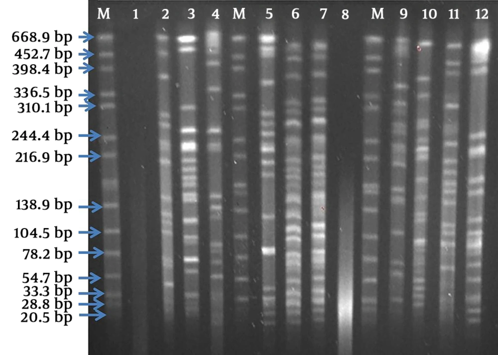
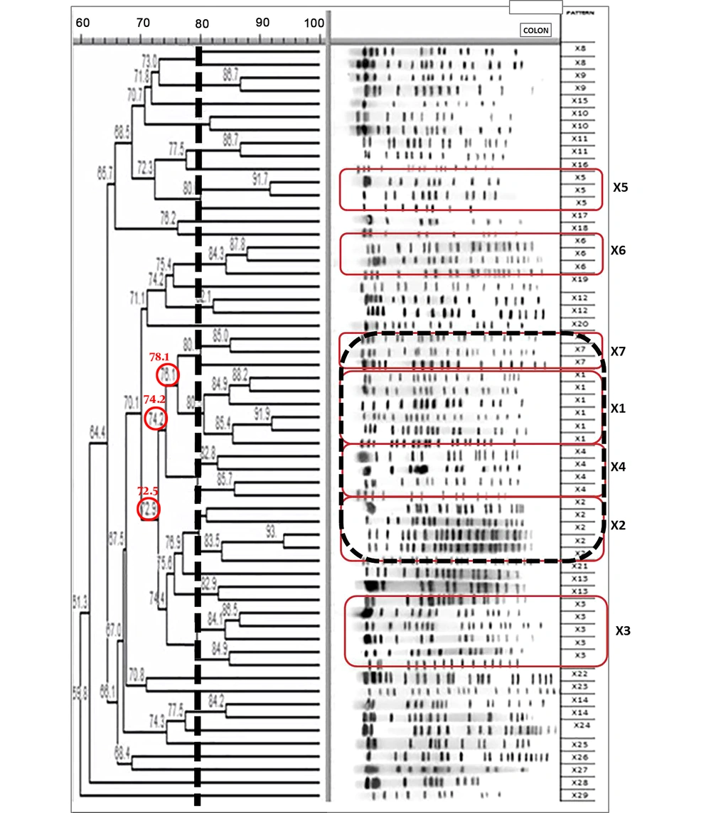
ESBL production was phenotypically determined in 36 (58%) isolates. The PFGE results showed the majority of isolates had 14-19 DNA bands (16 bands on average) (Figures 1 and 2). According to the DNA fingerprinting of isolates, 29 clusters (X1-29) were differentiated, which included 14 clones and 15 unique clusters. Twenty-seven strains from one hospital constituted 9 clustered (Figure 2). Each one of the X15 to X29 Clusters had a unique genotype and the majority of their strains (> 50%) were from the emergency ward. Four isolates with diverse origins were non-typable by PFGE.
The clusters with the genetic pattern of X1, X2, X4, and X7 (18 strains) were from Imam Khomeini Hospital, which showed the most genetic similarity. The majority of these strains were from ICU, as well as burning and infectious wards. They were presented as the prevalent clones. The highest genomic homology was for X1 and X7 clones associated with ICU and burning ward.
The ESBL producing isolates showed no significant homogenous genetic pattern clones (P > 0.05).
Clone X3 was comprised of isolates from the Emergency Department of Imam Reza Hospital and remarkably similar to the X1, X2, X4, and X7 clones from Imam Khomeini Hospital. The members of X6 clone were from the internal and surgery wards and ICU. There was no significant correlation between the resistance to beta-lactam antibiotics and clusters.
5. Discussion
The results of this study show that most of the K. pneumoniae isolates produce ESBL, which may lead to clonal dissemination of resistant strains. This is consistent with other studies, which have shown that the ESBL producing isolates in hospitalized patients are more likely to be exposed to antimicrobial agents, especially to the third generation Cephalosporins (14). Carbapenems still show good efficacy against ESBL producing K. pneumoniae isolates (2). However, emerging resistance to this group of antibiotics may limit their potency in the future. The tremendous increase in the prevalence of ESBLs among bacteria is a serious alarm since the majority of them are multi-drug resistant. In addition to observing the high diversity of isolates, we also observed similar strains. The genotypic polymorphism probably indicates the genetic diversity of isolates or the different origins of them. Given the various types of clinical samples and several medical centers for sampling, it is not unexpected that the isolates might be acquired from different sources. Similarly, several researches on K. pneumoniae isolates in Iran from 2008 to 2014 showed the genotype diversity between isolates of this bacterium. For example, studies on 35 and 54 K. pneumoniae isolates using PFGE (with 85% similarity of isolates), reported 21 and 42 different clusters, respectively (15, 16). Other studies on 71 and 31 of ESBL producing K. pneumoniae isolates using PFGE, have reported 62 and 26 genotypes, respectively (17, 18). These studies also used isolates from various clinical samples, including urine, lung secretions, blood, wounds, and sputum. The results of the above studies are consistent with our results for high genotype diversity among isolates. Furthermore, the research in Asian countries on K. pneumoniae, including studies on 256 and 93 isolates using PFGE, resulted in 136 and 41 different genotypes, respectively (19, 20). The results of these studies are also similar to our results in terms of the various genomic patterns of isolates. However, some studies have reported fewer varieties of K. pneumoniae isolates. For example, in a study conducted on 92 of ESBL-producing K. pneumoniae isolates mostly from neonatal units, only 13 genotype patterns were identified (21). One explanation could be the fact that the most isolates in this study were from limited sources in one hospital. Studies on 26, 66, and 57 isolates of K. pneumoniae in Canada, Bosnia and Herzegovina, and Jamaica using PFGE resulted in 3, 4, and 10 deferent genotypes, respectively (22-24). These studies also evaluated the isolates from limited sources, which may explain their genotypic similarity.
The strains with a similar genotypic pattern among our isolates may suggest the dissemination of these strains through the hospital environment, in particular, in ICU and burn ward. The hospitalized patients such as burned patients are at high risk of infection with various nosocomial pathogens due to the destruction of the skin barrier, suppression of the immune system, and invasive procedures (25). The transmission of resistant strains in the hospital environment may happen in various ways, including hospital staff or contaminated devices, equipment, and cleaners. These bacterial strains can cause nosocomial infections with a limited choice of effective antibiotics for their treatment.
There was no significant correlation between genotypes with the ESBL production and antibiotic resistance phenotypes. Since the most ESBL and antibiotic resistance genes on plasmid are small sequences (40 - 50 Kb), they may not be detected in PFGE bands (26). The average number of PFGE bands of our isolates was consistent with the results of other studies, indicating good genome digestion by the restriction enzyme in our study (17, 21, 22, 27-30).
5.1. Conclusions
Our results offer suitable data on molecular epidemiology of K. pneumoniae isolates in Kermanshah, especially hospital environment. The diversity patterns of K. pneumoniae genotypes indicate the various origins of strains isolated from hospitalized patients. However, the presence of some strains with similar genotypes in clusters can show a common source for these strains and may reflect the clonal spread through hospital environments, especially in ICU and burn ward of hospitals. Therefore, as a requirement, Kermanshah hospitals should implement a more effective system of infection control to reduce the dissemination of highly resistant strains of bacteria within the hospital environment.


