


1. Background
Echinococcosis is a helminthic zoonotic infection caused by adult or larval stages of tapeworm species belonging to the genus Echinococcus. The adult worm lives parasitically in the intestine of the carnivores, which act as definitive hosts (e.g., dog, wolf, dingo, etc.) and produces infective eggs. Either cestode segments containing eggs or free eggs are passed in the feces and environment. After oral uptake of the released eggs by the grazing animals, which act as intermediate hosts (e.g., sheep, goats, cattle, camels, etc.), the metacestode develops in internal organs. The aberrant hosts (e.g., humans) that do not play a role in the natural cycle are involved by accidentally ingesting the eggs. Whereas the infection of definitive hosts with immature or mature intestinal metacestode does not cause morbidity, the infected intermediate or aberrant hosts can be ended up with severe or even fatal outcomes (1).
Different species of Echinococcus cause four diseases. Cystic Echinococcosis (CE) is caused by the metacestode stage of E. granulosus, which initially emerges as fluid-filled bladders. This cyst gradually expands and forms connective tissues. Since the incubation period lasts several months to years, about 60% of CE cases remain asymptotic before the cysts rupture or exert a mass effect. Geographically, CE has a universal distribution, with an estimated mortality rate of 0.2 per 100,000 cases (2). Alveolar Echinococcosis (AE) is another well-known type caused by the larval stage of E. multilocularis and can metastasize as infiltrative parasitic lesions in different organs. Alveolar echinococcosis is confined to the northern hemisphere (3). Although AE is less common than CE, it poses a significant public health burden (especially in endemic areas) because the treatment process is more costly and complex (4). Other species are E. vogeli and E. oligarthus, known as polycystic echinococcosis, and are restricted to central and north America (4).
Living in sheep-raising areas, contact with free-roaming dogs, slaughtering at home, drinking non-boiled water, eating raw vegetables, and not washing hands before meals are likely to play a significant role in human infection (5, 6). A recent review showed that radical resection with pre- and post-operative anthelminthic treatment results in low recurrence and complication rates (7). However, early diagnosis using imaging and immunological tests in high-risk populations will result in a better outcome and decrease the long-term burden of disease (8).
Iran, with a 79.3 million livestock population (49.9 million sheep, 22.3 million goats, and 7.1 million cattle), is a potentially high-risk area where zoonotic pathogens and their putative threats to humans are of utmost medical and veterinary importance (especially in rural areas) (9). A recent review showed that the overall prevalence of CE in Iran is 4.2% (95% CI = 3.0 - 5.5%) (10). The first-degree family members who live with the infected patients seem to be at higher risk of transmission due to the common risk factors.
2. Objectives
This study was designed to investigate the prevalence of echinococcosis among family members of the infected patients who underwent surgical treatment.
3. Methods
3.1. Study Design
This cross-sectional study was conducted at Mashhad University of Medical Sciences. Ninety-six patients who underwent intervention due to echinococcosis at Imam Reza hospital were included from September 2016 to March 2017. All family members were invited by phone calls to perform three diagnostic tests, including anti-hydatid cyst Immunoglobulin G (IgG), ultrasound examination of the liver, and chest X-ray test.
The study protocol was according to the Declaration of Helsinki and approved by the Institutional Review Board of Mashhad University of Medical Sciences (IR.MUMS.fm.REC.1395.239). Written informed consent was obtained from all volunteers before data collection.
3.2. Data Collection
Different variables, including socioeconomic data and potential risk factors, were taken into account, including age, gender, occupation, education level, residence place (rural or urban area), type of water used (well or plumbing water), hazardous dietary habits like using unwashed vegetables, and history of contact with livestock and dog.
3.3. Statistical Analyses
Descriptive analysis was applied to collected variables, taking frequencies for categorical variables and central tendency measures for continuous variables. We used the Mann-Whitney U test and Fisher’s exact test to inspect the difference in continuous and binary variables. The significance level was set at P < 0.05. Statistical analyses were performed using SPSS version 21 (IBM SPSS Inc., Armonk, NY, United States).
4. Results
A total of 96 patients underwent surgical intervention due to echinococcosis infection during the study period. After making phone calls, 46 (47.9%) patients accepted to participate in the study. After obtaining informed consent from all family members living with the infected patient, 114 individuals were included to perform screening tests. The mean age of the study sample was 35.6 years (SD = 13.9) (range: 15 - 85 years), and 44.7% were male. Most participants lived in an urban area at the time of the study (89%), and only 22% had a history of living in a rural area. About 21% and 14% of participants reported a living history with livestock and dog, respectively.
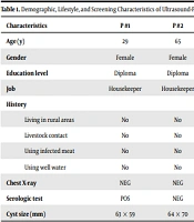
A total of seven participants from five families were diagnosed to be infected in ultrasound examination (Table 1). They were primarily female (N = 5) with low education levels. Only one patient reported a history of living in a rural area and contact with livestock. No pulmonary infection was found in chest X-ray images, and only one patient had a negative serologic test result. The cysts’ size varied from 20 × 48 mm to 207 × 146 mm. The comparison of collected variables between the infected and non-infected groups is shown in Table 2.
| Characteristics | P #1 | P #2 | P #3 | P #4 | P #5 | P #6 | P #7 |
|---|---|---|---|---|---|---|---|
| Age (y) | 29 | 65 | 51 | 21 | 28 | 27 | 33 |
| Gender | Female | Female | Male | Male | Female | Female | Female |
| Education level | Diploma | Diploma | Illiterate | Illiterate | Illiterate | BS | BS |
| Job | Housekeeper | Housekeeper | Businessman | N/A | Housekeeper | N/A | Employee |
| History | |||||||
| Living in rural areas | No | No | Yes | No | No | No | No |
| Livestock contact | No | No | Yes | No | No | No | No |
| Using infected meat | No | No | No | No | No | No | No |
| Using well water | No | No | No | No | No | No | No |
| Chest X-ray | NEG | NEG | NEG | NEG | NEG | NEG | NEG |
| Serologic test | POS | NEG | POS | POS | POS | POS | POS a |
| Cyst size (mm) | 63 × 59 | 64 × 70 | 40 × 42 | N/A | 110 × 103 | 20 × 48 | 207 × 146 |
Abbreviations: BS, Bachelor's degree; N/A, not available; NEG, negative; POS, positive.
a The test result was borderline positive.
| Characteristics | Infected (N = 7) | Non-infected (N = 107) | P Value |
|---|---|---|---|
| Age (y) | 36 ± 15.9 | 36 ± 13.8 | 0.921 a |
| Male gender | 2 (28.5) | 49 (45.8) | 0.457 b |
| Education level | N/A | ||
| Illiterate | 3 (42.9) | 32 (29.9) | |
| Diploma | 2 (28.5) | 54 (50.5) | |
| Bachelor’s degree | 2 (28.5) | 18 (16.8) | |
| Master | 0 (0) | 3 (2.8) | |
| Job | N/A | ||
| Housekeeper | 3 (42.9) | 38 (35.5) | |
| Businessman | 1 (14.3) | 32 (29.9) | |
| Employee | 1 (14.3) | 12 (11.2) | |
| Student | 0 (0) | 8 (7.5) | |
| Unemployed | 0 (0) | 7 (6.5) | |
| Worker | 0 (0) | 5 (4.7) | |
| Ranchman | 0 (0) | 1 (0.9) | |
| History | |||
| Living in rural areas | 1 (14.3) | 25 (23.4) | N/A |
| Livestock contact | 1 (14.3) | 24 (22.4) | N/A |
| Using infected meat | 0 (0) | 2 (1.9) | N/A |
| Using well water | 0 (0) | 2 (1.9) | N/A |
Abbreviation: N/A, not applicable.
a Analysis by independent-samples t test.
b Analysis by Fisher’s exact test.
5. Discussion
5.1. Main Findings
We found a prevalence rate of 6% for echinococcosis infection among family members living with the patients undergoing surgical resection. Serologic tests confirmed the findings of ultrasound examinations, except for one patient. The infection was extended to the lungs in none of the patients, which might be considered a benefit of the early detection of infection. However, it should be noted that the chest X-ray test can only distinguish uncomplicated pulmonary cysts, whereas complicated cysts may change the radiologic appearance of the tissue, undetectable in radiologic images (11). The patients reported no lifestyle risk factors, except for one patient who had a history of living in a rural area and contact with livestock. In addition, the patients' low to medium education level necessitates the need for educational interventions to increase the knowledge of risk factors of echinococcosis among individuals who are considered high-risk populations. Moreover, there were no significant differences between the infected and non-infected groups concerning age (P = 0.921) and gender (P = 0.457) distributions.
5.2. Comparison with Similar Studies
Similar screening studies were conducted in different parts of Iran from 2006 to 2011. The diagnostic tests (mostly Enzyme-linked Immunosorbent Assay, ELISA) were performed for the general population referring to clinics, laboratories, or blood transfusion centers. Two studies in Khuzestan and Ilam (western Iran) reported 13.8% and 1.2% prevalence rates, respectively (12, 13). In 2009, two studies in northwest Iran reported different prevalence rates (1.28% and 9.2% in Tabriz and Ardabil, respectively) (14, 15). Another study in central Iran (Kerman) found that 37 out of 451 (8.2%) individuals had CE infection (16). In 2006, the prevalence rate of echinococcosis was 1.6% in Tehran (the capital of Iran) (17). The latest screening study performed in Jahrom (Fras province in southwest Iran) showed that 6.3% of individuals referred to a clinical laboratory had CE infection (18). The prevalence rate varied between 1.2% and 13.8% in different geographical regions of Iran, which is in line with the results of our study.
A serological screening study was performed to estimate the prevalence rate of CE in a high-risk population from Cairo slaughterhouse employees and the households nearby. The researchers used the Indirect Haemagglutination (IHA), and Enzyme-linked Immunoelectrotransfer Blot (EITB) tests and found the prevalence of CE as 1.6% and 1.1% in Cairo slaughterhouse workers and the households nearby, respectively (19). Another similar study used ELISA to screen the household members of surgical cases of hydatidosis in central Tunisia. It was shown that eight out of 355 (2.2%) household members were serologically positive (20).
5.3. Limitations and Strengths
The small sample size, primarily due to the low participation rate (47.9%), may affect the generalizability of the results. The lack of a control group (family members of individuals without echinococcosis) prevented us from comparing groups. In contrast, performing three diagnostic tests on family members of patients who underwent surgical resection in the largest hospital in northeast Iran can be a study strength.
5.4. Conclusions
The screening of the first-degree family members of patients with confirmed echinococcosis results in the early diagnosis, lowers the disease's economic burden, and improves patients' outcomes and quality of life.
