Detection and Genotyping of Papillomavirus by Real-Time PCR in Iraqi Patients

authors:

avatar Zeina S M Al-Hadeithi 1 , avatar Saade Abdalkareem Jasim ORCID 2 , * , avatar Omar Dheyauldeen Salahldina 2

College of Pharmacy, Al-Nahrain University, Kadhimiya, Baghdad, Iraq
Medical Laboratory Techniques Department, Al-Maarif University College, Ramadi, Iraq

how to cite: Al-Hadeithi Z S M, Jasim S A, Salahldina O D. Detection and Genotyping of Papillomavirus by Real-Time PCR in Iraqi Patients. Arch Clin Infect Dis. 2022;17(3):e121143. https://doi.org/10.5812/archcid-121143.

Abstract

Background:

Cervical cancer (CC) is linked to human papillomavirus (HPV). Globally, the prevalence and genotype distribution differ significantly.

Objectives:

The goal of this study was to find HPV 14, 16, 18, and 45 genotypes in urogenital swabs by using a real-time PCR amplification test for quantitative genotyping of HPV DNA types 16, 18, and 45 and for simultaneous quantitative detection of HPV DNA types 31, 33, 35, 39, 51, 52, 56, 58, 59, 66, and 68, for a total of 14 HPV genotypes.

Methods:

This case-control study included 86 cervical swabs from Iraqi women referred by the Al-Yarmook teaching hospital in Baghdad, Iraq. The ages of cases varied from 23 to 70 years and specimens were obtained between March 2020 and March 2021. The DNA was extracted for molecular assay. Fourteen HPV genotypes were detected using real-time PCR (16, 18, 45, 31, 33, 35, 39, 51, 52, 56, 58, 59, 66, and 68). The detection protocol was based on the commercial Kit V31-100/F FRT as follows. For each sample reaction, 10х(N+1) μL of PCR-mix-1-FRT HPV 14 was added into a new tube. Then, 5.0х(N+1) μL of PCR-mix-2 buffer and 0.5х(N+1) μL of TaqF DNA polymerase were added. The tubes were vortexed. Finally, the prepared tubes added 10 μL of DNA samples from test or control samples. The statistical analysis was conducted using the statistical package for SPSS and Excel 2016 software.

Results:

Genotype 16 had the highest frequency, followed by genotypes 45 (22%), 18 (14%), 35 and 59 (6%), 52 and 58 (4%), and 31 (2%), while genotypes 33, 39, 51, 56, 66, and 68 had the lowest frequency (1%).

Conclusions:

The real-time PCR was efficient for detecting and genotyping HPV-DNA and could help in earlier detection and clinical care of HPV-infected patients by reducing costs and workload.

1. Background

Papillomaviruses sometimes cause infections of the skin and mucosal membrane, which leads to the emergence of different types of warts (cutaneous, plantar, flat, anal, genital, and laryngeal) or many cancers (uterus, penis, and vulva cancers) (1). The leading cause of human papillomavirus transmission is sexual contact. Various types of this virus are preferentially associated with some clinical lesions. One of the most common types of cancer worldwide is cervical cancer (1). Cervical cancer (CC) has the fourth highest incidence and fatality rate of any malignancy globally. On the other hand, it is the leading cause of female cancer and death in many low-resource nations. Cervical cancer develops slowly, taking years to decades (2). The finding that human papillomavirus (HPV) infection is intangible with CC development was a watershed moment in preventing this disease from causing CC development.

E6 and E7 are the two major carcinogenic proteins produced by the HPV cycle (3). These genes' products bind to and inactivate tumor suppressor genes, causing the host cell cycle to be disrupted. Twelve HPV genotypes have been designated as high risk (HR-HPV): 16, 18, 31, 33, 35, 39, 45, 51, 52, 56, 58, and 59. More than 60 genetic types of HPV have been described. There are strong associations between certain genotypes and clinical lesions (4, 5). Human papillomavirus is the most studied and well-known. Human papillomavirus has around 150 different types (6). Real-time PCR can distinguish nearly related sequences by combining simultaneous PCR amplification with extensive computer analysis of the collected kinetics data (7). Fluorescent dyes are used to detect the amplified product in real-time PCR. These dyes are linked to PCR primers and oligonucleotide probes, which selectively adhere to the amplified product during thermocycling, allowing for accurate detection of viral presence in the sample (8). As a result, real-time PCR has been used in screening programs because it provides a highly sensitive method for qualitative identification and genotyping of HR-HPV types in clinical samples (9). The cellular biology of HPV supports a primary role in the development of neoplasia (10). The viral types most closely associated with CC, particularly HPV-16 and 18, can transform cells in the culture to lose their normal growth control mechanisms. The DNA from these types integrates into host DNA (10, 11).

2. Objectives

The goal of this study was to find HPV 14, 16, 18, and 45 genotypes in urogenital swabs by using a real-time PCR amplification test for quantitative genotyping of HPV DNA types 16, 18, and 45 and for simultaneous quantitative detection of HPV DNA types 31, 33, 35, 39, 51, 52, 56, 58, 59, 66, and 68, for a total of 14 HPV genotypes.

3. Methods

Inclusion Criteria: Samples were used from women with CC confirmed by some tests recommended by the doctor.

Exclusion Criteria: Contaminated samples were excluded, as were individuals who received treatment before taking the sample.

This is a case-control study. Eighty-six cervical swabs from Iraqi women referred by the Al-Yarmook teaching hospital in Baghdad, Iraq, were included in the study. The ages of cases varied from 23 to 70 years, and specimens were obtained between March 2020 and March 2021. A doctor completed a case investigation form with information specific to each patient, including personal data, gynecological history, Pap smear, and histological examination results.

3.1. Sample Collection

Cervical swabs were taken per guidelines. For sampling, a cervical brush (1 - 1.5 cm) was inserted into the cervical canal after removing any excess mucus from the area around the ectocervix. The brush was then rotated three full turns in a counterclockwise direction before being removed from the canal and inserted into a 2 mL nuclease-free tube with 0.5 mL of the transport medium (Sacace), leaving the brush end inside the tube. Swab samples were frozen at -20 - 80°C or kept at 2 - 8°C for no more than 48 hours.

3.2. DNA Isolation

DNA was extracted from the vaginal swabs using the commercial kit DNA-Sorb-A (Sacace, REF K-1-1/A) following the manufacturer's instructions. After the DNA was successfully extracted, the eluted DNA was stored at -20°C (12).

3.3. Detection and Quantification of HPV

Detection protocol was done using the commercial Kit V31-100/F FRT and as follows: For each sample reaction (including clinical samples and controls), 10х(N+1) μL of PCR-mix-1-FRT HPV 14 was added into a new tube. Then, 5.0х(N+1) μL of PCR-mix-2 buffer and 0.5х(N+1) μL of TaqF DNA polymerase were added. The tubes were then vortexed. Finally, 10 μL of DNA sample was added from test or control samples to the prepared tubes (13).

3.4. Statistical Analysis

Statistical analysis was performed using SPSS and Excel 2016 software.

4. Results

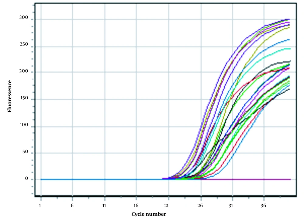
After the completion of the real-time PCR, the amplification results for all samples are shown in Figure 1. Each curve represents the amplification of the desired target region of the virus. The test was declared successful if the negative controls showed no positive fluorescence signal and the standards (Fam, Joe/Hex, Rox, Cy5, Cy5.5) showed positive signals in all channels.

Positive result of real-time PCR amplification

In this study, 14 HPV genotypes were assayed, and the resulting frequencies are shown in Table 1. Genotype 16 showed the highest frequency, followed by genotypes 45 (22%), 18 (14%), 35 (6%), 59 (6%), 52 (4%), and 58 (4%). Genotype 31 showed a 2% frequency, while genotypes 33, 39, 51, 56, 66, and 68 showed the lowest frequency (1%).

Table 1.

Frequency of Virus Infections

GenotypeNumber of CasesFrequency
162529.06977
181416.27907
451922.09302
3122.325581
3311.162791
3566.976744
3911.162791
5111.162791
5244.651163
5611.162791
5844.651163
5966.976744
6611.162791
6811.162791

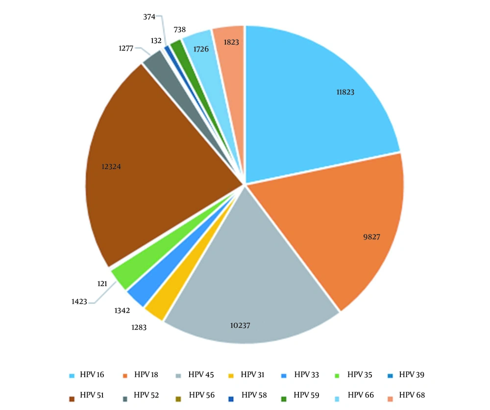
The sensitivity of the PCR-Tm analysis assay for each genotype and their corresponding mean copy numbers are seen in Table 2 and Figure 2.

Table 2.

Virus Genotypes and Their Mean Copy Numbers

Genotype (HPV)Copy Numbers
1611823
189827
4510237
311283
331342
351423
39121
5112324
521277
56132
58374
59738
661726
681823
Genotypes of HPV and their frequencies

5. Discussion

An estimated 90% of all HPV infections go away on their own within two years after they are contracted. In a small percentage of cases, certain HPV infections can persist and progress to cancer (14)

In research conducted by Alizi et al. (15), the prevalence of HPV among Iraqi women was very low (9%). These findings were consistent with an earlier study in Iraq by Faik et al., which showed 12.38% (16). Comparing Iraqi populations to those in industrialized countries, their Islamic nature and moral dedication to their country may explain the low incidence of HPV and other sexually transmitted illnesses. It has been determined via several examinations in Iraq that virus type 16 is the most prevalent variant in the country (17).

Other investigations used a Real Time amplification test for qualitative detection and genotyping of human papillomavirus (16, 18, 31, 33, 35, 39, 45, 51, 52, 56, 58, 59, 66, and 68) in urogenital swabs and biopsies (Sacace, Italy), and related methodologies yielded comparable results (18, 19).

A study from Iraq found that HPV 33 (18%) was the most common type, and the other found HPV 16 (58.3%) as the most common type, followed by HPV31/33 (25%) and HPV18 (16.7%), which are consistent with our findings (16, 20). The disparity in HPV genotype prevalence may be attributable to sample size, geographic differences, utilizing various techniques, and different primer pairs.

According to He and He (21), the overall rate of HR-HPV infection was 12.6% in 17,319 women, and the five most prevalent types were genotypes 52, 53, 58, 16, and 56, which agrees with our findings.

Similar frequencies were observed in Tianjin (12.5%) (22), Guangdong (7.3%) (23), and Beijing (7.0%) (24) in China. The most frequent HR-HPV types in Sichuan (52, 53, 58, 16, 56) did not appear to be the same as in other Chinese cities (13). In other countries, studies have found that HPV frequency is 12.2% in Canada (25), 34.5% in Peru, 10.3% in Iran, and 9.3% in Australia (26-28).

Aoyama-Kikawa et al. found that HPV infection was present in just 4.6% of Japanese women (29). These results confirm Becker et al. that HPV prevalence varies by geography and ethnicity (30). The only exception is HPV 66, classified as "possibly" carcinogenic (31-33). HPVs 16 and 18 are the most frequent high-risk genotypes worldwide, accounting for 75% of all cervical cancer cases and HPV45 (34, 35).

5.1. Conclusions

The real-time PCR method efficiently detected and genotyped HPV-DNA, which could help in earlier detection and clinical care of HPV-infected patients by reducing costs and workload.
Acknowledgements

References

  • 1.

    Lowy DR; Howley. Papillomaviruses. In: Knipe DM, Howley PM, editors. Fields' Virology, Volume 2. Philadelphia, PA: Lippincott Williams & Wilkins; 2007. p. 2299-354.

  • 2.

    Bray F, Ferlay J, Soerjomataram I, Siegel RL, Torre LA, Jemal A. Global cancer statistics 2018: GLOBOCAN estimates of incidence and mortality worldwide for 36 cancers in 185 countries. CA Cancer J Clin. 2018;68(6):394-424. [PubMed ID: 30207593]. https://doi.org/10.3322/caac.21492.

  • 3.

    Surriabre P, Torrico A, Vargas T, Ugarte F, Rodriguez P, Fontaine V. Assessment of a new low-cost, PCR-based strategy for high-risk human papillomavirus DNA detection for cervical cancer prevention. BMC Infect Dis. 2019;19(1):842. [PubMed ID: 31615443]. [PubMed Central ID: PMC6794773]. https://doi.org/10.1186/s12879-019-4527-9.

  • 4.

    Nemes JA, Deli L, Nemes Z, Marton IJ. Expression of p16INK4A, p53, and Rb proteins are independent from the presence of human papillomavirus genes in oral squamous cell carcinoma. Oral Surg Oral Med Oral Pathol Oral Radiol Endod. 2006;102(3):344-52. [PubMed ID: 16920543]. https://doi.org/10.1016/j.tripleo.2005.10.069.

  • 5.

    Al-Hadeithi ZSM. Using ISSR markers to build a phylogenetic of Barley Genotypes. Iraqi J Sci. 2015;56(2C):1682-8.

  • 6.

    Dias TC, Longatto-Filho A, Campanella NC. Human papillomavirus genotyping as a tool for cervical cancer prevention: from commercially available human papillomavirus DNA test to next-generation sequencing. Future Sci OA. 2020;6(9):FSO603. [PubMed ID: 33235804]. [PubMed Central ID: PMC7668120]. https://doi.org/10.2144/fsoa-2019-0159.

  • 7.

    Kandala NJ; Abdul-Samad. The Molecular Detection of HPV Infection in samples of Iraqi Women with Abnormal cervical Smears. Iraqi J Sci. 2018;59(4B):1995-2004.

  • 8.

    Rodriguez-Lazaro D, Cook N, Hernandez M. Real-time PCR in Food Science: PCR Diagnostics. Curr Issues Mol Biol. 2013;15:39-44. [PubMed ID: 23513039].

  • 9.

    Nsaif GS, Al-Mualm M, Faisal AJ. The effect of e-cigarettes smoking on expression and methylation of CYP1A1 and CYP1B1 genes and other biochemical parameters. Mater Today Proc. 2021;In Press. https://doi.org/10.1016/j.matpr.2021.07.403.

  • 10.

    Beutner KR, Tyring S. Human Papillomavirus and Human Disease. Am J Med. 1997;102(5):9-15. https://doi.org/10.1016/s0002-9343(97)00178-2.

  • 11.

    Maruyama T, McLachlan A, Iino S, Koike K, Kurokawa K, Milich DR. The serology of chronic hepatitis B infection revisited. J Clin Invest. 1993;91(6):2586-95. [PubMed ID: 8514870]. [PubMed Central ID: PMC443322]. https://doi.org/10.1172/JCI116497.

  • 12.

    Sambrook J. Molecular Cloning: A Laboratory manual. Plainview, New York: Cold Spring Harbor Laboratory Press; 1989.

  • 13.

    Jasim SA, Ahmed NS, Mousa AA, Hmed AA, Sofy AR. Correlation between both genetic polymorphism and serum level of toll-like receptor 4 with viral load and genotype of hepatitis C virus in Iraqi patients. Gene Rep. 2021;24:101287. https://doi.org/10.1016/j.genrep.2021.101287.

  • 14.

    Demarco M, Hyun N, Carter-Pokras O, Raine-Bennett TR, Cheung L, Chen X, et al. A study of type-specific HPV natural history and implications for contemporary cervical cancer screening programs. EClinicalMedicine. 2020;22:100293. [PubMed ID: 32510043]. [PubMed Central ID: PMC7264956]. https://doi.org/10.1016/j.eclinm.2020.100293.

  • 15.

    Alizi S, Jasim W, Sami H, Alkhafaji B. Detection and Genotyping of Human Papillomavirus DNA in Cervical Neoplasia of Iraqi Women Using Real Time PCR Technique. IOSR J Dent Med Sci. 2018;17(8):1-5.

  • 16.

    Faik AJ, Saber MQ, Mohammed WJ, Ibraheem BZ, Lateef KR, Areeg S. Hassen AS. Genotyping of High-risk Human Papilloma virus (HPV) among Iraqi women in Baghdad by Multiplex PCR. Journal of Biotechnology Research Center. 2015;9(1):38-45. https://doi.org/10.24126/jobrc.2015.9.1.402.

  • 17.

    Co-expression of E6 and E7 Genes of High Risk Types of Human Papillomavirus and P53 Gene in Malignant and Premalignant Cervical Lesions [dissertation]. Baghdad, Iraq: University of Al-Nahain; 2016.

  • 18.

    Venceslau EM, Bezerra MM, Lopes ACM, Souza ÉV, Onofre ASC, de Melo CM, et al. HPV detection using primers MY09/MY11 and GP5+/GP6+ in patients with cytologic and/or colposcopic changes. J Bras Patol Med Lab. 2014;50(4):280-5. https://doi.org/10.5935/1676-2444.20140028.

  • 19.

    Alwan NAS, Shalal MM; Mezaal; Aziz. Prevalence of HPV Genotype in Cervical Cells among Iraqi Patients with Abnormal Pap Smears. Iraqi Journal of Biotechnology. 2017;16(2):19-27.

  • 20.

    Ali SHM. Molecular biological studies of Human Papillomavirus infections in patient with cervical neoplasia [dissertation]. Baghdad, Iraq: University of Al-Nahrain; 2001.

  • 21.

    He L, He J. Distribution of high-risk HPV types among women in Sichuan province, China: a cross-sectional study. BMC Infect Dis. 2019;19(1):390. [PubMed ID: 31068141]. [PubMed Central ID: PMC6505120]. https://doi.org/10.1186/s12879-019-4038-8.

  • 22.

    Chen X, Wallin KL, Duan M, Gharizadeh B, Zheng B, Qu P. Prevalence and genotype distribution of cervical human papillomavirus (HPV) among women in urban Tianjin, China. J Med Virol. 2015;87(11):1966-72. [PubMed ID: 26073652]. https://doi.org/10.1002/jmv.24248.

  • 23.

    Jing L, Zhong X, Huang W, Liu Y, Wang M, Miao Z, et al. HPV genotypes and associated cervical cytological abnormalities in women from the Pearl River Delta region of Guangdong province, China: a cross-sectional study. BMC Infect Dis. 2014;14:388. [PubMed ID: 25016305]. [PubMed Central ID: PMC4226991]. https://doi.org/10.1186/1471-2334-14-388.

  • 24.

    Zhang D, Li T, Chen L, Zhang X, Zhao G, Liu Z. Epidemiological investigation of the relationship between common lower genital tract infections and high-risk human papillomavirus infections among women in Beijing, China. PLoS One. 2017;12(5). e0178033. [PubMed ID: 28531212]. [PubMed Central ID: PMC5439700]. https://doi.org/10.1371/journal.pone.0178033.

  • 25.

    Ogilvie GS, Cook DA, Taylor DL, Rank C, Kan L, Yu A, et al. Population-based evaluation of type-specific HPV prevalence among women in British Columbia, Canada. Vaccine. 2013;31(7):1129-33. [PubMed ID: 23273510]. https://doi.org/10.1016/j.vaccine.2012.09.085.

  • 26.

    Iwasaki R, Galvez-Philpott F, Arias-Stella J; Arias-Stella Jr. Prevalence of high-risk human papillomavirus by cobas 4800 HPV test in urban Peru. Braz J Infect Dis. 2014;18(5):469-72. [PubMed ID: 24835620]. [PubMed Central ID: PMC9428228]. https://doi.org/10.1016/j.bjid.2014.01.010.

  • 27.

    Jamdar F, Farzaneh F, Navidpour F, Younesi S, Balvayeh P, Hosseini M, et al. Prevalence of human papillomavirus infection among Iranian women using COBAS HPV DNA testing. Infect Agent Cancer. 2018;13:6. [PubMed ID: 29416557]. [PubMed Central ID: PMC5784531]. https://doi.org/10.1186/s13027-018-0178-5.

  • 28.

    Brotherton JM, Hawkes D, Sultana F, Malloy MJ, Machalek DA, Smith MA, et al. Age-specific HPV prevalence among 116,052 women in Australia's renewed cervical screening program: A new tool for monitoring vaccine impact. Vaccine. 2019;37(3):412-6. [PubMed ID: 30551987]. https://doi.org/10.1016/j.vaccine.2018.11.075.

  • 29.

    Aoyama-Kikawa S, Fujita H, Hanley SJB, Kasamo M, Kikuchi K, Torigoe T, et al. Comparison of human papillomavirus genotyping and cytology triage, COMPACT Study: Design, methods and baseline results in 14 642 women. Cancer Sci. 2018;109(6):2003-12. [PubMed ID: 29660849]. [PubMed Central ID: PMC5989866]. https://doi.org/10.1111/cas.13608.

  • 30.

    Becker TM, Wheeler CM, McGough NS, Parmenter CA, Jordan SW, Stidley CA, et al. Sexually transmitted diseases and other risk factors for cervical dysplasia among southwestern Hispanic and non-Hispanic white women. JAMA. 1994;271(15):1181-8. [PubMed ID: 8151876].

  • 31.

    Bouvard V, Baan R, Straif K, Grosse Y, Secretan B, El Ghissassi F, et al. A review of human carcinogens--Part B: biological agents. Lancet Oncol. 2009;10(4):321-2. [PubMed ID: 19350698]. https://doi.org/10.1016/s1470-2045(09)70096-8.

  • 32.

    de Martel C, Plummer M, Vignat J, Franceschi S. Worldwide burden of cancer attributable to HPV by site, country and HPV type. Int J Cancer. 2017;141(4):664-70. [PubMed ID: 28369882]. [PubMed Central ID: PMC5520228]. https://doi.org/10.1002/ijc.30716.

  • 33.

    Al-Hadeithi ZSM. Detection of genetic polymorphism in Iraqi barley using SSR-PCR Analysis. Iraqi J Sci. 2016;27(2B):1158-64.

  • 34.

    Molina-Pineda A, Lopez-Cardona MG, Limon-Toledo LP, Canton-Romero JC, Martinez-Silva MG, Ramos-Sanchez HV, et al. High frequency of HPV genotypes 59, 66, 52, 51, 39 and 56 in women from Western Mexico. BMC Infect Dis. 2020;20(1):889. [PubMed ID: 33238902]. [PubMed Central ID: PMC7690193]. https://doi.org/10.1186/s12879-020-05627-x.

  • 35.

    Pimenoff VN, Tous S, Benavente Y, Alemany L, Quint W, Bosch FX, et al. Distinct geographic clustering of oncogenic human papillomaviruses multiple infections in cervical cancers: Results from a worldwide cross-sectional study. Int J Cancer. 2019;144(10):2478-88. [PubMed ID: 30387873]. https://doi.org/10.1002/ijc.31964.