1. Background
Food-borne parasitic zoonoses (FBPZs) caused by helminths or protozoans are responsible for many diseases in humans and animals. These types of diseases are caused by the consumption of infected meat, plants, and water, and they arouse considerable public health concerns and can pose socioeconomic problems. Two liver flukes, Fasciola hepatica and Fasciola gigantica, as the main FBPZs, account for fascioliasis, a severe zoonotic global disease in human and animal hosts (1, 2). Nearly 180 million people are at risk of fascioliasis, while 2.4 - 17 million people are infected worldwide, according to World Health Organization (WHO) reports (1, 3). The global prevalence of fascioliasis is increasing among humans. Moreover, Fasciola species (spp.) affect livestock, causing considerable problems to industry and farmers in developed and developing countries (4).
The geographical distribution of Fasciola spp. is linked to climate and environmental conditions, including the presence of pastures, wetlands, and water bodies. Such conditions create a suitable environment for transmitting the parasite's infecting stages and the growth and reproduction of snails as intermediate hosts. In addition to climatic and environmental factors, some other variants, such as anthropogenic environmental changes, people traveling, and importing and exporting livestock, are responsible for the occurrence of fascioliasis (5). Iran is located in the Middle East and borders the Caspian Sea by the north and the Persian Gulf and Oman Sea by the south. This country has favorable conditions for developing various parasites because of its climate, geographical location, and biological and cultural characteristics (6). WHO has included Iran among the six countries with serious fascioliasis problems (7). In recent years, the prevalence of Fasciola spp. has increased in Iran, and new foci of the disease have been reported from some provinces (6). Fascioliasis is an emerging or re-emerging disease in Guilan province and is endemic in the Mazandaran Province, both located in the north of Iran near the Caspian Sea, and some cases have been reported in other provinces. The greatest outbreak of fascioliasis in history occurred in Guilin. This province has experienced two waves of fascioliasis epidemics. The first wave of the epidemic was in 1987, while the second wave occurred ten years later, and thousands of individuals were involved (8). Recently, some new cases have been reported in these provinces.
Many articles reported the Fasciola epidemiology in Guilan Province, but new studies are needed in other provinces. Mazandaran Province, an endemic region of Fasciola spp., has the same climatic conditions and agricultural and farming traditions as Guilan. The knowledge of fascioliasis in cities of Mazandaran Province may facilitate our understanding of the disease in northern Iran (9).
2. Objectives
The purpose of this study was to perform a one-year study of Fasciola seroepidemiology in order to evaluate the extent and epidemiological characteristics of human fascioliasis in Qaemshahr, Mazandaran Province, Iran, for the first time.
3. Methods
3.1. Ethical Considerations
This research is carried out following the ethical principles and standards for conducting medical research. Ethical approval of this study was given by the Ethics Committee of Mazandaran University of Medical Sciences, Sari, Iran (IR.MAZUMS.REC.1398.772). Informed consent forms were obtained from the participants or their guardians.
3.2. Study Type
This descriptive observational study was conducted between 2018 - 2019 in health centers belonging to Mazandaran University of Medical Sciences in Qadikola, Sarookola, Alamshir, Qarakheil, and Arate towns of Qaemshahr County, Mazandaran Province, Iran.
3.3. Period of Study
The study was carried out for 24 months, from January 2018 to December 2019.
3.4. Sample Size
The statistical population of this research (n = 2,418) was selected by cluster sampling method from affiliated outpatients referring to the clinical laboratories of hospitals or health centers of Mazandaran University of Medical Sciences from 5 different localities of Qaemshahr county, Mazandaran province. The samples were collected in accordance with the population of each town.
3.5. Sample Collection
All participants were informed about the objectives and procedures of the study and were collected after informed consent was obtained from them or their guardians.
Five milliliter of venous blood specimens were obtained from all participants. These blood samples were referred to the Parasitology Laboratory of the Department of Parasitology, School of Medicine, Mazandaran University of Medical Sciences, Sari, Iran, and then were centrifuged at 2,500 g for 15 min to obtain serum. hyperlipidemic and hemolyzed serums were excluded, and other collected sera were stored at -20°C until further use.
All individuals filled out a questionnaire containing demographic information, including age, gender, and occupation, as well as a history of consuming raw vegetables, elaborated olive, wild or traditional vegetables, tap or spring water, and anthelminthic drugs, and a history of abdominal pain, gastrointestinal symptoms, and jaundice.
3.6. The Technique Used In the Study
The specific anti-Fasciola hepatica immunoglobulin G (IgG) antibodies in serum samples were analyzed using the commercial enzyme-linked immunosorbent assay (ELISA) kit (Fasciola ELISA Kit, Pishtazteb, Tehran, Iran) according to the manufacturer's instructions. Finally, the absorbance was measured by an ELISA reader at 450 nm. The cut-off point was set at 0.282. The positive and suspicious cases were re-checked. Positive serologic test results underwent clinical and paraclinical evaluation.
3.7. Statistical Analysis
Frequency (n) and percentage (%) values were used to describe quality variables and mean scores for quantity ones. In the first step, each variable was separately entered into a univariate logistic regression model (unadjusted method). Afterward, the variables with a P-value of less than 0.2 were selected for a multiple logistic regression model using the enter method (adjusted method). The results of logistic regression were reported with odds ratio, 95% confidence intervals, and p-value. The Hosmer-Lemeshow test was applied to evaluate the goodness-of-fit of the logistic regression model in each step (P > 0.05). All statistical analyses were performed in SPSS software (version 24). A P-value of less than 0.05 was considered significant.
4. Results
4.1. Prevalence Rate or Positivity Rate
Anti-Fasciola antibodies were detected in 60 (2.48%) individuals by ELISA test, while 2,358 (97.51%) participants were seronegative. Seropositivity of fascioliasis was found among 20% (12/60) female and 80% (48/60) male participants (95% CI: 14.7, 8.02 - 29.2).
4.2. Risk Factors Assessment
Out of 2418 patients, 1866 (77.2%) were female, and 552 (22.8 %) were male, aged 7 - 77 years old. Among this population, 426 and 1,992 participants lived in urban and rural areas, respectively.
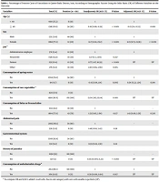
A significant relationship was observed between the seropositivity of Fasciola infection and gender (P < 0.001). The results of the univariate analysis, using the adjusted method, showed that anti-Fasciola antibodies were significantly 14.7 times higher in women than in men, and the results of the adjusting model showed that it is 12.9 times (12.9, 95%CI: 6.73 - 23.36, P < 0.001). Considering the age of participants, the prevalence of anti-Fasciola antibodies was higher in the group of > 40-year-old (90%) than in the group of < 40-year-old (10%), (95% CI: 0.18, 0.06 - 0.39, P < 0.001) Based on the results of the multiple logistic regression model, the probability of Fasciola contamination rate was 82% lower in the group of > 40-year-old than in the other group. Accordingly, the result was 77% based on the estimation performed by adjusted method estimates (OR = 0.23, 95%CI: 0.1 - 0.55, P = 0.001).
However, it was revealed that age (P = 0.001) and gender (P < 0.001) had a significant relationship with the presence of antibodies against Fasciola infection. The Fasciola infection was observed higher in Sarookola (30%, 18/294) and Alamashir (30%, 18/530), while a low seroprevalence was found in Qadikola (0%). It was also revealed that the prevalence of Fasciola infection was significantly higher in rural communities (80%).
Among various job groups, the incidence of Fasciola infection among administrator employees (OR = 8.77, 95%CI: 4.17 - 18.39; P < 0.001), housewives (OR = 1.37, 95%CI: 0.7 - 2.69, P = 0.357), and other jobs (OR = 1.40, 95%CI: 0.66 - 2.96, P = 0.385) were not significantly different. Nevertheless, a significant relationship was observed between farmers and Fasciola seropositivity (OR = 8.77, 95%CI: 4.17 - 18.39; P < 0.001). In the statistical analysis of the epidemiological data, the relationship of seropositivity to Fasciola spp. with the consumption of spring water (OR = 0.43, 95%CI: 0.19 - 1.14, P = 0.005), raw vegetables (OR = 13.24, 95%CI: 1.83 - 95.14, P = 0.001), anthelminthic drugs (OR = 0.58, 95%CI: 0.018 - 0.94, P = 0.017), and history of jaundice (OR = 0.03, 95%CI: 0.004 - 0.23, P < 0.001) were statistically significant.
The findings revealed that Fasciola IgG positivity had no significant relationship with the 247 consumption of traditional appetizers, including Dalar and elaborated olive (OR = 1.53, 95%CI: 248 0.858 - 2.64, P = 0.137), abdominal pain (OR = 1.46, 95%CI: 0.62 - 3.43, P = 0.38), and gastrointestinal symptoms (OR = 0.59, 95%CI: 0.25 - 1.38, P = 0.22). All demographic factors and variables are summarized in Table 1.
| Variables | Total (%) | Positive (n = 60) | Unadjusted; OR (95% CI) | P-Value | Adjusted; OR (95% CI) | P-Value |
|---|---|---|---|---|---|---|
| Age (y) | ||||||
| < 40 | 900 (37.2) | 6 (10) | 1 | 1 | ||
| ≥ 40 | 1518 (62.8) | 54 (90) | 0.18 (0.06, 0.39) | < 0.001 | 0.23 (0.1, 0.55) | 0.001 |
| Sex | ||||||
| Male | 552 (22.8) | 48 (80) | 1 | 1 | ||
| Female | 1866 (77.2) | 12 (20) | 14.7 (8.02, 29.2) | < 0.001 | 12.9 (6.73, 23.36) | < 0.001 |
| Job a | ||||||
| Administrator employee | 372 (15.4) | 12 (20) | 1 | 1 | ||
| Housewife | 1518 (62.8) | 18 (30) | 1.37 (0.7, 2.69) | 0.357 | ||
| Farmer | 252 (10.4) | 12 (20) | 8.77 (4.17, 18.39) | < 0.001 | UP | UP |
| Other | 276 (11.4) | 18 (30) | 1.40 (0.66, 2.96) | 0.385 | ||
| Consumption of spring water | ||||||
| No | 2304 (95.3) | 54 (90) | 1 | 1 | ||
| Yes | 114 (4.7) | 6 (10) | 0.43 (0.19, 1.14) | 0.005 | 0.54 (0.22, 1.34) | 0.184 |
| Consumption of raw vegetables a | ||||||
| No | 420 (17.4) | 0 (0) | ||||
| Yes | 1998 (82.6) | 60 (100) | 13.24 (1.83, 95.14) | 0.001 | UP | UP |
| Consumption of Delar or elaborated olive | ||||||
| No | 534 (22.1) | 18 (30) | 1 | - | - | |
| Yes | 1884 (77.9) | 42 (70) | 1.53 (0.85, 2.64) | 0.137 | 1.43 (0.80, 2.56) | 0.226 |
| Abdominal pain | ||||||
| No | 2082 (86.1) | 54 (90) | 1 | - | - | |
| Yes | 336 (13.9) | 6 (10) | 1.46 (0.62, 3.43) | 0.38 | ||
| Gastrointestinal system | ||||||
| No | 2268 (93.8) | 54 (90) | ||||
| Yes | 150 (6.2) | 6 (10) | 0.59 (0.25, 1.38) | 0.22 | ||
| History of jaundice | ||||||
| No | 2298 (95) | 60 (100) | - | - | ||
| Yes | 120 (5) | 0 (0) | 0.03 (0.004, 0.23) | < 0.001 | UP | UP |
| Consumption of anthelminthic drugs a | ||||||
| No | 2154 (89.1) | 60 (100) | 1 | 1 | ||
| Yes | 264 (10.9) | 0 (0) | 0.58 (0.018, 0.94) | 0.017 | UP | UP |
a To compute OR and 95%CI, added 1 to all cells. Due to exit category with zero cells unable to perform (UP).
5. Discussion
According to the WHO report, fascioliasis is included in the list of neglected tropical diseases among the food-borne trematodiases group (10, 11). It is estimated that around 17 million people are infected with fascioliasis globally. Extreme pathogenicity conditions, such as neurological and ophthalmological situations leading to permanent sequelae and even fatal cases, express the public health importance of this disease (12). The wide distribution of Fasciola human and livestock infection was considered of only secondary importance until 1990. Human infection with Fasciola spp. began to show its importance in the 90s, with the progressive description of many human endemic areas and an increase in human infection reports (11). Human fascioliasis is most important in the Middle East and North Africa (MENA) region, which includes more than 20 countries. Human fascioliasis is prevalent mostly in northern areas of Iran, as one of the MENA countries with two epidemics of high incidence rate (13).
In this study, 2,418 individuals and 60 IgG seropositive cases of Fasciola spp. were enrolled. There was a wide variety of seroprevalence estimates provided by different research. According to Aryaeipour et al., the rate of seropositivity for human fascioliasis was 24.8% in 13 provinces of Iran (14). In Guilan and Mazandaran provinces, the seroprevalence rate was estimated at 37.3%, while a lower seropositivity rate was found in Markazi, Kordestan, Hamedan, Fars, and Azerbaijan provinces, Iran (2%). In another study, the overall seroprevalence rate of infection was obtained as 1.36% in Anzali, Guilan Province, Iran (15). The results of a study conducted by Sarkari et al. Showed that the seroprevalence rate of human fascioliasis in Yasuj, Iran, was relatively high (1.86%), and this area could be considered a newly emerging focus of the disease (16).
Human fascioliasis transmission dynamics are strongly influenced by the climate, farming practices, dietary habits, wandering animals, diversity, and the multiplicity of intermediate hosts (13). All these risk factors are observed in the MENA region to varying degrees, especially in Iran. Livestock husbandry is also an integral part of farming among agricultural communities in the fascioliasis endemic areas, including Iran. Livestock is considered the origin of infection in humans. In addition, a suitable climate and farming practices that favor the survival of the snail intermediate hosts are often common throughout the region. Several species of Lymnaea, including Galba truncatula and Lymnaea gedrosiana, have been confirmed to be important in the transmission of F. hepatica and F. gigantica, respectively (13). In northern endemic areas of Iran, a number of favorable conditions, including numerous irrigation canals, agricultural crop traditions (primarily rice), high temperatures above 20°C, high rainfall, and short dry seasons, facilitate the transmission of fascioliasis and lymnaeid populations (17). In a one-year study of abattoirs in Amol, Mazandaran Province, Iran, the prevalence of Fasciola spp. among sheep and goats was calculated at 7.7% and 5.4%, respectively (18).
According to the findings of this research, the positivity rate of Fasciola infection varied by age and gender in the Qaemshahr district. Our data showed that Fasciola IgG positivity against Fasciola was predominantly observed in the older age group of > 40 years old (90%) than in the age group of > 40 years old (10%). The results of our study are in agreement with those reported by Rokni et al. in Guilan province (15) and Asadian in Meshkin Shahr, Ardabil Province, Iran (19). They showed that the older age group (i.e., > 40 years old) had a significantly higher seroprevalence than the younger age group (i.e., < 40 years old). The researchers of this study suggest that the higher IgG positivity rate among the older age groups might be due to their longer exposure to the risk factors and one of the transmission routes. Based on the results of the current study, the IgG seropositivity rate was higher in males (80%) than in females (20%) in Qaemshahr County.
Metacercaria of Fasciola spp. remain suspended in water used for cooking and drinking. Contaminated water and raw water plants contaminated with metacercariae can be a source of Fasciola infection in humans (20). Our data revealed a significant difference between drinking water and the seropositivity rate of Fasciola infection. The results of 10% of participants with a history of drinking spring water were IgG seropositive (90% of the subjects did not have a history of drinking spring water).
Traditional and raw vegetables are another important source of infection in the northern part of Iran. Two very important Fasciola sources in these endemic regions are green salad (local name: Dalal) and elaborated olive (local name: Zeytoon-parvardeh) as appetizers (13). Ground aquatic plants, such as Mentha pulegium (local name: Khlivash), Mentha piperita (local name: Bineh), and Eryngium coucasicum (local name: Choochagh), are used for the aforementioned appetizers. Metacercariae maintains two weeks in Zeytoon-parvardeh or Dalal (made of these plants), with survival rates of 66.6% and 57.8%, respectively (21). Our research showed that 100% of participants with a history of raw vegetable consumption and 70% of individuals with a history of Dalal or elaborated olive consumption had IgG antibodies against Fasciola. These findings are consistent with those of a study conducted by Sarkari et al. in Yasuj (16).
Fascioliasis may cause various clinical symptoms ranging from asymptomatic infection to severe diseases. Our results indicated no significant difference between IgG seropositivity rate and a history of abdominal pain or gastrointestinal symptoms. These results disagreed with those reported in a study performed by Sarkari et al. in Yasuj (16).
5.1. Conclusions
To the best of our knowledge, this was the first report of the human Fasciola IgG seropositive rate in Qaemshahr, Mazandaran Province. We found that 2% of Qaemshahr residents are seropositive for Fasciola infection, while 98% are seronegative. As a result of our results and the WHO epidemiological classification, human fascioliasis is hypoendemic in Mazandaran Province (22). Identifying fasciolosis risk factors can lead to adopting several effective strategies to prevent Fasciola infection. Health education combined with factors such as patients’ remedy, vector control, livestock and grazing land management, and animal treatment can significantly decrease the prevalence of fasciolosis. The most effective solution recommended for the prevention of disease re-emergence is the application of integrated preventive and control strategies.
