1. Introduction
Angiostrongylus cantonensis is a parasitic worm commonly found in rats and mollusks in the Asia-Pacific region. Human infection can occur through the consumption of raw or undercooked contaminated hosts or food. While cases are rare, there have been roughly 2800 cases reported in 30 countries. The parasite primarily affects the central nervous system, causing angiostrongylus meningitis, characterized by neck stiffness and headaches (1, 2). However, specific symptoms may vary depending on the location of the worm's inflammatory response. This case report presents a patient whose initial presentation was neuropathic pain.
2. Case Presentation
2.1. Patient Information
A 27-year-old female patient was admitted to the ICU for sedation monitoring due to progressive pain that had persisted for 2 weeks prior to hospital admission. The pain was most intense in the back and radiated to both arms and legs, with an intensity rating of 8/10 on the numerical scale. The patient also experienced allodynia, hyperesthesia with tingling and numbing sensations, and upper motor neuron tetraparesis in all extremities. The initial assessment showed preserved autonomic function and no sign of cranial nerve palsy.
Thorough evaluations were conducted to examine potential immune system defects in the patient. HIV testing, including CD4, CD8, and CD20 cell counts, was conducted with normal results. Serum aquaporin testing revealed negative results for anti-aquaporin 4 antibodies, suggesting no autoimmune involvement in the central nervous system.
Although the spine MRI scan did not reveal any lesions, the lumbar puncture revealed yellow, cloudy CSF with notable abnormalities. The CSF analysis showed a high cell count of 215 cells/mm³, composed of polymorphonuclear cells (96/mm³) and mononuclear cells (119/mm³). The patient was initially diagnosed with bacterial meningitis and promptly treated with a regimen consisting of 2 g of meropenem administered every 8 hours, along with methylprednisolone at a daily dosage of 500 mg.
To manage the pain, the patient was given high doses of sedative analgesics. This included continuous morphine at 15 mg over 24 hours with intermittent 2 mg intravenous morphine during episodes of breakthrough pain. Other sedative-analgesics were also used, such as a combination of propofol up to 100 mg/hour, dexmedetomidine up to 0.8 micrograms/kilogram body weight/hour, intermittent fentanyl and ketamine, pregabalin 75 mg every 12 hours, tramadol 100 mg every 8 hours, celecoxib 100 mg every 12 hours, and neurotransmitter modulating drugs such as duloxetine and risperidone. Due to the combination of all these medications, the patient required close ICU monitoring and was placed on a ventilator.
2.2. Clinical Findings
Upon admission to the ICU, the patient presented with neurological symptoms that worsened during the first week of their stay. Bilateral VI cranial nerve and left VII cranial nerve palsy were observed, along with paresis of all four extremities. Although the patient's consciousness was altered, it was likely due to the high dose of sedatives administered. A brain MRI scan and a second lumbar puncture were conducted due to the patient's worsening condition. The MRI results showed multiple hyperintense lesions in the periaqueductal gray matter, white matter, cerebellum, left side of the pons, and medulla oblongata, indicating encephalitis caused by infection. The lumbar puncture revealed a high opening pressure of 49 cmH2O, which was relieved by draining 25 mL of CSF to reduce the pressure to 14 cmH2O. To manage high intracranial pressure, continuous lumbar drainage was inserted, and the patient was kept under deep sedation.
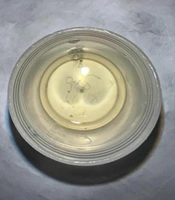
Although high doses of meropenem were administered, the CSF analysis showed no improvement in the yellowish and cloudy appearance of the fluid, nor did it improve the cell counts. The CSF protein concentration was measured at 60 mg/dL, with glucose levels of 66 mg/dL. The India ink staining did not show the presence of Cryptococcus, and the gram staining of the CSF did not show any specific microbial presence. Additionally, there was no indication of tuberculosis, viral pathogens, or fungal growth in the CSF. The CSF analysis revealed the presence of multiple worms, later identified as A. cantonensis, which prompted the administration of albendazole 400 mg twice daily for 21 days (Figure 1). Despite the finding, the patient exhibited no recollection of consuming any uncooked or partially cooked meals during the preceding months.
During the course of treatment, the patient developed pneumonia, critical illness polyneuropathy, dysphagia, insomnia, and organic mental disturbances. To support recovery, the patient underwent bronchial toilet via bronchoscopy, tracheostomy, feeding tube insertion, physical rehabilitation, acupuncture, and supportive psycho-cognitive therapy.
2.3. Follow-up and Outcomes
The patient was discharged from the ICU after a 34-day stay and subsequently admitted to the hospital ward for an additional month. The patient's response to treatment was evaluated through a series of clinical and laboratory parameters. Clinically, the patient's neurological status, including motor function and cranial nerve deficits, was closely monitored for any signs of improvement or deterioration. Laboratory parameters such as inflammatory markers, complete blood count, and metabolic panels were regularly assessed to track systemic response to treatment and identify any complications.
During the outpatient clinic visit at the one-month follow-up, the tracheostomy and feeding tube were removed. An endoscopic evaluation confirmed the resolution of dysphonia and dysphagia. However, hemiparesis and cranial nerve palsy persisted despite treatment.
3. Discussion
The primary objective of intensive care unit (ICU) management is to sustain life and organ function while identifying and treating the underlying disease. We recently treated a patient exhibiting a rare presentation of a parasitic central nervous system infection. The patient had severe back pain that required high doses of sedative-analgesics and thus needed ventilator support to counteract the respiratory depressive effects of the drugs administered. The initial treatment focused on relieving neurological symptoms and managing the patient's pain.
During the first week of the ICU stay, the patient developed several new neurologic symptoms despite treatment, including cranial nerve palsy. Consequently, a lumbar puncture and brain MRI were reconducted. The suspicion of parasitic helminth infection arose due to the new onset of neurological symptoms, CSF eosinophilia, increased CSF opening pressure, and protein levels, which were later confirmed by parasite identification in the CSF. Early treatment is crucial, as A. cantonensis brain infection can be life-threatening (1). There has been a case reported where a larva was directly observed coming out of the spinal needle during lumbar puncture (2). Serology tests such as ELISA or western blot and PCR-based tests are more sensitive and can detect the infection earlier (3, 4). However, these tests were not available at our center and thus were not performed (3, 4). We suspect that the initial lumbar puncture and MRI were inconclusive as the parasite was still in its early larval phase.
The choice of initial empirical treatment comprising meropenem and methylprednisolone was guided by several factors, particularly considering the likelihood of the more common acute bacterial meningitis. Meropenem, a broad-spectrum antibiotic, was selected to provide coverage against a wide range of potential bacterial pathogens commonly associated with meningitis, including Streptococcus pneumoniae,Neisseriameningitidis, and Haemophilus influenzae, among others. This empirical approach aims to promptly target the suspected bacterial etiology while awaiting specific microbiological identification from cerebrospinal fluid cultures, which can take several days.
The adjunctive use of methylprednisolone was initiated to mitigate potential neuroinflammatory responses and reduce the risk of complications associated with bacterial meningitis, such as cerebral edema and intracranial hypertension. Corticosteroids have been shown to improve outcomes in select cases of bacterial meningitis, particularly those with evidence of cerebral inflammation or vasculitis. Therefore, the combination of meropenem and methylprednisolone was deemed appropriate in this clinical scenario to address the suspected infectious etiology and mitigate neurologic sequelae associated with bacterial meningitis. High-dose sedative-analgesics were helpful to supplement steroids and continuous lumbar drainage for managing increased intracranial pressure.
After the confirmed identification of the parasite, antihelminthic albendazole treatment was initiated. Albendazole is the drug of choice for this infection. However, there is a risk that it may aggravate the inflammatory response from antigens released by dead parasites (5). Despite this risk, we administered albendazole as the patient's clinical presentation was deteriorating and she was already on high-dose steroids to suppress inflammation.
Unfortunately, due to prolonged ICU care and mechanical ventilation, the patient developed ventilator-associated pneumonia and critical illness polyneuropathy. Additionally, the patient experienced mood disturbances and sleeping difficulties, which may be attributed to both the central nervous system infection and prolonged use of analgosedatives. To address these issues, a multidisciplinary team involving a neurologist, pulmonologist, otolaryngologist, physical rehabilitation physician, psychiatrist, and acupuncture physician provided comprehensive care.
In April 2024, a literature search was conducted through four electronic databases, namely PubMed, Medline, Scopus, and ProQuest. The search focused on the medical subject headings (MeSH) of "Angiostrongylus cantonensis" and "intensive care unit". The search was restricted by article type and language. A detailed search strategy is presented in Table 1. The preliminary search using the databases produced a total of 43 relevant studies. Removal of duplicates and further assessment of the full-text articles led to the identification of 3 studies that were eligible to be included (6-8). The patient characteristics for all reported cases are summarized in Table 2.
| Search Terms | Limits | Number of Records |
|---|---|---|
| PubMed | Article type: Case reports, language: English | 2 |
| 1. "Angiostrongylus cantonensis" | 1,314 | |
| 2. "Intensive care unit" | 182,174 | |
| 1 AND 2 | 3 | |
| Medline | Article type: Case reports, language: English | 2 |
| 1. "Angiostrongylus cantonensis" | 1,322 | |
| 2. "Intensive care unit" | 195,306 | |
| 1 AND 2 | 3 | |
| Scopus | Document type: Article, language: English | 19 |
| 1. "Angiostrongylus cantonensis" | 4,628 | |
| 2. "Intensive care unit" | 659,813 | |
| 1 AND 2 | 41 | |
| ProQuest | Document type: Article, case study, report, language: English | 20 |
| 1. "Angiostrongylus cantonensis" | 1,526 | |
| 2. "Intensive care unit" | 452,878 | |
| 1 AND 2 | 93 |
Search Strategy
| Case Report | Liu et al. (6) | Feng et al. (7) | Chiong et al. (8) |
|---|---|---|---|
| Published year | 2022 | 2022 | 2019 |
| Country | China | China | Australia |
| Age | 8 | 27 | 36 |
| Sex | Male | Male | Male |
| Clinical presentation | Low-grade fever, paroxysmal headache, mental fatigue, loss of appetite, loss of consciousness | Skin itching, rashes, emesis, myalgia, quadriparesis, fever, generalized flaccid paralysis, gatism, mental aberrations, mild headache, stomachache, papilloedema | Drowsiness, fever, agitation, unsteady gait, urinary incontinence, reduced lateral gaze of the right eye with dysconjugate eye movements, headache |
| Diagnostic test | Complete blood count CSF analysis (cell count, glucose, protein, culture), electroencephalogram (EEG), brain MRI, autoimmune and antibody tests, enzyme-linked immunosorbent assay (ELISA), metagenomic next-generation sequencing (mNGS) | Complete blood count CSF analysis (opening pressure, cell count, glucose, protein, gram stain, fungal stain, cytology), brain CT, mNGS, ELISA, abdominal CT, quantitative polymerase chain reaction (qPCR), brain MRI | Complete blood count renal function tests, liver function tests CSF analysis (opening pressure, cell count, glucose, protein, bacterial culture, India ink stain, Giemsa stain, cryptococcal antigen, flow cytometry, PCR), brain CT, brain MRI, EEG, brain biopsy, chest CT, bronchoscopy |
| Treatment regimens | Acyclovir 250 mg Q8H dexamethasone 0.3 mg/kg vancomycin 0.65 g Q8H meropenem 1.3 g Q8H albendazole 0.2 g BID methylprednisolone 20 - 40 mg QD doxycycline 0.064 g BID | Methylprednisolone 40 - 500 mg QD immunoglobulin 0.4 g/kg QD ceftriaxone linezolid meropenem dexamethasone 10 mg QD albendazole 400 mg BID | Empirical antibacterial and antiviral therapy albendazole 15 mg/kg QD prednisolone 50 mg QD |
| Treatment duration (days) | 57 | 31 | 8 |
| Outcome | On discharge: No fever and no nervous system symptoms GCS score 14 mildly high peripheral blood and CSF WBC levels alight A. cantonensis DNA coverage by CSF mNGS 1-year follow-up: No neurological consequences | Died | On discharge (against medical advice): No residual neurological deficits resolved peripheral eosinophilia 7-month follow-up: No neurological consequences |
Case Report Comparison
The comparison of reported cases is a critical component in advancing our understanding and management of A. cantonensis infection, particularly within the ICU context. Compared to previous case reports, ours was the only case with an initial chief complaint of severe back pain (Table 2). This adds to the variability of neurological symptoms that A. cantonensis can trigger. Despite the fact that all cases eventually received the gold standard albendazole, outcomes varied from complete recovery to death. All cases also administered other antibiotics and/or antiviral treatments, highlighting the challenges in establishing a diagnosis (6-8).
In conclusion, our report emphasized the vital role of ICU management in providing care for patients with a progressively declining condition without an initial definitive diagnosis. Early analgesia and sedation, steroids, antibiotics, mechanical ventilation, and lumbar drainage were central to alleviating symptoms while establishing a diagnosis. Follow-up treatments such as bronchoscopy, tracheostomy, and rehabilitation programs helped the patient achieve a more favorable outcome.

