1. Introduction
Myiasis is the infestation of animal or human tissues and organs, caused by fly larvae. Although the most common sites of infestation are skin wounds, less common sites include the eyes, nose, paranasal sinuses, throat, and urogenital tract (1-3). Myiasis occurs in overcrowded areas with poor hygienic conditions, although it may also occur in hospitals and nursing homes (4). Most cases of myiasis in humans are only temporary, as humans are regarded as unsuitable hosts. In general, this infection occurs mostly in rural areas, where man lives in close contact with animals (5).
Intestinal myiasis in humans is probably an accidental infestation, related to the ingestion of contaminated uncooked food or water containing fly larvae. It can lead to the development of signs and symptoms, similar to those associated with intestinal parasites (6). Most larvae are destroyed by the digestive juice, whereas others can live in the intestinal tract and produce intestinal distress. Moreover, the larvae can exceptionally reach the intestinal tube through the anus (rectal myiasis) (6). Laboratory findings in cases of intestinal myiasis have indicated the presence of dipteran larvae in one or more consecutive stool specimens. In fact, microscopic examination of the stool is diagnostic.
The green bottle fly, Lucilia illustris, is a fly of the Calliphoridae family. The adult fly typically feeds on flowers, while the female needs some sort of carrion protein in order to breed and lay eggs. Due to the predictable nature of its development, L. illustris is often used by forensic entomologists to determine the time and place of death. Medically, L. illustris is often used for maggot debridement therapy, as it only causes myiasis of necrotic tissues and is a rare cause of myiasis in humans (7).
2. Case Presentation
A 45-year-old female, residing in Kurdistan province (west of Iran), presented to Shahid Ghazi hospital (Kurdistan, Iran) with specific abdominal pain and loose stool. The complementary studies, including blood analysis, abdominal X-ray films, and abdominal ultrasonography, were normal. She had always lived in an urban habitat, and there was no suspicion of contact with unclean water or contaminated food ingestion. Fecal samples from the patient, sent to the parasitology laboratory for routine testing, revealed the presence of L. illustris larva.
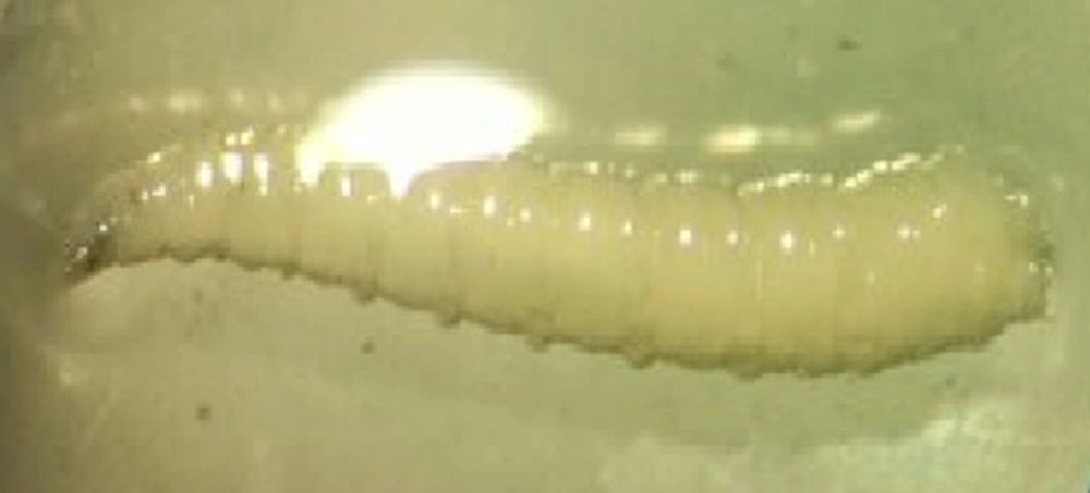
The maggot was removed from the patient's stool and placed in 10% neutral-buffered formalin. A cylindrical vermiform maggot, measuring 9 mm in length and 3 mm in diameter, was observed under the dissecting microscope (Figure 1). The specimen was gently washed in phosphate-buffered saline (pH: 7.4) and cleared in graded solutions of glycerol (up to 80%) (8).
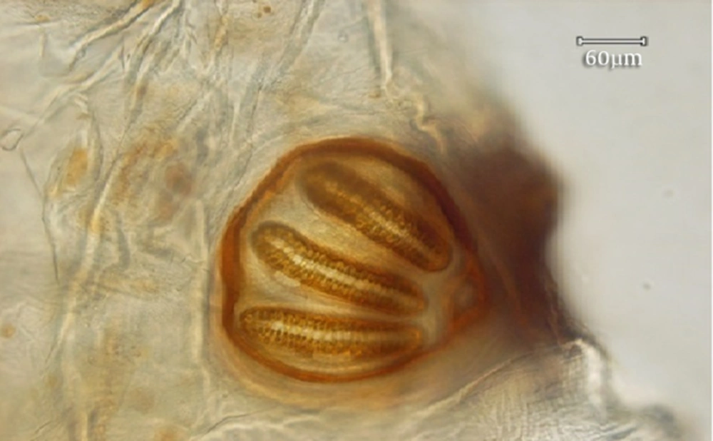
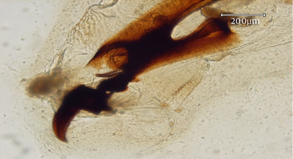
The larva was identified as the third instar of L. illustris (Diptera: Calliphoridae). The body of larva in necrophagous Calliphoridae follows the general pattern of Calyptrata, i.e., three thoracic segments, seven abdominal segments, and an anal division (Figure 1). The third instar of L. illustris was easily distinguishable from other instars by the presence of anterior spiracles (Figure 2). The larva had a pair of strong mandibles for eating food substrates and a cephaloskeleton without a sclerotised area below the posterior tip of the ventral cornua (Figure 3) (9). Following the excretion of larva, the symptoms subsided within a few hours, and the patient remained asymptomatic several weeks later.
3. Discussion
Myiasis is a term used for the infestation of living human or vertebrate animal tissues with larvae of various dipterous flies, which feed on the host’s dead or living tissues, liquid body substances, or ingested food at least for a certain period of time (10). Overall, more than 50 fly species have been reported to cause myiasis in humans (11).
There are few reports of intestinal infestation, especially from developing countries, such as India (10), Africa (12), North America (13), Thailand (14), Japan (15), China (16), Taiwan (17), and Iran (18). Clinical presentation is variable, including abdominal pain, nausea and vomiting, or anal pruritus; also, in some cases, it can be asymptomatic (19).
Some families of myiatic flies (including the most common myiatic flies from Calliphoridae, Sarcophagidae, and Muscidae families) can cause the development of traumatic myiasis in Iran. In fact, members of these families are present and active in various parts of Iran (18). Knowledge of the fauna of medically important flies is useful for determining the risk of fly attacks as myiasis agents. The first well documented case of myiasis in Iran was reported by Minar (1976). Although many researchers have reported a history of throat myiasis, in Iran, myiasis history is not well documented. To the best of our knowledge, the present case is the first report of myiasis in Iran in a human, caused by L. illustris.
As previously mentioned, myiasis of this type has not been previously reported in Iran, and the present case is the first report of human intestinal myiasis, caused by L. illustris in the country. Although this type of myiasis does not cause any significant diseases and has a very low occurrence rate, it is important to report such cases, as patients become concerned when they discover larvae in their stool. Moreover, health professionals should be aware that this infestation may happen regardless of the socioeconomic conditions.
3.1. Conclusions
Although intestinal myiasis has a very low occurrence rate, awareness of intestinal larvae in rural areas, especially during spring and summer, leads to a more prompt diagnosis and application of a specific therapy for the disease.


