Calcium Metabolism Disorders in Patients with COVID-19

authors:

avatar Mojgan Sanjari ORCID 1 , avatar Sedigheh Barzekar ORCID 2 , * , avatar Mostafa Kashani ORCID 2

Endocrinology and Metabolism Research Center, Institute of Basic and Clinical Physiology Sciences, Kerman University of Medical Sciences, Kerman, Iran
Sirjan School of Medical Sciences, Sirjan, Iran

How To Cite Sanjari M, Barzekar S, Kashani M. Calcium Metabolism Disorders in Patients with COVID-19. Arch Clin Infect Dis. 2025;20(1):e148828. https://doi.org/10.5812/archcid-148828.

Abstract

Background:

We aimed to evaluate calcium (Ca) metabolism disorders in patients with COVID-19, a novel virus with numerous unknown aspects and potential complications. This study was conducted due to the scarcity of evidence on this subject and the crucial importance of conducting a comprehensive assessment.

Objectives:

We hypothesized that this research would shed light on this previously unexplored phenomenon.

Methods:

This study was a descriptive cross-sectional study conducted on COVID-19 patients admitted to Afzalipur Kerman Medical Center, Iran, in 2021. Data collection involved demographic characteristics and laboratory results. A 5 cc blood sample was collected in a clot test tube to perform total Ca tests using the photometric method (Arsenazo III kit), ionized Ca tests via the ISE method, magnesium measurements with the photometric method using the Xylidyl blue kit, iPTH (intact parathyroid hormone) assessments, and 25-OH-VITD3 measurements using the ELISA technique with the Monobind kit.

Results:

The present study included 162 participants, comprising 59% males and 41% females, with an average age of 49 years. Among the patients, the most prevalent Ca metabolism disorders were low vitamin D levels, including both deficiency and insufficiency (59%), as well as elevated ionized Ca levels (43%). Conversely, the least common Ca metabolism disorders in these patients were hyperparathyroidism (7%) and excessive Ca adjustment (8%).

Conclusions:

The findings suggest the possibility of Ca metabolism disorders, particularly hypocalcemia, in COVID-19 patients. Given the nature of this study, we recommend conducting longitudinal and more comprehensive research in this field to investigate contributing factors in more detail and establish a cause-and-effect relationship.

1. Background

Calcium (Ca) is one of the key elements responsible for numerous functions in the body (1); its levels can be influenced by parameters such as parathyroid hormone (PTH), vitamin D, phosphorus, and magnesium levels. Any imbalance in these factors alters Ca levels. While hypercalcemia can be associated with increased PTH or vitamin D, conditions such as hypoparathyroidism, nephrosis, and pancreatitis can induce hypocalcemia (2). Moreover, vitamin D, as a fat-soluble vitamin, commonly enhances the absorption of Ca, phosphate, and magnesium, which are essential for the body’s functions (3).

The outbreak of COVID-19 was first identified in 2019 in Wuhan, China (4). Although fever, cough, and fatigue are the primary clinical manifestations, previous investigations have indicated the importance of assessing endocrine parameters in these patients (5-8). It has been noted that this novel virus is associated with abnormalities in various vitamins, electrolytes, and trace elements (9-11). In patients with COVID-19, hypocalcemia is one of the significant biochemical abnormalities and a common problem that may be linked to poor prognosis (12). Measuring ionized Ca levels in these patients revealed hypocalcemia in 82% of cases (13). Earlier studies have shown an association between vitamin D deficiency and viral conditions (14, 15). It has been suggested that vitamin D deficiency may affect infection and disease expression and that it plays a fundamental role in regulating the COVID-19 endocrine phenotype (16). Furthermore, vitamin D has a significant physiological role in maintaining appropriate immunity, reducing pro-inflammatory factors, and boosting anti-inflammatory cytokine production, which helps patients with COVID-19 (17). Despite speculation that COVID-19 might cause hypoparathyroidism (18), magnesium deficiency is widely considered to mimic the risk factors for its occurrence (18, 19).

Although the effect of viral infections on Ca metabolism is not a new concept, previous investigations focused on viruses such as SARS and Ebola during past outbreaks, highlighting hypocalcemia as a predominant biochemical abnormality (20, 21). It was even noted that 75% of patients affected by the Ebola virus had hypocalcemia during hospitalization (21).

2. Objectives

The present study, however, aims to assess Ca metabolism disorders in patients with COVID-19, a novel virus with distinct unknown aspects and complications. This study was conducted due to the scarcity of evidence on this issue and the importance of a thorough assessment. We hypothesize that our findings will shed light on this unexplored phenomenon.

3. Methods

3.1. Patients and Settings

This descriptive cross-sectional study was conducted in 2021 at Afzalipur Kerman Medical Center in Iran, focusing on patients hospitalized with COVID-19. The inclusion criteria consisted of individuals aged 18 to 60 years with a confirmed diagnosis of COVID-19 based on a positive PCR test, provided their clinical symptoms had manifested within the last 14 days. The exclusion criteria included a history of parathyroid diseases, chronic kidney disease, malignancies, known chronic medical conditions (such as cardiovascular, pulmonary, metabolic, rheumatological, hematological, neurological, or psychological disorders), pregnancy, breastfeeding, recent consumption of Ca and vitamin D supplements within the past month, and lack of consent to participate in the study.

In 2021, a total of 280 COVID-19 patients with positive test results were hospitalized at Afzalipur Hospital in Kerman. Using Cochran's sampling formula with an error level of d = 0.05, 162 of these patients were randomly selected to participate in this study.

3.2. Data Gathering

Data were collected in a format that included demographic characteristics and laboratory results.

3.3. Laboratory Tests

A 5 cc blood sample was obtained and collected in a clot test tube for the purpose of conducting various tests, including total Ca measurements using the photometric method with the Arsenazo III kit, ionized Ca assessments via the ISE method, magnesium level determination using the Xylidyl blue kit, intact parathyroid hormone (iPTH) analysis, and 25-OH-VITD3 measurement using the ELISA technique with the Monobind kit. The serum sample was carefully separated at the Afzalipur Hospital laboratory in Kerman and stored at -70°C. Subsequently, the sample was sent to the laboratory at Mehrgan Hospital in Kerman for further analysis.

The normal range is as follows:

Total calcium = 6.8 - 5.10 mg/dL

Ionized calcium = 1.12 - 1.32 mmol/L

Albumin = 5.3 - 2.5 g/dL

Magnesium = 8.1 - 6.2 mg/dL

iPTH = 15 - 65 pg/mL

25OHvitD ≤ 10 ng/mL: Deficiency

11 - 29 ng/mL: Insufficiency

30 ≤ ng/mL: Sufficiency

3.4. Ethics Approval and Consent to Participate

A written informed consent form was obtained from each patient, and they consented to participate in this study. This investigation was approved by the ethics committee of Kerman University of Medical Sciences (Code: IR.KMU.AH.REC.1400.216, Date: 2021-12-05). All experiments were conducted in accordance with the relevant guidelines and regulations of the Declaration of Helsinki.

3.5. Statistical Analysis

Data were gathered and reported as mean, standard deviation, number, and percentage using IBM SPSS version 21 software (SPSS Inc., Chicago, IL).

4. Results

The present study was conducted on 162 patients, comprising 59% males and 41% females, with a mean age of 49 years (Table 1).

Table 1.

Demographic Characteristics of Patients

VariablesNo. (%)
Gender
Female 67 (41)
Male 95 (59)
Age groups (y)
< 20 3 (2)
21 - 30 11 (7)
31 - 40 20 (12)
41 - 50 26 (16)
51 - 60 100 (62)
> 60 2 (1)
Underlying disease
High blood pressure 27 (17)
Diabetes 43 (27)

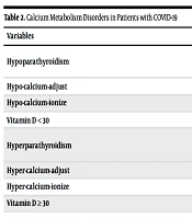
As indicated in Table 2, the most common Ca metabolism disorders in patients were low vitamin D, including deficiency and insufficiency (59%), hypercalcemia (43%), hypocalcemia (40%), vitamin D insufficiency (38%), hypomagnesemia (37%), and Ca adjustment abnormalities (35%). Moreover, the lowest prevalence of Ca metabolism disorders in these patients was hyperparathyroidism (7%) and Ca adjustment abnormalities (8%).

Table 2.

Calcium Metabolism Disorders in Patients with COVID-19

VariablesNo. (%)Confidence Interval
Hypoparathyroidism40 (25)
Ca7.3 (7.1 - 7.4)
iPTH26.4 (20 - 33)
Hypo-calcium-adjust56 (35)7.60 (7.5 - 7.7)
Hypo-calcium-ionize65 (40)0.92 (0.89 - 0.95)
Vitamin D < 3097 (59)49 (46 - 51.8)
Hyperparathyroidism12 (7)
Ca11.2 (10.5 - 11.8)
iPTH42 (20 - 64)
Hyper-calcium-adjust13 (8)11.6 (10.9 - 12.4)
Hyper-calcium-ionize69 (43)1.9 (1.7 - 2)
Vitamin D ≥ 3065 (40)49 (46 - 51.8)

5. Discussion

This research aimed to assess the prevalence of Ca metabolism disorders among patients hospitalized with COVID-19 at Afzalipur Kerman Medical Center. In this investigation, the most frequently observed Ca metabolism disorders among patients included low levels of vitamin D, encompassing both deficiency and insufficiency (59%), elevated ionized Ca (43%), diminished ionized Ca (40%), vitamin D insufficiency (38%), hypomagnesemia (37%), and reduced Ca adjustment (35%). Conversely, the least commonly found Ca metabolism disorders in these patients were hyperparathyroidism (7%) and excessive Ca adjustment (8%).

Depending on the definition of hypocalcemia, numerous studies have established a surprisingly high incidence of hypocalcemia, ranging from 62.6% to 87.2%. Studies that used total serum Ca levels to define hypocalcemia, such as 2.2 mmol/L (8.8 mg/dL), 2.15 mmol/L (8.6 mg/dL), or 2.12 mmol/L (8.5 mg/dL), observed hypocalcemia prevalence ranging from 62.6% to 74.7% (22-25). However, a higher prevalence (> 80%) was noted by those who defined it based on ionized serum Ca levels (12, 13, 26, 27). Although we found a consistent trend for hypocalcemia based on both total and ionized levels, our study showed lower levels of hypocalcemia (35% vs. 43%, respectively), which may be attributed to different sample sizes, measurement methods, and inclusion criteria for patients. Notably, Cappellini et al. observed lower total and ionized Ca levels in patients with positive nasopharyngeal swabs compared to patients admitted to emergency departments with similar signs and symptoms but negative nasopharyngeal tests (26). Although we included only patients with positive PCR results, and based on the above-mentioned investigation, higher hypocalcemia may have been expected, the findings still underscore the importance of measuring these parameters in patients with COVID-19.

The present work indicated that 59% of patients had vitamin D levels < 30, a high prevalence that has been confirmed by other similar investigations (22-25, 27-31). It is noteworthy that chronic hypovitaminosis D alters Ca metabolism and reduces the absorption of Ca and phosphorus from the intestinal tract. Additionally, COVID-19 may exacerbate hypocalcemia, particularly in those with pre-existing vitamin D deficiency and severe infections (29). However, there is ongoing debate regarding the treatment of vitamin D deficiency in patients with COVID-19 (32-35). The results of a systematic review and meta-analysis demonstrated that vitamin D supplementation decreased the risk of acute respiratory infections, primarily in individuals treated with daily or weekly doses, but not with bolus doses (36). In a single-center study, 36.7% and 86.7% of COVID-19 patients had hypocalcemia (Caadj < 2.15 mmol/L) and vitamin D deficiency (< 20 ng/mL), respectively. The study noted that these abnormalities were more common in patients with severe infections who required hospitalization, and they recommended further research to determine the effect of these impairments on improving treatment strategies (37). di Filippo et al., who retrospectively assessed 118 patients hospitalized for COVID-19, reported that 76.6% had hypocalcemia (total serum Ca < 2.2 mmol/L), with only 6.7% exhibiting hypervitaminosis D (> 30 ng/mL) (13). Moreover, Bossoni et al. reported a case of a hospitalized 72-year-old female with a positive nasopharyngeal swab for COVID-19. Her chief complaints for hospitalization were mild fever, headache, dysarthria, and perioral paresthesia. The patient was found to have total and ionized hypocalcemia, hyperphosphatemia, and hypoparathyroidism (38).

Although there are limited case reports on the effect of COVID-19 on the parathyroid gland, our results indicated that 25% of patients had hypoparathyroidism. A case report has presented COVID-19 as the cause of hypoparathyroidism in a 46-year-old male admitted for hypoxemia secondary to this viral infection, after excluding other known causes of hypoparathyroidism, such as genetic factors or malignancies. Due to the nature of their report, only limited conclusions could be drawn, but they emphasized the need for further studies to assess the link between COVID-19 and parathyroid dysfunction (39). Additionally, other case reports have noted this relationship (40, 41). These limited case reports, along with our substantial results, underscore the importance of a thorough understanding of the mechanisms through which COVID-19 may trigger parathyroid dysfunctions.

While normal magnesium levels exert a protective effect against viral infections, magnesium deficiency can be linked to these infections. Hypomagnesemia reduces the cytotoxicity of NK and T-cells, increases NF-κB expression, and promotes proinflammatory activities via the upregulation of cytokine production in monocytes (42). The results of the present investigation indicated that 60 patients (37% of all) had hypomagnesemia. Similarly, previous studies have highlighted the critical role of hypomagnesemia in the severe outcomes of COVID-19 infection. Quilliot et al., who assessed serum magnesium levels in 300 patients upon admission, demonstrated that 48% had hypomagnesemia. Furthermore, Guerrero-Romero et al. reported hypomagnesemia in 44.1% of patients (43, 44).

The first limitation of the present study is the absence of a control group. Further research can enhance the generalizability of the findings by addressing this limitation. Another limitation of this study is its single-center design, which may affect the results. This factor could not be controlled by the researchers, and thus it is recommended that multi-center studies be conducted to eliminate other confounding factors. Due to socio-economic differences across the country, similar research should be conducted in other regions of Iran, and the obtained results should be compared.

5.1. Conclusions

The results of this research indicated that there is a possibility of Ca metabolism disorders, especially hypocalcemia, in patients with COVID-19. Since this study was cross-sectional, it is recommended to conduct longitudinal and more comprehensive studies in this field for a more detailed investigation of the contributing factors and to establish a cause-and-effect relationship.
References

  • 1.

    Wawrzyniak N, Suliburska J. Nutritional and health factors affecting the bioavailability of calcium: a narrative review. Nutr Rev. 2021;79(12):1307-20. [PubMed ID: 33491051]. https://doi.org/10.1093/nutrit/nuaa138.

  • 2.

    Endres DB. Tietz Textbook of Clinical Chemistry and Molecular Diagnostics. Philadelphia: Elsevier Health Sciences; 2006. p. 1891-924.

  • 3.

    Luciani F, Caroleo MC, Cannataro R, Mirra D, D’Agostino B, Gallelli L, et al. Immunological Response to SARS-CoV-2 Is Sustained by Vitamin D: A Case Presentation of One-Year Follow-Up. Reports. 2021;4(2). https://doi.org/10.3390/reports4020018.

  • 4.

    Guan WJ, Ni ZY, Hu Y, Liang WH, Ou CQ, He JX, et al. Clinical Characteristics of Coronavirus Disease 2019 in China. N Engl J Med. 2020;382(18):1708-20. [PubMed ID: 32109013]. [PubMed Central ID: PMC7092819]. https://doi.org/10.1056/NEJMoa2002032.

  • 5.

    Harapan H, Itoh N, Yufika A, Winardi W, Keam S, Te H, et al. Coronavirus disease 2019 (COVID-19): A literature review. J Infect Public Health. 2020;13(5):667-73. [PubMed ID: 32340833]. [PubMed Central ID: PMC7142680]. https://doi.org/10.1016/j.jiph.2020.03.019.

  • 6.

    Zhang C, Wu Z, Li JW, Zhao H, Wang GQ. Cytokine release syndrome in severe COVID-19: interleukin-6 receptor antagonist tocilizumab may be the key to reduce mortality. Int J Antimicrob Agents. 2020;55(5):105954. [PubMed ID: 32234467]. [PubMed Central ID: PMC7118634]. https://doi.org/10.1016/j.ijantimicag.2020.105954.

  • 7.

    Ponti G, Maccaferri M, Ruini C, Tomasi A, Ozben T. Biomarkers associated with COVID-19 disease progression. Crit Rev Clin Lab Sci. 2020;57(6):389-99. [PubMed ID: 32503382]. [PubMed Central ID: PMC7284147]. https://doi.org/10.1080/10408363.2020.1770685.

  • 8.

    Hu B, Guo H, Zhou P, Shi ZL. Characteristics of SARS-CoV-2 and COVID-19. Nat Rev Microbiol. 2021;19(3):141-54. [PubMed ID: 33024307]. [PubMed Central ID: PMC7537588]. https://doi.org/10.1038/s41579-020-00459-7.

  • 9.

    Lippi G, South AM, Henry BM. Electrolyte imbalances in patients with severe coronavirus disease 2019 (COVID-19). Ann Clin Biochem. 2020;57(3):262-5. [PubMed ID: 32266828]. [PubMed Central ID: PMC8173320]. https://doi.org/10.1177/0004563220922255.

  • 10.

    Mercola J, Grant WB, Wagner CL. Evidence Regarding Vitamin D and Risk of COVID-19 and Its Severity. Nutr J. 2020;12(11). [PubMed ID: 33142828]. [PubMed Central ID: PMC7692080]. https://doi.org/10.3390/nu12113361.

  • 11.

    Fakhrolmobasheri M, Mazaheri-Tehrani S, Kieliszek M, Zeinalian M, Abbasi M, Karimi F, et al. COVID-19 and Selenium Deficiency: a Systematic Review. Biol Trace Elem Res. 2022;200(9):3945-56. [PubMed ID: 34739678]. [PubMed Central ID: PMC8569840]. https://doi.org/10.1007/s12011-021-02997-4.

  • 12.

    di Filippo L, Formenti AM, Doga M, Frara S, Rovere-Querini P, Bosi E, et al. Hypocalcemia is a distinctive biochemical feature of hospitalized COVID-19 patients. Endocrin J. 2021;71(1):9-13. [PubMed ID: 33165763]. [PubMed Central ID: PMC7649576]. https://doi.org/10.1007/s12020-020-02541-9.

  • 13.

    di Filippo L, Doga M, Frara S, Giustina A. Hypocalcemia in COVID-19: Prevalence, clinical significance and therapeutic implications. Rev Endocr Metab Disord. 2022;23(2):299-308. [PubMed ID: 33846867]. [PubMed Central ID: PMC8041474]. https://doi.org/10.1007/s11154-021-09655-z.

  • 14.

    Holick MF. High prevalence of vitamin D inadequacy and implications for health. Mayo Clin Proc. 2006;81(3):353-73. [PubMed ID: 16529140]. https://doi.org/10.4065/81.3.353.

  • 15.

    Ginde AA, Mansbach JM, Camargo CA. Association between serum 25-hydroxyvitamin D level and upper respiratory tract infection in the Third National Health and Nutrition Examination Survey. Arch Intern Med. 2009;169(4):384-90. [PubMed ID: 19237723]. [PubMed Central ID: PMC3447082]. https://doi.org/10.1001/archinternmed.2008.560.

  • 16.

    Puig-Domingo M, Marazuela M, Giustina A. COVID-19 and endocrine diseases. A statement from the European Society of Endocrinology. Endocrin J. 2020;68(1):2-5. [PubMed ID: 32279224]. [PubMed Central ID: PMC7150529]. https://doi.org/10.1007/s12020-020-02294-5.

  • 17.

    Gallelli L, Mannino GC, Luciani F, de Sire A, Mancuso E, Gangemi P, et al. Vitamin D Serum Levels in Subjects Tested for SARS-CoV-2: What Are the Differences among Acute, Healed, and Negative COVID-19 Patients? A Multicenter Real-Practice Study. Nutr J. 2021;13(11). [PubMed ID: 34836187]. [PubMed Central ID: PMC8625490]. https://doi.org/10.3390/nu13113932.

  • 18.

    Wolf FI, Maier JA, Rosanoff A, Barbagallo M, Baniasadi S, Castiglioni S, et al. The magnesium global network (MaGNet) to promote research on magnesium in diseases focusing on covid-19. Magnes Res J. 2021;34(2):90-2. [PubMed ID: 34524085]. [PubMed Central ID: PMC10617598]. https://doi.org/10.1684/mrh.2021.0479.

  • 19.

    Iotti S, Wolf F, Mazur A, Maier JA. The COVID-19 pandemic: is there a role for magnesium? Hypotheses and perspectives. Magnes Res J. 2020;33(2):21-7. [PubMed ID: 32554340]. https://doi.org/10.1684/mrh.2020.0465.

  • 20.

    Booth CM, Matukas LM, Tomlinson GA, Rachlis AR, Rose DB, Dwosh HA, et al. Clinical features and short-term outcomes of 144 patients with SARS in the greater Toronto area. JAMA. 2003;289(21):2801-9. [PubMed ID: 12734147]. https://doi.org/10.1001/jama.289.21.JOC30885.

  • 21.

    Uyeki TM, Mehta AK, Davey RJ, Liddell AM, Wolf T, Vetter P, et al. Clinical Management of Ebola Virus Disease in the United States and Europe. N Engl J Med. 2016;374(7):636-46. [PubMed ID: 26886522]. [PubMed Central ID: PMC4972324]. https://doi.org/10.1056/NEJMoa1504874.

  • 22.

    Bennouar S, Cherif AB, Kessira A, Bennouar DE, Abdi S. Vitamin D Deficiency and Low Serum Calcium as Predictors of Poor Prognosis in Patients with Severe COVID-19. J Am Coll Nutr. 2021;40(2):104-10. [PubMed ID: 33434117]. [PubMed Central ID: PMC7814570]. https://doi.org/10.1080/07315724.2020.1856013.

  • 23.

    Hernández JL, Nan D, Fernandez-Ayala M, García-Unzueta M, Hernández-Hernández MA, López-Hoyos M, et al. Vitamin D Status in Hospitalized Patients with SARS-CoV-2 Infection. J Clinic Endocrinol Metab. 2021;106(3):e1343-53. https://doi.org/10.1210/clinem/dgaa733.

  • 24.

    Linli Z, Chen Y, Guo S. Identifying and quantifying robust risk factors for mortality in critically ill patients with COVID-19 using quantile regression. Am J Emerg Med. 2020;20. https://doi.org/10.21203/rs.3.rs-28676/v1.

  • 25.

    Liu J, Han P, Wu J, Gong J, Tian D. Prevalence and predictive value of hypocalcemia in severe COVID-19 patients. J Infect Public Health. 2020;13(9):1224-8. [PubMed ID: 32622796]. [PubMed Central ID: PMC7306733]. https://doi.org/10.1016/j.jiph.2020.05.029.

  • 26.

    Cappellini F, Brivio R, Casati M, Cavallero A, Contro E, Brambilla P. Low levels of total and ionized calcium in blood of COVID-19 patients. Clin Chem Lab Med. 2020;58(9):e171-3. [PubMed ID: 32459190]. https://doi.org/10.1515/cclm-2020-0611.

  • 27.

    Elezagic D, Johannis W, Burst V, Klein F, Streichert T. Venous blood gas analysis in patients with COVID-19 symptoms in the early assessment of virus positivity. De Gruyter. 2020;45(1):27-30. https://doi.org/10.1515/labmed-2020-0126.

  • 28.

    Pal R, Ram S, Zohmangaihi D, Biswas I, Suri V, Yaddanapudi LN, et al. High Prevalence of Hypocalcemia in Non-severe COVID-19 Patients: A Retrospective Case-Control Study. Front Med (Lausanne). 2020;7:590805. [PubMed ID: 33490095]. [PubMed Central ID: PMC7817940]. https://doi.org/10.3389/fmed.2020.590805.

  • 29.

    Sun JK, Zhang WH, Zou L, Liu Y, Li JJ, Kan XH, et al. Serum calcium as a biomarker of clinical severity and prognosis in patients with coronavirus disease 2019. Aging J (Albany NY). 2020;12(12):11287-95. [PubMed ID: 32589164]. [PubMed Central ID: PMC7343468]. https://doi.org/10.18632/aging.103526.

  • 30.

    Tezcan ME, Dogan Gokce G, Sen N, Zorlutuna Kaymak N, Ozer RS. Baseline electrolyte abnormalities would be related to poor prognosis in hospitalized coronavirus disease 2019 patients. New Microbes New Infect. 2020;37:100753. [PubMed ID: 32904987]. [PubMed Central ID: PMC7462442]. https://doi.org/10.1016/j.nmni.2020.100753.

  • 31.

    Torres B, Alcubilla P, Gonzalez-Cordon A, Inciarte A, Chumbita M, Cardozo C, et al. Impact of low serum calcium at hospital admission on SARS-CoV-2 infection outcome. Int J Infect Dis. 2020;104:164-8. [PubMed ID: 33278624]. [PubMed Central ID: PMC7709580]. https://doi.org/10.1016/j.ijid.2020.11.207.

  • 32.

    Bouillon R, Marcocci C, Carmeliet G, Bikle D, White JH, Dawson-Hughes B, et al. Skeletal and Extraskeletal Actions of Vitamin D: Current Evidence and Outstanding Questions. Endocr Rev. 2019;40(4):1109-51. [PubMed ID: 30321335]. [PubMed Central ID: PMC6626501]. https://doi.org/10.1210/er.2018-00126.

  • 33.

    Giustina A, Adler RA, Binkley N, Bollerslev J, Bouillon R, Dawson-Hughes B, et al. Consensus statement from 2(nd) International Conference on Controversies in Vitamin D. Rev Endocr Metab Disord. 2020;21(1):89-116. [PubMed ID: 32180081]. [PubMed Central ID: PMC7113202]. https://doi.org/10.1007/s11154-019-09532-w.

  • 34.

    Giustina A, Bouillon R, Binkley N, Sempos C, Adler RA, Bollerslev J, et al. Controversies in Vitamin D: A Statement From the Third International Conference. J Bone Mineral Res Plus. 2020;4(12). e10417. [PubMed ID: 33354643]. [PubMed Central ID: PMC7745884]. https://doi.org/10.1002/jbm4.10417.

  • 35.

    Yamshchikov AV, Desai NS, Blumberg HM, Ziegler TR, Tangpricha V. Vitamin D for treatment and prevention of infectious diseases: a systematic review of randomized controlled trials. Endocr Pract. 2009;15(5):438-49. [PubMed ID: 19491064]. [PubMed Central ID: PMC2855046]. https://doi.org/10.4158/EP09101.ORR.

  • 36.

    Martineau AR, Jolliffe DA, Hooper RL, Greenberg L, Aloia JF, Bergman P, et al. Vitamin D supplementation to prevent acute respiratory tract infections: systematic review and meta-analysis of individual participant data. BMJ. 2017;356. i6583. [PubMed ID: 28202713]. [PubMed Central ID: PMC5310969]. https://doi.org/10.1136/bmj.i6583.

  • 37.

    Maganeva I, Eremkina A, Bibik E, Gorbacheva A, Kovaleva E, Aynetdinova A, et al. COVID-19 and impaired calcium metabolism upon admission to the hospital. Endocrin Abstract J. 2021;73. https://doi.org/10.1530/endoabs.73.AEP128.

  • 38.

    Bossoni S, Chiesa L, Giustina A. Severe hypocalcemia in a thyroidectomized woman with Covid-19 infection. Endocrin J. 2020;68(2):253-4. [PubMed ID: 32346814]. [PubMed Central ID: PMC7188455]. https://doi.org/10.1007/s12020-020-02326-0.

  • 39.

    Elkattawy S, Alyacoub R, Ayad S, Pandya M, Eckman A. A Novel Case of Hypoparathyroidism Secondary to SARS-CoV-2 Infection. Cureus. 2020;12. https://doi.org/10.7759/cureus.10097.

  • 40.

    Dianatfar M, Sanjari M, Dalfardi B. Hypoparathyroidism After COVID-19 Pneumonia. Shiraz E-Med J. 2021;22(12). https://doi.org/10.5812/semj.115832.

  • 41.

    Georgakopoulou VE, Avramopoulos P, Papalexis P, Bitsani A, Damaskos C, Garmpi A, et al. COVID-19 induced hypoparathyroidism: A case report. Exp Ther Med. 2022;23(5):346. [PubMed ID: 35401797]. [PubMed Central ID: PMC8988155]. https://doi.org/10.3892/etm.2022.11276.

  • 42.

    Weglicki WB. Hypomagnesemia and inflammation: clinical and basic aspects. Annu Rev Nutr. 2012;32:55-71. [PubMed ID: 22404119]. https://doi.org/10.1146/annurev-nutr-071811-150656.

  • 43.

    Quilliot D, Bonsack O, Jaussaud R, Mazur A. Dysmagnesemia in Covid-19 cohort patients: prevalence and associated factors. Magnes Res. 2020;33(4):114-22. [PubMed ID: 33678604]. https://doi.org/10.1684/mrh.2021.0476.

  • 44.

    Guerrero-Romero F, Mercado M, Rodriguez-Moran M, Ramirez-Renteria C, Martinez-Aguilar G, Marrero-Rodriguez D, et al. Magnesium-to-Calcium Ratio and Mortality from COVID-19. Nutr J. 2022;14(9). [PubMed ID: 35565654]. [PubMed Central ID: PMC9101802]. https://doi.org/10.3390/nu14091686.