Dear Editor,
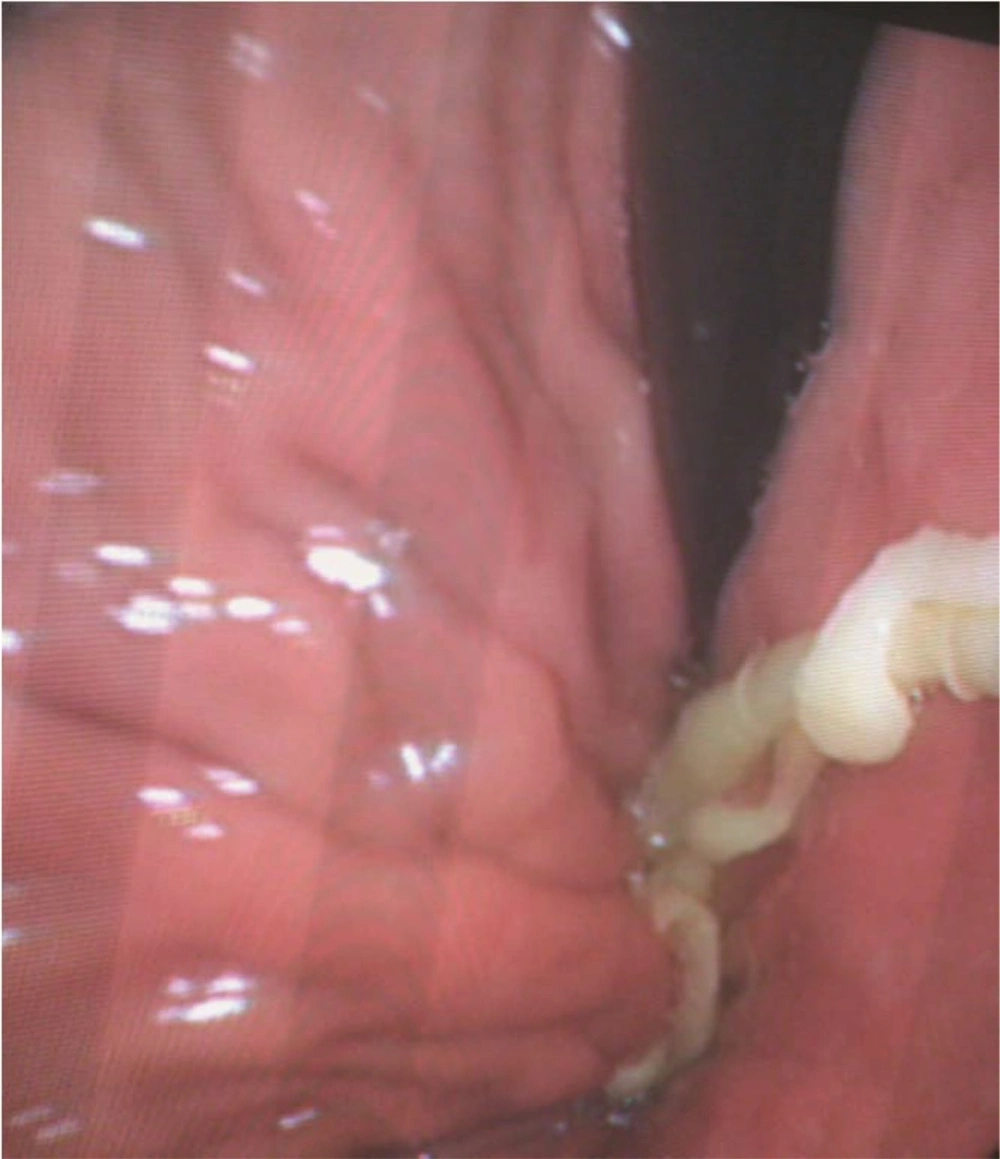
A fifty-four-year-old woman was admitted to emergency department with a two-week history of dyspepsia, abdominal pain, and nausea. For two months she had experienced intermittent episodes of dyspepsia, nausea and vomiting. Her abdominal pain was described as epigastric, non-radiating, worse after eating, and not relieved by defecation. She never had jaundice, hematemesis, melena, hematochezia, or tenesmus. During general physical examination, her vital signs were stable. No oropharyngeal lesion was observed. The respiratory and cardiovascular examinations were unremarkable. Her abdomen was soft and not tender in epigastrium. No free fluid or organomegaly was appreciated. No peritoneal signs were elicited. Her digital rectal exam was also inconspicuous. Given routine lab tests, the patient was found to be anemic with borderline microcytosis. Her white blood cell and differential count were normal. Her urinalysis did not show any sign of urinary tract infection. Amylase and lipase were within normal limits. Transaminases were normal. An abdominal X-ray was taken and showed no evidence of obstruction or perforation. Stool analysis (e g, stool cultures, ova and parasite examination) was negative. The patient later underwent esophagogastroduodenoscopy, as shown in the image which is taken from her stomach (Figure 1).
Humans are the only definitive hosts for Taenia saginata (T. saginata). Eggs or gravid proglottids are passed through human stool. Cattles become infected by ingesting vegetation, contaminated with eggs or gravid proglottids. In animal’s intestine, the oncospheres hatch, invade the intestinal wall, and migrate hematogenously to striated muscles, where they develop in cysticerci. Humans become infected by ingesting raw or undercooked infected meat containing cysticerci. In human intestine, protoscolices are released from the cysts and attached to the intestinal wall via suckers and hooks (1). The prevalence of taeniasis in Iran has been disregarded in recent years - estimated to be about 0.5% in Mazandaran province - where it appears to have the highest prevalence rate (2). Tapeworm infections are usually asymptomatic, however mild abdominal pain or discomfort, nausea, change in appetite, weakness, and weight loss can also occur (3). It is usually diagnosed by detection of the eggs or proglottids in stool. Only one other report of taeniasis diagnosed on upper gastrointestinal endoscopy was found (4). A single dose of Praziquantel (10 mg/kg) is highly effective for the treatment of taeniasis. The patient was prescribed Praziquantel 600 mg and a month later at the follow-up visit, she was reported to have no symptoms.
