1. Introduction
The distribution of tuberculosis is not uniform across the globe; about 80% of the population in many Asian and African countries prove to have positive tuberculin tests, while only 5 - 10% of the US population test positive for tuberculin (1). Ocular complications of TB are rare, estimated at 1.4% among the patients referred to eye clinics in Boston sanatorium (2). This number might vary in different regions due to variable prevalence of TB worldwide. It is of high importance to point out that not only TB may develop with no sign of systemic disease but also most of the patients have no history of TB and the chest radiography for half of them appears normal. Additionally, eye pathology is also rarely advised because of visual loss risk of biopsies. Hence, the diagnosis would be almost presumptive (3).
2. Case Presentation
The patient was a fifty-six-year-old retired teacher who has experienced gradual visual loss for six years and blurry vision, non-productive coughs for months in the time of the study. In addition, she had lost 3 to 4 Kg for two weeks before the admission and 38 to 39 Kg for seven years. Besides, she suffered from anorexia and insomnia. She had severe pain in her eyes while looking at something and purulent discharge of medial canthus of both eyes existed due to conjnctivitis. Notably, she had the habit of smoking four packs of cigarettes per day for years. Her visual acuity estimated light perception in right and count finger at 1.5 m in left eye and her eyes were painful on movements. She had one palpable lymph-node, 1 cm x 1 cm, non-mobile and non-tender in left submandibular space and her lungs were normal and clear on ascultation. The paraclinical results are available in Table 1.
aAbbreviations: CBC, complete blood count; WBC, white blood cells; RBC, red blood cells; Hb, hemoglobin; Hct, hematocrit; Plt, plaquetas; ESR, erythrocyte sedimentation rate; CRP, C-reactive protein
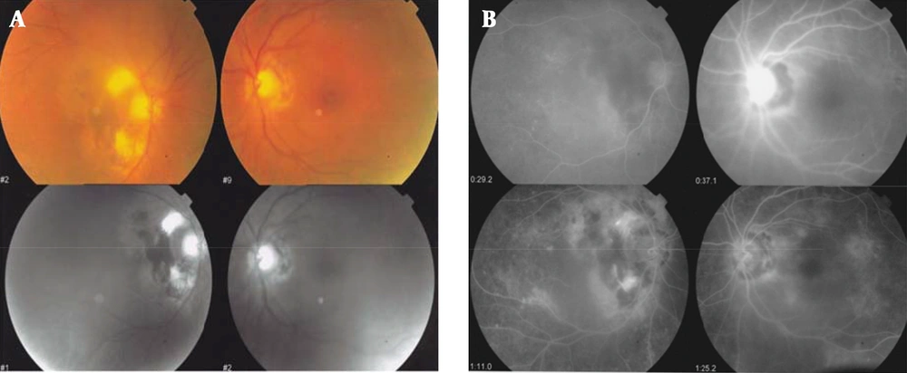
The indurations of PPD test was 20 mm. spiral CT-scan without contrast showed multiple nodules in both lungs majority of which were sub-pleural, suggestive for secondary TB. Being suspected of ocular TB, ophthalmology consultation was recommended. Her BCVA of right eye had light perception and the left eye is 0.5 m count finger (0.5 mfc). Slit lamp examination showed 2 + cell and flair in anterior chamber with granulomatose keratit percipitances (kps). Mild cataract was observed in both eyes. In fundal examination, choroidal granuloma with peri-papillary pigmentation was seen in right eye, suggesting TB induced granuloma and multiple peri-papillary scars was also seen in left one (Figure 1).
Systemic anti-tuberculosis therapy was begun immediately (with INH 300 mg + RIF 600 mg + PZA 1000 mg + ETP 800 mg per day). The patient was discharged with final diagnosis of ocular TB (Choroiditis). She was referred to ophthalmologist for angiography and was advised to pursue medical follow-up. The diagnosis was confirmed by response to therapy and patient completed anti TB medication. BCVA at right eye was improved to 0.5 mfc and the left one to 6 mfc. Reaction of Ac resolved after six months of anti TB therapy, but KPS deteriorated. Cataract operation was performed 3 months after anti TB therapy, but BCVA had no significant improvement after cataract operation.
3. Conclusions
Ocular TB was described first in 1711 by Maitre-Jan (4) and it was in 1855 that Eduard von Jaeger first detected the Choroidal tubercles (5) which appeared to be similar microscopically to tubercles elsewhere in the body by Cohnheim in 1867 (6). In 1882, Julius van Michel identified tubercle bacillus in the eye (7). Mechanisms which may cause eye infection by TB are hematogenous spread as the most common form of ocular involvement, primary exogenous infection when the involved part is conjunctiva or eye lid, direct extension, and hypersensitivity reaction which is assumed to be responsible for Phlyctenular or Eales diseases (8-10).
TB can invade one eye or both in any part, but focal or multifocal choroiditis is the most frequent form, estimated at 78% and 94% (11, 12). Differential diagnosis of TB choroiditis based on lesion appearance includes Sarcoidosis, Syphilis and rarely metastatic diseases (13). Chronic anterior uveitis, usually granulomatous and retinal vasculitis or periphlebitis, are the second and third common manifestations. Other manifestations include scleritis, interstitial keratitis and optic neuritis (14). Eales` disease, which is mostly common in patients from India, Pakistan and Afghanistan typically, manifests itself with vascular sheathing of retinal veins (periphlebitis), vitreous inflammation and peripheral retinal capillary occlusion. Noteworthily, the last one leads to neovascularization and subsequent retinal hemorrhage (14). Before the discovery of Polymerase Chain Reactions (PCR) technology, the majority of cases were considered to have "presumed ocular TB”, because (15):
- Biopsies and culture are practical in cases of lid involvement, keratitis or Orbital tuberculosis although the majority of cases present with intraocular involvement.
- Aqueous and vitreous paracentesis generally fails to result positive bacterial cultures (16).
- Isolating the microorganism is difficult technically.
- The features are similar to other granulomatous inflammations of eye.
- And the ocular manifestations of TB are extremely variable.
The initial evaluation of suspected patient (which the ocular involvement usually accompanies with systemic disease) requires the complete history including exposure, physical examination, sputum smear and culture, PPD test, chest X-radiography and ruling out the other causes for granulomatous inflammations (15). Recently, PCR and ELISA play important roles in detection of mycobacterial DNA or antigens in ocular tissues including eyelid skin, conjunctiva, aqueous and vitreous with unproven specificity and sensitivity (17-24). The main treatment of ocular TB is the same systemic anti-Tuberculosis therapy which can lead to remission of inflammation and improvement in visual acuity to nearly premorbid levels. Topical ointment and subconjunctival injection can contribute to adequate intraocular drug levels especially in anterior segments. Furthermore, parenteral administration is the method of choice in posterior intraocular TB which leads to higher vitreous drug levels (15).
TB in endemic areas rare manifestations are more likely to be seen due to high prevalence of infection. Hence, TB should be considered as a differential diagnosis, especially when the symptoms are progressing gradually and response to treatment is not satisfactory. In ocular involvement the important issue is the possibility of accompanying no systemic manifestations, and other difficulties in diagnosis as mentioned before. All in all, ocular TB must not be ruled out rapidly if history and/or early paraclinic findings are not in favor of TB infection, especially in endemic areas.
