1. Introduction
Although the prevalence of tuberculosis decreased quickly worldwide after the widespread use of anti-tuberculosis drugs in the 1940s, the incidence rates have increased in the recent years owing to government complacency regarding the tuberculosis problem, inadequate public health measures, HIV coinfection, intravenous drug abuse, multidrug resistance, and an increased number of immunocompromised patients (1). Unfortunately, many patients, particularly the elderly, succumb to miliary tuberculosis before the chest radiograph becomes abnormal (2). Diagnosis of atypical tuberculosis is difficult. Tuberculosis from a bone marrow specimen is an indication of disseminated disease, which carries a high mortality rate (3). Tuberculosis involvement of liver as a part of disseminated tuberculosis is seen in up to 50 - 80% of cases, with the increasing resurgence of tuberculosis. The incidence of hepatic tuberculosis has also been increasing; hepatic tuberculosis lacks typical clinical symptoms and imaging diagnosis, so can easily be misdiagnosed and delayed treatment in clinical settings (1). In this report, we described a case of tuberculosis in an old male patient with atypical presentation and multi-organ involvement including liver, bone marrow, sacroiliac joint and psoas abscess. The purpose of this paper was to draw attention to the importance of an uncommon presentation of a commonly encountered condition. However, treatment should be initiated immediately based on strong clinical suspicion, because mortality from miliary tuberculosis is most often due to delays in treatment. Therefore, it is important for physicians to be aware of rare presentations of tuberculosis to avoid diagnostic delays.
2. Case Presentation
During October 2014, a 68-year-old male, living in Khoram Abad, was admitted to our hospital in Tehran (capital of Iran) with chief complaint of fever. He had shaking chills, night sweats and anorexia for the past two months. He didn’t have any complain of cough, sputum, abdominal pain or musculoskeletal disorder. He had lost about 15 kg in two months. He did not mention a history of liver disease, and wasn’t immunosuppressed. Family history of tuberculosis was negative. He didn’t consume alcohol and denied intravenous drug use. He had been managed symptomatically with antipyretics and short course of various antibiotic combinations.
Upon admission, he had a body temperature of 39°C, blood pressure of 110/70 mmHg, a pulse rate of 87/minute and a respiratory rate of 19/minute. On physical examination we noted a splenomegaly of 15 cm and shifting dullness suggestive of intra-peritoneal free fluid. Other physical exams were within the normal range.
Laboratory tests revealed hypochromic microcytic anemia (Hemoglobin (Hb): 9.7 gr/dL, mean cell volume (MCV): 76 fl), white blood count (WBC) count of 4400/UI (neutrophils at 76.5%, lymphocytes at 15.6%), platelets count of 169000/μL, C-reactive protein (CRP) of 56 mg/dL [normal range < 6] and erythrocyte sedimentation rate (ESR) of 60 mm/hour [normal range < 12]. His blood culture revealed no growth. Blood testing for malaria parasites, borrelia and brucellosis were all negative. Tuberculin test was positive: PPD 30 mm. Liver function tests were abnormal. Aspartate aminotransferase (AST): 60 U/L [< 37], alanine aminotransferase (ALT): 33 U/L [< 41], alkaline phosphatase (ALP): 854 U/L [< 300], gamma-glutamyl transpeptidase (GGT): 270 IU/L [< 32]. Furthermore, serum bilirubin was 1.4 mg/dL [< 1.2], total protein level: 5.8 g/dL [6.6 - 8.8], albumin level: 2.9 g/dL [3.5 - 5.3], prothrombin time (PT): 12 seconds [10 - 14], creatinine level: 1.3 mg/dL [0.8 - 1.3], iron: 45 μg/mL [50 - 120], total iron binding capacity (TIBC): 310 μg/mL [250 - 450], and his ferritin level was 623 ng/mL [200 - 600]. Enzyme linked immunosorbent assay (ELISA) for HIV and serology of viral hepatitis (B and C) were negative. Antinuclear antibody (ANA), anti-smooth muscle antibody (ASMA), anti mitochondrial antibody (AMA), anti-neutrophil cytoplasmic antibody (ANCA) and liver kidney microsome Type 1 antibodies (anti LKM1) were negative. Serum protein electrophoresis was normal. From the tumor markers; carcinoembryonic antigen (CEA), alpha feto protein (Afp) and CA19-9 were negative. Upper gastrointestinal (GI) endoscopy and colonoscopy findings were not suspicious for malignancy. His serum-ascites albumin gradient was calculated to be 1.6 g/dL and protein level was 2 gr/dL. The polymerase chain reaction (PCR) of TB in ascites fluid was negative.
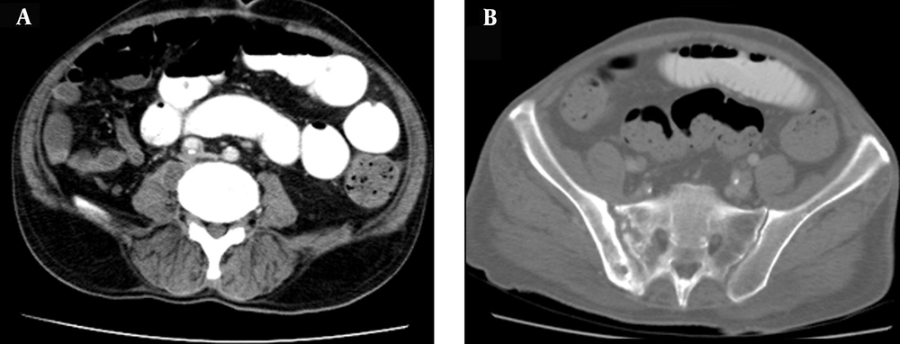
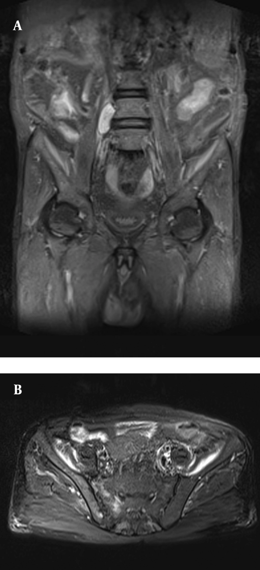
Chest X-ray was normal. The echocardiography revealed normal findings. Abdominal sonography revealed increased echogenicity of the liver and two small hyper echoic lesions in liver segments one and eight, and multiple small stones in the gallbladder and ascites. No evidence of biliary obstruction was detected and common bile duct (CBD) was of normal diameter. Color Doppler sonography of the portal vein and hepatic veins were normal. Chest CT scan revealed non-significant mediastinal lymph nodes with short axis of less than 10 mm and a calcified granuloma, measuring 5 mm, in the right middle lobe. An abdominopelvic CT scan with IV and oral contrast was performed and depicted an oval hypodense lesion with ring enhancement measuring 40 × 20 mm in the medial aspect of the right psoas muscle at the level of the L5 vertebra (Figure 1 A). Adjacent disco-vertebral structures from L4 through S1 were unremarkable; however, there were unilateral erosive and sclerotic changes in the right sacroiliac joint (Figure 1 B). There were also two hypodense lesions in segments one and eight of the liver, measuring 8 mL in favor of benign lesions. The caudate lobe was prominent associated with periportal hypodensity suggesting underlying parenchymal disease. The abdominopelvic magnetic resonance imaging (MRI) depicted a hyper-signal area in the right iliac bone and adjacent gluteal muscle in favor of sacroiliitis and associated periarticular inflammatory process (Figure 2). Magnetic resonance cholangiopancreatography (MRCP) images depicted multiple filling defects due to a gallstone. No evidence of biliary obstruction was detected and the common bile duct was of normal diameter. Furthermore, CT-guided aspiration of the right psoas collection was performed. Analysis of aspirated pus was positive for acid-fast bacilli and the culture depicted mycobacterial growth. Gram staining and cytology for malignancy were negative. After confirmation of the diagnosis, an anti-tuberculosis regimen was started. Liver light microscopy revealed multiple portal and parenchymal microgranulomas forming from epithelioid histiocytic, giant cells and lymphoplasma cells surrounded by delicate fibrosis and accompanied by mild portal inflammation, fibrosis and macrovesicular steatosis and biliary pigment in some hepatocytes.
(A) Coronal short tau inversion recovery (STIR) image depicts a hyper-intense oval mass in the medial aspect or right psoas muscle with normal adjacent disco-vertebral structures. (B) Axial fat-suppression T2W image shows increased signal in the right iliac and sacral wings in favor of active sacroiliitis.
During the hospital stay, laboratory test revealed pancytopenia; WBC count 1500 × 10 3u/L (PMN 82%, Lymp 11.9%), red blood count (RBC) count: 3.75 × 10 6u/L, Hb: 7.9 gr/dL, MCV: 73 fl, platelet count: 115000/μL.
The bone marrow biopsy also revealed multiple epithelioid granuloma and Langhans-type giant cells with small foci of necrosis. Both samples were negative on Ziehl-Neelsen staining, which has low sensitivity to detect mycobacterial bacilli.
With diagnosis of extra pulmonary TB, an anti- tuberculosis regimen of four drugs was started. After four days, the patient’s general condition improved significantly and after two weeks he became afebrile. During admission, generalized edema occurred and laboratory test revealed total protein level of 5 g/dL [6.6 - 8.8] and albumin (Alb) level of 3.1 g/dL [3.5 - 3.3]. His urine analysis showed +1 proteinuria and RBC count: 1 - 2/high power field (HPF) and WBC count: 8 - 10/HPF. His urine culture was negative. His 24-hour urine protein level was 170 mg/day. Tuberculosis PCR of urine was negative. Diuretic and IV albumin were started. After three days, his edema was resolved and the patient was discharged with an anti-tuberculosis regimen of four drugs (isoniazid, rifampicin, pyrazinamide and ethambutol combination for two months plus isoniazid and rifampicin combination for the remainder). Our patient remained symptom free on follow up, two months after completion of treatment with no subsequent flare-ups.
3. Discussion
Tuberculosis is one of the oldest and most commonly encountered diseases. Although there is a significant steady decline in the incidence of active pulmonary tuberculosis due to early diagnosis and prompt treatment, the incidence of extra-pulmonary tuberculosis has remained constant particularly due to a delay in recognizing the condition when the clinical scenario consists mostly of nonspecific extra-pulmonary symptoms. Extrapulmonary tuberculosis is considered as a treatable disease with good outcome, requiring strict compliance. When it presents with bone marrow involvement, the outcome depends largely on timely diagnosis and early initiation of treatment (4). The primary diagnosis for our patient was psoas abscess. Psoas abscesses are usually secondary to the extension of infection from an adjacent site and primary psoas abscesses are seldom hematogenous (2). Psoas abscess in our case was not associated with adjacent spondylodiscitis, which made the primary diagnosis of tuberculosis belated. The patient did not have lower abdominal or back pain, or referred pain to the hip or knee, and physical exam of sacroiliac joint and hip was normal. The MRI was performed to find a relationship with adjacent tissue and showed ipsilateral sacroiliitis with periarticular inflammatory changes in the gluteal muscle. Bony changes of right sacroiliac joint seemed to be chronic on CT scan. Lack of associated clinical symptoms strengthened this view. However, signal alterations of respective areas on MRI suggested active inflammation. Osteomyelitis of the ilium or septic arthritis of the sacroiliac joint can penetrate the sheaths of either or both muscles in this location, producing an iliacus or psoas abscess (2). Our patient had asymptomatic ipsilateral sacroiliitis, and the psoas abscess was mostly because of direct expansion of the tuberculous sacroiliitis. Previous studies have reported on a total of 35 patients that have been diagnosed with sacroiliac tuberculosis. These patients had persistent lower back pain and difficulty in walking. These specifications were noted in all patients. Most of the patients (91.4%) had unilateral disease (5). Detection of acid-fast bacilli in aspirated pus of psoas abscess confirmed the diagnosis of tuberculosis, thus anti-tuberculosis regimen was started. Our patient had two hypodense lesions in the liver with abnormal liver function tests. From the tumor markers; CEA, Afp and CA 19-9 were negative. In rare instances, miliary tuberculosis may mimic cholangitis, with fever and liver function test abnormalities suggestive of obstructive disease, and little evidence of hepatocellular disease. Diagnosis is made by liver biopsy (2). According to the cholestatic pattern of the liver test, CBD with a normal size and negative result of AMA; liver biopsy for excluding intrahepatic cholestasis was considered. Based on the level of AST being more than ALT, high serum-Ascites albumin gradient (SAAG) ascites, negative result of viral hepatitis, metabolic disorder and autoimmune hepatitis, liver biopsy was performed to establish an etiological diagnosis and exclude of cirrhosis. Liver biopsy showed non-necrotizing granuloma combined with biliary pigment concentration in some hepatocytes, and portal fibrosis, that confirmed hepatic tuberculous and intrahepatic cholestasis yet were not compatible with cirrhosis. Hepatitis and fibrosis due to mycobacterial infection were demonstrated by periportal hypodensity and relative enlargement of caudate lobe on CT images of our case. The two hypodense lesions in the liver could be due to tuberculosis granulomas since they lack the typical appearance of liver abscess. Visceral tuberculous abscesses include hepatic, pancreatic and splenic abscesses that often occur in HIV positive patients (6), while our patient was HIV-negative. Inflammatory pseudo-tumors of the liver are rare and difficult to diagnose, mimicking malignant tumors. Three patients have been found with hepatic tuberculosis by various studies that were treated by major hepatic surgery because of the lack of definite diagnosis (1, 7). Also, from previous studies, two patients were misdiagnosed preoperatively as having cholangiocarcinoma yet were confirmed to have hepatic tuberculosis by postoperative pathology (1). Tuberculous peritonitis can cause low SAAG ascites and remains undiagnosed in patients with concomitant cirrhosis with ascites (2). In this patient, high SAAG ascites were detected and TB PCR was negative with no evidence of gross peritoneal involvement on CT scan or MRI. Different studies have shown a high sensitivity and specificity for multiplex PCR in tuberculous peritonitis, while in combination with adenosine deaminase (ADA) levels in cases of peritoneal tuberculosis, the specificity of diagnosis of peritoneal tuberculosis was increased to 95% (8). The sensitivity of a computed tomography scan in the prediction of tuberculosis is 69%. Patients with tuberculosis were likely to show mesenteric changes, macronodules (>5 mm in diameter), splenomegaly, and splenic calcification on CT imaging (9). The SAAG of > 1.1 g/dL reflects the presence of portal hypertension and indicates that the ascites is from an increased pressure in the hepatic sinusoids. An ascitic protein level of < 2.5 g/dL indicates that the hepatic sinusoids have been damaged and scarred, and no longer allow passage of protein, as it occurs with cirrhosis, late Budd-Chiari syndrome, or massive liver metastases (10). In this case, the mentioned causes were not detected and hepatic tuberculosis accompanied by fibrosis led to high SAAG ascites and reversion of AST to ALT without cirrhosis.
Disseminated tuberculosis should be considered when pancytopenia is associated with fever and weight loss or as a cause of other obscure hematological disorders. Late generalized tuberculosis, nonreactive tuberculosis and tuberculosis in complicating myeloproliferative disorders can cause aplastic anemia, thrombocytopenia, leukopenia and leukemoid reactions (2). The patient had pancytopenia and bone marrow biopsy, which showed multiple granulomas. A previous study reported on a patient with a six-month history of on and off moderate- to high-grade fever with bicytopenia, who was diagnosed to have tuberculosis of bone marrow (4). Our patient had tuberculosis with multi-organ involvement including liver, bone marrow, sacroiliac joint and psoas abscess, iliac bone and gluteal muscle. With diagnosis of extra-pulmonary tuberculosis, an antituberculosis regimen of four drugs was commenced. He responded well to our therapeutic protocol. Resolution of symptoms with anti-TB therapy supports the diagnosis of tuberculosis in our patient.
This case demonstrates the different presentations and diagnostic difficulties posed by atypical manifestations of tuberculosis. Tuberculosis is still a diagnostic challenge, especially when the presentation is atypical and extra-pulmonary unless a high degree of suspicion is maintained. In endemic areas it may be justifiable to treat for tuberculosis empirically without microbiological evidence when the clinical, histological and other circumstantial evidence are in favor.

