


1. Introduction
Ecthyma gangrenosum is a skin lesion presenting an invasive infection in the skin that commonly caused by Pseudomonas. Also, these lesions may rarely present in patients without bacteremia (1, 2). They usually show themselves as one or several macular lesions or papules with a red and inflamed area and then change into a hemorrhagic bulla and finally become necrotic (1, 3). The first case of Ecthyma gangrenosum was explained in 1987 and first named in 1930. Pathogenesis is mostly microbial invasion in cutaneous tissues caused by microorganism produced bacteremia. That microorganism is usually a Gram-negative and mostly Pseudomonas, which infiltrates through perivascular area. In case of no bacteremia, mostly cutaneous infiltration of microorganism is known as the cause (2). Although Ecthyma gangrenosum was pathognomonic for Pseudomonas-induced septicemia in the past, today other kinds of microorganisms, including a variety of bacteria and fungi and viruses are known as the triggering factor. One of the important, but rare causes of Ecthyma gangrenosum after Pseudomonas is E. coli, which can be as a result of bacteremia, cellulitis, and urinary tract infection (2, 4). E. coli-induced soft tissue infections are rare and are usually seen in patients with severe immunodeficiency (5).
2. Case Presentation
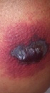
A 45-year-old woman known as the case of acute myeloid leukemia (AML-M5) who went into remission due to chemotherapy with 7-3 regimen (7 days cytarabine and 3 days daunorubicin). On the 21st day after chemotherapy, she was febrile and septic. A physical examination revealed an erythematous round lesion (3 × 3 cm) that had developed on the posterior aspect of the right thigh with central bolus necrosis (Figure 1). Absolute neutrophil count (ANC) was fewer than 200 cell/mm3 at that time. Sepsis work-up included 2 sets of blood culture for 30 minutes. Then imipenem, vancomycin, and amphotericin B started. After 48 hours, E. coli detection on both blood cultures that were sensitive to imipenem and amikacin. Vancomycin and amphotericin B were stopped and amikacin added to imipenem. Systemic symptoms subsided after a few days and Ecthyma gangernusom completely improved 2 weeks later.

Erythematous round lesion with central bolus necrosis
3. Discussion
Ecthyma gangrenosum is specific skin lesion, which is usually accompanied by Pseudomonas septicemia and rarely can be seen with other microorganisms such as Streptococcus, Aeromonas hydrophila, Staphylococcus aureus, Serratia marcescens, Pseudomonas maltophilia, Citrobacter freundii, E. coli, Candida albicans, Aspergillus, and Mucor species (3). In a review done by Vaiman et al. (4), 197 patients suffering from Ecthyma gangrenosum infection were studied between 1975 to 2014. In this study, they found out that in 123 patients, Pseudomonas caused 73.65% and other agents caused 17.35% and the remaining 9% was induced by fungal agents.
Ecthyma gangrenosum is mostly seen in patients with severe immunodeficiency, including Agammaglobulinemia, aplastic anemia, hematologic malignancies, especially patients with leukemia after chemotherapy, and also in HIV patients. However, it was also reported in patients without defect in the immune system (2-4). Ecthyma gangrenosum is usually diagnosed based on the patient’s history, underlying diseases, blood culture, and tissue culture. Early recognition and rapid antibiotic therapy for these lesions are very important in decreasing the mortality rate (2).
Nowadays, general infections caused by E. coli are of the important problems in patients with hematological malignancies, especially after chemotherapy, which is usually followed by bad prognoses (5). Cellulitis and different types of soft tissue infections caused by E. coli, are rare and usually need background risk factors such as reduced number or dysfunction of neutrophils (6). The first E. coli-induced Ecthyma gangrenosum was reported in 1979 in a 1-year-old baby with cellulitis on the left nostril without bacteremia and E. coli was isolated from the lesion and the nose (2, 5).
In a literature review by Patel et al. (2) in 2009, which was published in Cutis journal, there was 7 cases of Ecthyma gangrenosum caused by E. coli, and the first E. coli-induced Ecthyma gangrenosum was reported in a patient suffering from AML. We reported the second Ecthyma gangrenosum case that caused by E. coli in a woman who was an AML patient. Based on a search of the literature using PubMed/MEDLINE for EG/E. coli, until Nov. 24th in 2017, we have added 5 new cases to the table designed in Patel et al.’s article (Table 1) (2). So far, in 11 patients reported in the literature, at least 8 cases reported lesions on the lower limb, which seems is the most probable anatomic area for E. coli-induced Ecthyma gangrenosum.
| Case Report | Year | Number of Case | Presenting Site | Preceding Bacteremia | Underlying Disease |
|---|---|---|---|---|---|
| Anderson (7) | 1979 | 1 | Left nostril | no | Gastroenteritis |
| Rajan (8) | 1982 | 1 | Upper extremity and lower extremity | yes | Alcoholic cirrhosis |
| Edelstein and Cutting (9) | 1986 | 3 | Lower extremity and perianal skin | Yes | Lung cancer |
| Fuchshuber et al. (10) | 1998 | 1 | Lower extremity | Yes | Multiple myeloma |
| Patel et al. (2) | 2009 | 1 | Upper extremity | Yes | AML |
| Gomes et al. (11) | 2012 | 1 | Lower extremity | Yes | No |
| Pathak et al. (1) | 2013 | 1 | Lower extremity | Yes | No |
| Mouna et al. (3) | 2015 | 1 | Lower extremity | No | No |
| Salehi | 2018 | 1 | Lower extremity | Yes | AML |
Ecthyma gangrenosum Caused by Escherichia coli; Reports in the Literature (2)
About 81.8% of the patients equal to 9 out of 11 reported E. coli-induced bacteremia accompanying their disease, which reminds the importance of sending blood culture in patients with Ecthyma gangrenosum. In our case, the isolated E. coli was an extended spectrum β lactamase (ESBL) producer, which is an enzyme providing resistance to most beta-lactam antibiotics and makes the treatment more difficult. Given underlying diseases, 6 of the patients (54.4%) had a history of malignancy (3 pulmonary cancers, 2 AML, and 1 Multiple Myeloma), 3 patients (27.2 %) had no specific previous disease, 1 patient had cirrhosis, and 1 patient had Gastroenteritis. It seems the malignancy history is the most important risk factor in developing E. coli-induced Ecthyma gangrenosum.
3.1. Conclusions
Although E. coli-induced Ecthyma gangrenosum is a rare infectious lesion, especially in patients with a malignancy history, lower extremity lesions should be considered carefully. In these patients sending blood culture, antibiogram, and rapid appropriate antibiotic therapy can be effective in adopting good management and saving patients’ lives.
