Full Text
Full text is available in PDF
Archives of Clinical Infectious Diseases
Infectious Diseases and Tropical Medicine Research Center, SBUMS
Authors
Brain abscess is a focal intracerebral infection, which begins as a localized area of cerebritis and develops into a collection of pus surrounded by a well-vascularized capsule. In the present study, we studied all patients with brain abscess admitted in Ardabil Fatemi hospital.
In a retrospective hospital-based study, 24 patients with brain abscess entered the study. Medical records of patients were reviewed from January 2004 to January 2006. Brain abscess was defined as one or more localized lesions with the following characteristics in brain imaging (CT scan): hypodense center with a peripheral uniform ring enhancement following the injection of contrast material, or affected region surrounded by variable hypodense area of brain edema or nodular enhancement or area of low attenuation without enhancement. Predisposing factors for brain abscess were surveyed by reviewing medical records.
The study population included 17 males and 7 females with the mean age ( standard deviation) of 27.021.3 years. The following predisposing factors were noted in 22 (91.6%) patients; contiguous focus of infection (sinusitis and chronic otitis media) in 10 (41.6%), congenital heart disease in 5 (20.8%), post traumatic complications in 3 (12.5%), super infected hydatid cyst in 2 (8.3%) and neurosurgical complications in 2 cases (8.3%).
Ear in cerebellar and temporal lobe, frontal sinus in frontal lobe and heart in parietal lobe abscesses should be evaluated for the possible source of infection. Meanwhile, hydatid cyst may be an important risk for brain abscess in endemic areas like Ardabil
© 2008, Author(s). This open-access article is available under the Creative Commons Attribution 4.0 (CC BY 4.0) International License (https://creativecommons.org/licenses/by/4.0/), which allows for unrestricted use, distribution, and reproduction in any medium, provided that the original work is properly cited.
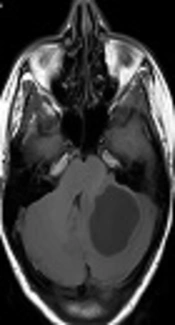
Tfifha M, Abdallah AB, Saadaoui K, Helal KB, Gueddiche MN, et al. Pyogenic Brain Abscess in Children: A Tunisian Multi-Centric Experience. Arch Pediatr Infect Dis. 2018;6(3):e55012. doi: https://doi.org/10.5812/pedinfect.55012
Rasras S, Teimouri A, Ranjbar F, Zaeim I, Ardeshiri M. Unusual Presentation of Hydatid Cyst: Case Reports for Neurosurgery (Three Cases). Jentashapir J Cell Mol Biol. 2015;6(5):e32024. doi: https://doi.org/10.17795/jjhr-32024
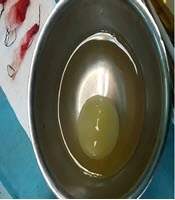
Nashibi M, Tafrishinejad A, Hussain Khan Z. Deep Seated Cerebral Hydatid Cyst and Its Anesthetic Considerations: A Case Report. Arch Neurosci. 2020;7(1):e100044. doi: https://doi.org/10.5812/ans.100044
Sabzghabaei A, Aeinechian S, Shojaee M, Kashani P, Manouchehrifar M. Epidemiological Features of Ischemic Brain Stroke; a Cross-Sectional Hospital-Based Study. Arch Neurosci. 2019;6(2):e74362. doi: https://doi.org/10.5812/ans.74362
Rostami M, Emadoleslami M, Karimi I. Tuberculous brain abscess in a child wit cystic fibrosis; a case report. Arch Clin Infect Dis. 2006;1(4):. doi:
Last Update: 3 days ago
Last Update: 3 days ago
Last Update: 3 days ago
Last Update: 14 hours ago