The First Identification of Nanobacteria-Like Structures in Vascular Plaques of Atherosclerosis Patients in Iran

authors:

avatar Ali Majidpour 1 , avatar Samira Rasouli 1 , * , avatar Hadi Sardarabadi 2 , avatar Morteza Noaparast 3 , avatar Masoud Khedmatgozar 1 , avatar Sara Fathizadeh 1 , avatar Maryam Ghooshchian 1 , avatar Mina Boustanshenas 1 , avatar Sayed Ali Mohammad Sadat 4

Antimicrobial Resistance Research Center, Institute of Immunology and Infectious Diseases, Iran University of Medical Sciences, Tehran, Iran
Department of Biomedical Engineering and Medical Physics, Shahid Beheshti University of Medical Sciences, Tehran, Iran
Vascular and Endovascular Department, Imam Khomeini Hospital, Tehran University of Medical Sciences, Tehran, Iran
Vascular and Endovascular Department, Yasuj University of Medical Sciences, Yasuj, Iran

how to cite: Majidpour A, Rasouli S, Sardarabadi H, Noaparast M , Khedmatgozar M , et al. The First Identification of Nanobacteria-Like Structures in Vascular Plaques of Atherosclerosis Patients in Iran. Arch Clin Infect Dis. 2019;14(4):e82826. https://doi.org/10.5812/archcid.82826.

Abstract

Background:

Nanobacteria-like structures (NLSs) or calcifying nanoscale particles (CNPs) are novel ambiguous entities. As their names imply, they are tiny creatures (100 to 500 nanometers). Compared to ordinary bacteria, they can exhibit some unbelievable properties such as resistance to conventional antibacterial procedures. Form a scale point of view, these microorganisms are much smaller than ordinary bacteria and are preserved from the outer environment by their rigid apatite shell. The above-mentioned protective properties of nanobacteria are responsible for a dangerous in vivo calcification procedure causing pathological infectious diseases.

Objectives:

The main goal of this study was to identify and isolate the nanobacteria from patients with atherosclerosis.

Methods:

Human vascular tissues were assessed for the presence of NLSs in atherosclerotic plaques at Sina Hospital, Tehran, Iran. All 40 samples obtained from patients as the wastes of surgery showed crystallization in vitro after about 10 days. Crystallization was seen also in filtered media. The precipitates of media were investigated by scanning electron microscopy (SEM).

Results:

NLSs were seen in all samples by SEM. After confirming the NLSs presence, they were characterized by TEM, XRD, and SEM/EDAX.

Conclusions:

The NLSs are observed in patients with atherosclerosis.

1. Background

It is hypothesized that nanoparticles in calcified specimens may be a kind of bacteria called “Nanobacteria”. Some calcified structures in the human body can cause various diseases including gallstones and gallbladder inflammation (1), kidney and urinary stone formation (2-5), Alzheimer’s disease (6), vascular plaque, arterial heart disease (7), polycystic kidney disease (PKD) (8), dental calculus (9), prostatitis (10), psammoma bodies in ovarian cancer (11), calciphylaxis (12), and other diseases (13, 14). These nanobacteria can cause such important diseases due to the unbelievably sturdy apatite shells of these organisms. In many studies, researchers claimed that nanobacteria could accumulate different minerals as nidi under in vivo and in vitro conditions. Nanobacteria by these processes can create a protecting layer on the cell membrane that inhibits the penetration of any antinanobacterial agent into its structure. The lack of developing methods for growth inhibition has led these novel nanoscale bacteria to cause many problems such as increased resistance to antibacterial agents and increased associated diseases.

Calcification is one of the main causes of atherosclerotic plaque formation (15). It has been suggested that regulatory mechanisms such as osteogenesis (16, 17), stimulation of vesicle-calcification by lectins (18), and mast cell and macrophage-induced calcification (19) are involved in vascular calcification. Some studies have shown that high-molecular-weight calcified particles can act as nidi and accumulate minerals such as calcium to ultimately cause the calcification of soft tissue and develop diseases such as atherosclerosis (20, 21). Concerning the pathologic factors involved in atherosclerosis, it has been suggested that nanobacteria are possibly the candidate infectious agents that could form a hard apatite shell at physiological levels to cause plaque formation in vessels.

2. Objectives

The main purpose of this study was to isolate and identify nanobacteria in patients with atherosclerosis.

3. Methods

We collected 14 vascular plaques from patients with atherosclerosis who had undergone surgery at Sina Hospital, Tehran, Iran, from February 2016 to June 2016. For the isolation of nanobacteria-like structures (NLSs), the homogenized vascular tissue samples were filtered through 0.22-μm membrane filters. Then, Dulbecco’s modified eagle medium (DMEM; Gibco) was added to the precipitates obtained from centrifugation (at 20000 g for 40 minutes). The suspension was cultured in six-well plates with each well containing 5 mL of DMEM supplemented with 10% fetal bovine serum (FBS). Culturing was carried out using humidified air at 37°C with 5% CO2 and 95% air (LAB-LINE BI65, USA). A medium containing 5 mL of DMEM and 10% FBS (FBS) but without vascular plaques was considered as the negative control. Calcifying nanoscale particles (CNPs) growth was assessed by turbidity analysis at the 650-nm wavelength. Based on optical observation obtained from CNPs growth in 60 days, the positive plates were selected and samples containing white-colored sediments in the bottom of the wells were used for electron microscopic analyses including SEM (LEO 1450 VP, Germany) and TEM (LEO 912 AB, Germany). In the SEM study, floating and adherent materials in a culture plate were examined separately. Before microscopic examinations, the samples were prepared by methods suitable for biological samples. Then, the samples were analyzed by XRD and SEM/EDAX.

4. Results

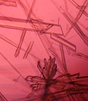
The investigation of CNPs growth was performed via changes in the optical properties of the CNPs once every 10 days of incubation time at the wavelength of 650 nm. The results indicated that the nanobacteria growth was too slow in 10 days of incubation. The pH variation was measured during the incubation time. The pH changed in the range of 7 to 8. To determine whether any other pathogen is present in the nanobacteria culture medium after filtering, the samples were cultured in current nutrient microbiological culture media every three days of incubation, showing no bacterial and fungal growth as the sign of contamination at 25, 30, 37, and 50°C. After about 10 days, the results obtained from inverted optical microscopy showed that the CNPs initiated a crystallization phenomenon and adhered to the bottom of the plates (Figure 1). After around two months, the samples were centrifuged and the two obtained phases of samples including the pellet and the supernatant were investigated using scanning electron microscopy. The evidence showed that the CNPs had spherical shapes. The SEM study showed a spherical shape for nanobacteria (Figure 2). In the culture without vascular plaques, no nanobacteria were observed. TEM results showed that the CNPs had a nanometer shell (Figure 3). Energy-dispersive spectroscopy (EDS) analysis of samples showed two elemental sharp peaks (carbon and oxygen) rather than other components (Figure 4).

Cultures of CNPs observed by inverted optical microscopy (200x magnification)
Scanning electron microscopy micrographs of spherical nanobacteria (scale bar 500 nm)
TEM micrograph of nanobacteria showing shells around the nanobacteria (scale bar 200 nm). This cover is a reason for the impenetrable properties of nanobacteria.
Energy dispersive X-ray analysis of nanobacteria; nanobacteria can cause in vitro biomineralization via the accumulation of minerals, especially calcium and phosphate.

5. Discussion

In atherosclerosis, plaques are formed inside the arteries. The plaques contain fat, cholesterol, calcium, and other substances found in the blood. Over time, plaques are hardened to narrow the arteries. Then, the flow of oxygen-rich blood is limited to the organs. Atherosclerosis can lead to serious problems, including heart attack, stroke, or even death. The existence of nanobacteria as nidi in any part of the body can lead to the accumulation of minerals. Hence, the nanobacteria precipitation in the interior of arteries as a spark can initiate reactions that eventually occlude the arteries, so caused atherosclerosis (22, 23). So far, different studies have been conducted on nanobacteria as a controversial issue. Confirming the results obtained from previous research, our results showed that nanobacteria were found in most extracted specimens from patients with atherosclerosis. Based on previous investigations and the results of the current study, minerals are accumulated on the cell membrane by nanobacteria to ultimately create hard apatite shells (7, 24). The ability to create these structures was confirmed using some approaches such as monoclonal antibody and turbidity assays. In the current study, we observed that the optical absorption increased with increasing the size of white sedimentations (containing nanobacteria based on SEM micrographs). Moreover, our results showed that with increasing the nanobacteria incubation time, the coating thickness of these organisms increased due to the accumulation of mineral components. Their protecting cover is so impenetrable that it is not readily accessible with anti-nanobacterial agents (24). To check whether the results obtained from nanobacteria culture are not attributed to the growth of any other organisms, it is very necessary to apply two approaches in experiments. First, all components should be sterilized. For sterilization and restricting the penetration of foreign entities, we used 0.2-μm filters. Hence, we ensured that the culture medium had minimum pathogen interference. Second, the existence of any organisms should be shown by electron microscopy. In this study, no contamination was seen in microscopy and staining methods.

This study opens new opportunities for the investigation of the pathogenesis of nanobacteria in atherosclerosis. Nonetheless, achieving more details on CNPs pathological properties needs more research.

Acknowledgements

References

  • 1.

    Wang L, Shen W, Wen J, An X, Cao L, Wang B. An animal model of black pigment gallstones caused by nanobacteria. Dig Dis Sci. 2006;51(6):1126-32. [PubMed ID: 16865581]. https://doi.org/10.1007/s10620-006-8019-6.

  • 2.

    Hong X, Wang X, Wang T, Yu C, Li H. Role of nanobacteria in the pathogenesis of kidney stone formation. Am J Transl Res. 2016;8(7):3227-34. [PubMed ID: 27508044]. [PubMed Central ID: PMC4969460].

  • 3.

    Ansari H, Sepahi AA, Sepahi MA. Different approaches to detect "nanobacteria" in patients with kidney stones: An infectious cause or a subset of life? Urol J. 2017;14(5):5001-7. https://doi.org/10.22037/uj.v14i5.4051.

  • 4.

    Kramer G, Klingler HC, Steiner GE. Role of bacteria in the development of kidney stones. Curr Opin Urol. 2000;10(1):35-8. [PubMed ID: 10650513].

  • 5.

    Bjorklund M, Ciftcioglu N, Kajander EO. Extraordinary survival of nanobacteria under extreme conditions.In: Hoover RB, Levin GV, Rozanov AY, Davies PCW, editors. Instruments, Methods, and Missions for Astrobiology. International Society for Optics and Photonics; 1998. p. 123-9.

  • 6.

    Ciftcioglu N, Bjorklund M, Kajander EO. Stone formation and calcification by nanobacteria in the human body.In: Hoover RB, Levin GV, Rozanov AY, Davies PCW, editors. Instruments, Methods, and Missions for Astrobiology. International Society for Optics and Photonics; 1998. p. 105-11.

  • 7.

    Ciftcioglu N, Bjorklund M, Kuorikoski K, Bergstrom K, Kajander EO. Nanobacteria: An infectious cause for kidney stone formation. Kidney Int. 1999;56(5):1893-8. [PubMed ID: 10571799]. https://doi.org/10.1046/j.1523-1755.1999.00755.x.

  • 8.

    Hjelle JT, Miller-Hjelle MA, Poxton IR, Kajander EO, Ciftcioglu N, Jones ML, et al. Endotoxin and nanobacteria in polycystic kidney disease. Kidney Int. 2000;57(6):2360-74. [PubMed ID: 10844606]. https://doi.org/10.1046/j.1523-1755.2000.00096.x.

  • 9.

    Zhang SM, Tian F, Jiang XQ, Li J, Xu C, Guo XK, et al. Evidence for calcifying nanoparticles in gingival crevicular fluid and dental calculus in periodontitis. J Periodontol. 2009;80(9):1462-70. [PubMed ID: 19722797]. https://doi.org/10.1902/jop.2009.080659.

  • 10.

    Shoskes DA, Katz E. Multimodal therapy for chronic prostatitis/chronic pelvic pain syndrome. Curr Urol Rep. 2005;6(4):296-9. [PubMed ID: 15978233].

  • 11.

    Wu JH, Deng YL, Liu Q, Yu JC, Liu YL, He ZQ, et al. Induction of apoptosis and autophagy by calcifying nanoparticles in human bladder cancer cells. Tumour Biol. 2017;39(6):1010428317707700. [PubMed ID: 28635397]. https://doi.org/10.1177/1010428317707688.

  • 12.

    Jelic TM, Malas AM, Groves SS, Jin B, Mellen PF, Osborne G, et al. Nanobacteria-caused mitral valve calciphylaxis in a man with diabetic renal failure. South Med J. 2004;97(2):194-8. [PubMed ID: 14982274]. https://doi.org/10.1097/01.SMJ.0000077067.44311.F0.

  • 13.

    Lin XC, Gao X, Lu GS, Song B, Zhang QH. Role of calcifying nanoparticles in the development of testicular microlithiasis in vivo. BMC Urol. 2017;17(1):99. [PubMed ID: 29084530]. [PubMed Central ID: PMC5663083]. https://doi.org/10.1186/s12894-017-0289-0.

  • 14.

    Hudelist G, Singer CF, Kubista E, Manavi M, Mueller R, Pischinger K, et al. Presence of nanobacteria in psammoma bodies of ovarian cancer: Evidence for pathogenetic role in intratumoral biomineralization. Histopathology. 2004;45(6):633-7. [PubMed ID: 15569055]. https://doi.org/10.1111/j.1365-2559.2004.02030.x.

  • 15.

    Huang H, Virmani R, Younis H, Burke AP, Kamm RD, Lee RT. The impact of calcification on the biomechanical stability of atherosclerotic plaques. Circulation. 2001;103(8):1051-6. [PubMed ID: 11222465]. https://doi.org/10.1161/01.cir.103.8.1051.

  • 16.

    Shioi A, Mori K, Jono S, Wakikawa T, Hiura Y, Koyama H, et al. Mechanism of atherosclerotic calcification. Z Kardiol. 2000;89 Suppl 2:75-9. [PubMed ID: 10769407].

  • 17.

    Shanahan CM, Proudfoot D, Tyson KL, Cary NR, Edmonds M, Weissberg PL. Expression of mineralisation-regulating proteins in association with human vascular calcification. Z Kardiol. 2000;89 Suppl 2:63-8. [PubMed ID: 10769405].

  • 18.

    Hsu HH, Tawfik O, Sun F. Effects of lectins on calcification by vesicles isolated from aortas of cholesterol-fed rabbits. Biochim Biophys Acta. 2000;1464(2):262-72. [PubMed ID: 10727613]. https://doi.org/10.1016/s0005-2736(00)00150-4.

  • 19.

    Jeziorska M, McCollum C, Woolley DE. Calcification in atherosclerotic plaque of human carotid arteries: Associations with mast cells and macrophages. J Pathol. 1998;185(1):10-7. [PubMed ID: 9713354]. https://doi.org/10.1002/(SICI)1096-9896(199805)185:1<10::AID-PATH71>3.0.CO;2-0.

  • 20.

    Price PA, Thomas GR, Pardini AW, Figueira WF, Caputo JM, Williamson MK. Discovery of a high molecular weight complex of calcium, phosphate, fetuin, and matrix gamma-carboxyglutamic acid protein in the serum of etidronate-treated rats. J Biol Chem. 2002;277(6):3926-34. [PubMed ID: 11724771]. https://doi.org/10.1074/jbc.M106366200.

  • 21.

    Price PA, Lim JE. The inhibition of calcium phosphate precipitation by fetuin is accompanied by the formation of a fetuin-mineral complex. J Biol Chem. 2003;278(24):22144-52. [PubMed ID: 12676929]. https://doi.org/10.1074/jbc.M300744200.

  • 22.

    Miller VM, Rodgers G, Charlesworth JA, Kirkland B, Severson SR, Rasmussen TE, et al. Evidence of nanobacterial-like structures in calcified human arteries and cardiac valves. Am J Physiol Heart Circ Physiol. 2004;287(3):H1115-24. [PubMed ID: 15142839]. https://doi.org/10.1152/ajpheart.00075.2004.

  • 23.

    Puskas LG, Tiszlavicz L, Razga Z, Torday LL, Krenacs T, Papp JG. Detection of nanobacteria-like particles in human atherosclerotic plaques. Acta Biol Hung. 2005;56(3-4):233-45. [PubMed ID: 16196199]. https://doi.org/10.1556/ABiol.56.2005.3-4.7.

  • 24.

    Sardarabadi H, Mashreghi M, Jamialahmadi K, Dianat T. Resistance of nanobacteria isolated from urinary and kidney stones to broad-spectrum antibiotics. Iran J Microbiol. 2014;6(4):230-3. [PubMed ID: 25802705]. [PubMed Central ID: PMC4367938].