1. Background
Clostridium perfringens (CP) is a Gram-positive, spore-forming, anaerobic bacteria. It is ubiquitous in the environment that can be found in the intestines of humans and livestock. CP is a causative agent of gas gangrene (myonecrosis) and gastrointestinal diseases in both humans and animals (1). The virulence of this organism is largely dependent on its ~17 toxins. However, C. perfringens strains have been classified into five types of (based on the production of alpha, beta, epsilon, and iota major toxins) A, B, C, D, and E (2), which all of them can produce the alpha-toxin (CPA). CPA is the main product in C. perfringens type A and is declared as a zinc metallophospholipase C that has phospholipase C, lecithinase, and sphingomyelinase activities, which are important for hemolysis and necrosis. Crystallographic studies of the alpha-toxin model showed that the N-terminal and the C-terminal domains are biologically active domains.
The N-terminal and the C-terminal domains contain a single active site of the enzyme and are essential for toxic and cytolysis activities, respectively (3, 4). The N-terminal and the C-terminal domains are immunogenic, but only the C-terminal domain instigates a protective immune response (3, 5). It is well established that both CPA and theta toxin (perfringolysin O) are responsible for C. perfringens-mediated myonecrotic infection (3). The cpa gene is located on a chromosome (197 bp in length) and converted into a 398 amino acids (aa) complete protein with a N-terminal signal peptide sequence of 28 aa, which (if separated) produces an active protein with a molecular weight of 43 kDa (6, 7).
Clostridium septicum (CS) is a Gram-positive, spore-forming, anaerobic bacteria. CS is a causative agent of non-traumatic and traumatic myonecrosis. Additionally, this organism is reported to be associated with large colon cancer, immunosuppression, neutropenia, and diabetes mellitus (8). CS can produce several toxins such as alpha, beta, gamma, and delta. The organism alpha-toxin (AT) is one of the most important lethal virulence factora that is responsible for gas gangrene (9). The csa gene is 1332 bp in length and is located on a chromosome, and can be converted into an inactive protoxin. The secreted protoxin is a 46.55 kDa protein with a signal peptide of 31 aa.
This protein binds to the cell membrane and activates by proteolytic hydrolyses, such as furin (in vivo) and trypsin (in vitro), which separate a C-terminal 45 aa peptide. The activated toxin forms an oligomer pre-pore complex that results in the production of pores with a diameter of about 1.3 - 1.6 nm, which then induces rapid cell necrosis (9-11). Uppalapati et al. (12) produced a synthetic construct recombinant fusion protein αCS, including C. perfringens and S. aureus alpha toxins, that were bind via a glycine linker fragment using fusion PCR.
2. Objectives
The current study aimed to investigate molecular cloning of a new bi-functional fusion protein of cpa and csa genes in a suitable host cell (E. coli TOP10).
3. Methods
3.1. Bacterial Strains
Clostridium perfringens type A strain PTCC1765 (ATCC13124) was obtained from Iran industrial and scientific research organization, and C. septicum strain CN913 were retrieved from Razi Serum and Vaccine Research Institute.
3.2. The Designing of Alpha-Alpha Gene Fusion
Nucleotide sequences of cpa (KY584046.1) and csa (JN783989.2) genes were retrieved from the GenBank. Both cpa and csa genes were linked via the linker fragment AEAAAKEAAAKA (13). NdeI and XhoI restriction sites and their flanking regions were added at the 5’ end of cpa and the 3’ end of csa, respectively. In silico analysis of the chimeric fusion protein structural prediction was determined.
3.3. Fusion Protein Tertiary Structural Prediction and its Validation
Tertiary structural prediction of the fusion protein was performed by the I-TASSER server (14). This server generates three-dimension models based on multiple threading alignments (15). UCSF Chimera software was used for visualization of the uploaded models (16). In order to minimize the consumed energy, the best model from Swiss-PDB Viewer 4.10 (SPDBV) was used (17), and subsequently, Ramachandran plot analysis was validated with the help of the Rampage server (18).
3.4. Primers
The designed primers (5’ → 3’) used in the present study (flanking regions, restriction enzyme sites, primer, and linker) (19-21) were as follows:
cpa fp: TGGCATATGAAAAGAAAGATTTGTAAG = 27
cpa rp: GCCCTCGAGTTTTATATTATAAGTTGA = 27
csa fp: GAGCATATGTCAAAAAAATCTTTTGCT = 27
csa rp: GCCCTCGAGACGTTTTCCTCTTCTCTT’ = 27
linker+ cpa rp: TTTCGCCGCCGCTTCCGCTTTTATATTATAAGTTGA = 36
Linker+ csa fp: GCGGAAGCGGCGGCGAAAGAAGCGGCGGCGAAAGCGCTTACAAATCTTGA
AGAG = 54
cpa fp for nested PCR: AGACTTTTGCAGAGGAAAGA = 20
csa rp for nested PCR: TATCTTTTGCACGATACCCA = 20
pUC57 fp: GCCATTCGCCATTCAGG = 17
pUC57 rp: TAGCTCACTCATTAGGCAC = 19 (according to the manufacturer’s recommendations).
3.5. Cultivation and Isolation of Genomic DNA
C. perfringens type A and C. septicum strains were cultured anaerobically in the liver extraction media at 37°C for 18 - 24 hours. Afterward, the supernatant was removed from the pellet by centrifugation for 15 minutes at 4000 rpm. C. perfringens type A and csa genes DNA were extracted following the Bozorgkhoo et al. (19).
3.6. PCR Amplification
Amplification of cpa and csa genes were performed independently in a 25 µL reaction mix containing 1X PCR buffer (with 1.5 mM MgSO4) (Thermo Fisher Scientific, Lithuania), 200 µM dNTPs mix (Sinaclon, Iran), 0.5 µM each primer (Sinaclon, Iran),1unit Pfu DNA polymerase (Thermo Fisher Scientific, Lithuania), 100 ng template DNA, and sterile DNase free water (Sinaclon, Iran) in 200 µL micro tube. Thirty-five cycles of DNA, for both toxin genes, were performed (denaturation at 95°C for 1 minute, annealing at 58°C for 1 minute, and extension at 72°C for 3 minutes). Then, having the amplified cpa and csa gene fragments, fusion PCR method were performed as stated by Uppalapati et al. (20).
3.7. Analyzing of Amplified PCR Products
After amplification by PCR reaction, all amplicons were electrophoresed for their qualification in gel electrophoresis (1.0% agarose (Sinaclon, Iran)) (20). The resulting PCR products (about 1.2 kb) were purified by Gel Extraction Kit (Vivantis, Malaysia) according to the manufacturer’s recommendations (21, 22).
3.8. Chimeric Fusion Protein Gene Construct and Verification
In a two steps PCR reaction, purified cpa and csa genes were bind by the fusion PCR method as stated earlier (21), In summary, A 3 cycles reaction (as the first step reaction) was performed by primer free PCR in a 20 µL reaction. Afterward, cpa forward and csa reverse primers were added to the same reaction and continued for 35 cycles. On the extracted and purified fusion PCR product, the nested PCR was performed for verification of α-α fusion gene construction by cpa forward and csa reverse internal primers.
3.9. Cloning Vector Construction and Transformation
Using a ligation reaction, the purified fusion gene was inserted in a linearized pUC57 cloning vector consisting of an antibiotic resistant gene (ampicillin). Briefly, 1 µL pUC57 (Bio Basic, Canada), 13 µL purified fusion gene, 1 µL T4 ligase DNA (Thermo Fisher Scientific, Lithuania), 6 µL 5X reaction buffer (Bio Basic, Canada), and 4 µL sterile DNase free water (Sinaclon, Iran) were combined in a 200 µL microtube and incubated at 16°C for 16 - 18 hours. E. coli TOP10 strain was used to produce competent cells. The ligation mixture was utilized directly for transformation (10/100 µL) of E. coli strains TOP10 (21). The strains were cultured on an LB agar medium containing 50 µg/mL ampicillin (Bio Basic, Canada) and incubated at 37°C for one night as described previously (23).
3.10. Confirmation of the Synthetic Fusion Gene
Colony PCR was performed for ten colonies containing pUC57 recombinant vector (pUC57/αα) using internal and pUC57 forward and reverse primers according to the no. 3.4. One non-recombinant vector (pUC57) was also exposed to the same PCR method as the negative control. One colony of E. coli TOP10 strain competent, but not transformed, was also cultured on the same medium. Plasmid extraction was performed by Plasmid Extraction Kit (Vivantis, Malaysia) at the next step. Then, plasmid digestion was performed using Nde1 and Xho1 restriction enzymes (Thermo Fisher Scientific, Lithuania). Accordingly, two sets of PCR procedures were performed by original and nested cpa forward and csa reverse primers for α-α gene fusion construction with the purified plasmid. Moreover, a 1.0% agarose gel electrophoresis method was performed for all of the abovementioned PCR products and recombinant plasmids. Additionally, the purified and extracted pUC57/αα was sequenced to validate ligation and transformation in Bio Basic Company, Canada (19).
3.11. Statistical Analysis
In silico analyses were performed by Swiss-PDB Viewer (spdv) version 4.10 software. Graphical illustrations were constructed using Microsoft PowerPoint 2016 and UCSF chimera software.
4. Results
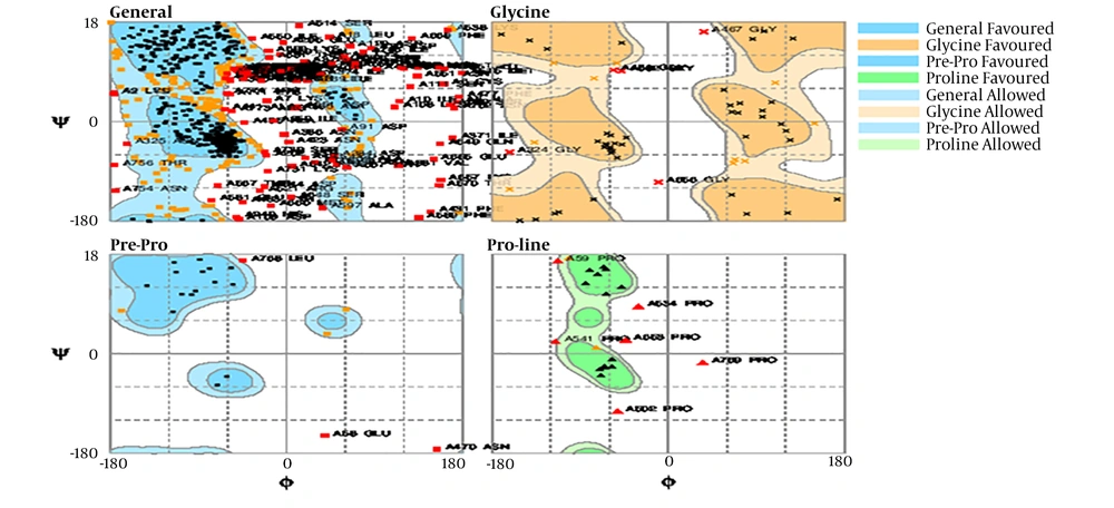
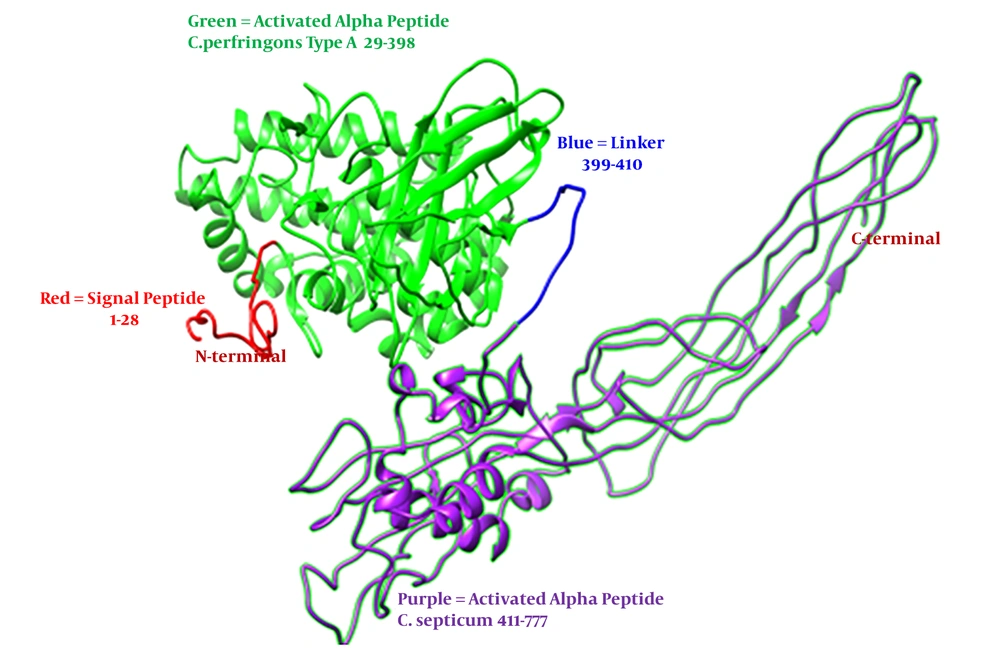
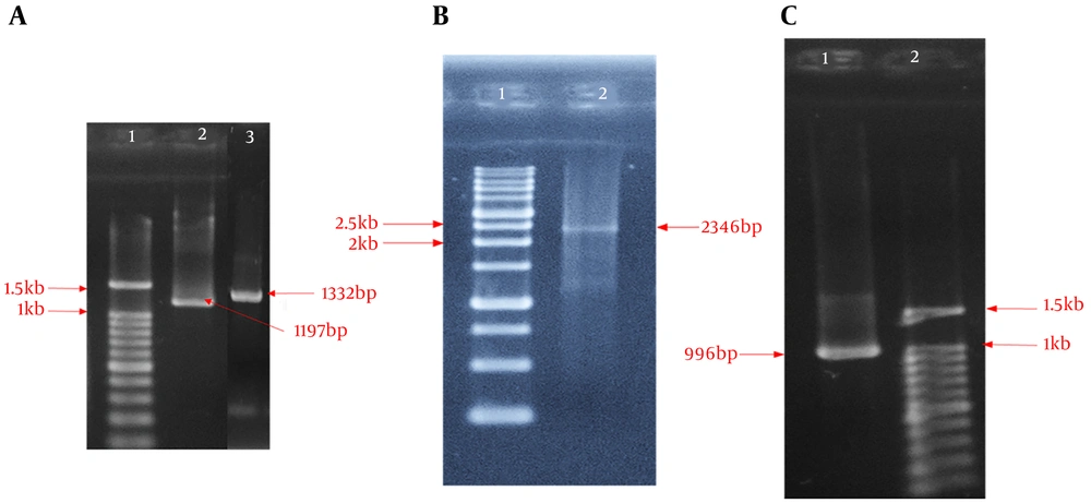
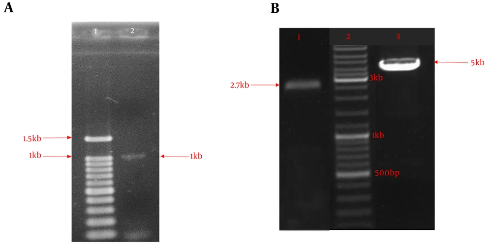
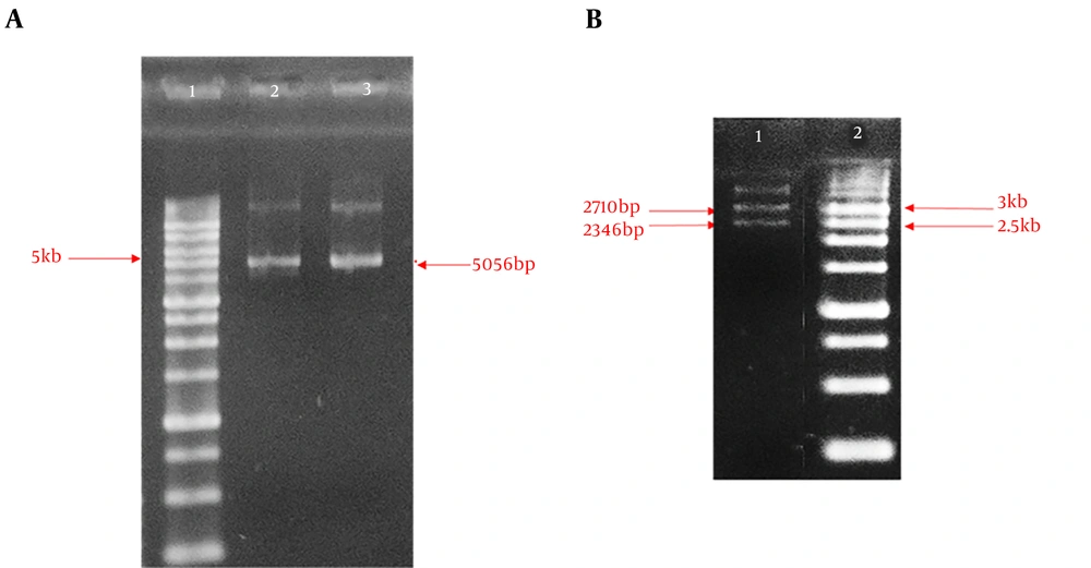
This experimental research intended to design a new bi-functional protein fusion for cpa and csa genes in E. coli TOP10. The I-TASSER server was used to predict the alpha-alpha fusion protein. The alpha-alpha protein fusion analysis was performed by the I-TASSER server: confidence score (C-score), estimated template modeling score (TM-score), and estimated root mean square deviation (RMSD) were -2.68, 0.41 ± 0.14, and 15.1 ± 3.5, respectively. Then, the form was exposed by the SPDBV-4.10 software for energy minimizing, and Rampage or Ramachandran plot analysis was performed to validate geometrical structure as a natural like protein. As shown in Figure 1, amino acid residues percent in the synthetic model for favored, allowed, and outlier regions was 70.5%, 17.9%, and 11.6%, respectively. So, the tertiary structure of the fusion protein construction was confirmed and visualized with the UCSF Chimera tool (Figure 2). The sequence of the synthetic construct (α-α fusion gene) was submitted to the NCBI database (accession number MK908396). After extracting the DNA of cpa and csa genes, PCR procedures were performed by two pairs of special primers for their gene amplification. Figure 3A show the result on agarose gel electrophoresis of PCR products. Subsequently, the synthetic construct of the alpha-alpha fusion gene was produced by the original cpa forward and csa reverse primers (Figure 3B). The synthesis construct using nested PCR (internal cpa forward and csa reverse primers) showed acceptable amplification of the desired fragment (Figure 3C). One 16 hours’ culture of E. coli TOP10 showed colonies on the LB-AMP agar medium, in which ligation product was transformed directly, while the culture of one non-transformed competent E. coli TOP10 colony did not grow on the LB-AMP agar plate. Screening gel electrophoresis (colony PCR) using internal cpa forward and csa reveres primers showed ~1 kb DNA fragment, which our designed linker fragment was included (Figure 4A). Colony PCR using pUC57 forward and reverse primers, as stated by the manufacturer’s suggestions, showed that 5 kb DNA fragment and negative control PCR product were one 2.7 kb bond (Figure 4B). Recombinant pUC57/αα vector was extracted from E. coli/TOP10/pUC57/αα (Figure 5A) and then digested with NdeI and XhoI restriction endonucleases (Figure 5B). Sequencing results of extracted and purified recombinant pUC57/αα confirmed a synthetic construct of the α-α chimeric fusion gene.
A, 1% agarose gel electrophoresis of PCR product. Lane 1: 100 bp DNA size marker, lane 2: C. perfringens type A alpha toxin gene, lane 3: C. septicum alpha toxin gene; B, fusion PCR product. Lane 1: 1 kb DNA size marker, lane 2: C. perfringens alpha-C. septicum alpha(α-α) fusion toxin gene; and C, nested PCR product. Lane 1: Nested PCR product for confirmation fusion gene, lane 2: 100 bp DNA size marker.
1% agarose gel electrophoresis for the screening of α-α fusion gene construction by colony PCR. A, Lane 1: 100 bp DNA size marker, lane 2: PCR product of recombinant E. coli/ Top10/pUC57/αα colony PCR with internal primer; B, Lane 1: Negative control PCR product (non-recombinant E. coli/ Top10/pUC57), Lane 2: 1 kb DNA Size marker, lane 3: PCR product of recombinant E. coli/ Top10/pUC57/αα colony PCR with pUC57 primer.
1% agarose gel electrophoresis for the screening of α-α fusion gene construction by enzymatic digestion. A, Lane 1: 1 kb DNA Size marker, lanes 2 and 3: uncut purified recombinant cloning vector (pUC57/αα); B, Lane 1: *digested purified recombinant cloning vector, lane 2: 1 kb DNA size marker. *Enzymatic digestion using NdeI and XhoI restriction endonuclease.
5. Discussion
A bi-functional protein fusion contains two protein domains or proteins bind by a fragment. Designing and producing a proper fusion protein has several difficulties. The fusion protein is designed to achieve better characterize and new functionality. Therefore, it is essential to have a proper cell for cloning. Then, the next step would be expression (21). In this study, the DNA sequence of cpa and csa genes was retrieved from the GenBank (7, 11). Before producing synthetic fusion gene, in silico analysis was used to design the alpha-alpha fusion protein structure. According to the results of the in silico analysis (C-score and TM-score of -2.68 and 0.41 ± 0.14, respectively), the designed model had an appropriate topology.
C-score and TM-scores are usually in the range of -5 to 2 and > 0.17, respectively. The Ramachandran plot (amino acid residues in favored and allowed regions = 88.4%) indicated that the geometrical structure of alpha-alpha fusion protein is acceptable as a natural like protein.
Because the percentage of amino acid residues is more than 85% in favored and allowed regions (17, 18). Genomic DNA of cpa and csa genes was used for amplification by employing forward and reverse primers designed from the available sequence. At the 5’ and 3’ends of the cpa gene, NdeI cleavage site and the first part of the designed linker were inserted via PCR method, respectively. At the 5’ and 3’ ends of the csa gene, respectively, were inserted in the second part of the designed linker and XhoI cleavage site via PCR method. In the present study, amplified and purified products were analyzed using the PCR method. By comparing the obtained sequences with previous reports obtained from the GenBank, the sequence identities of the PCR products were confirmed. The sequence of the amplified cpa gene was 1218 bp in length with 5 domains. These domains contain a flanking place (nucleotides 1 to 3), NdeI restriction site (nucleotides 4 to 9), alpha signal peptide (nucleotides 7 to 90), alpha active peptide (nucleotides 91 to 1200), and linker (nucleotides 1201 to 1218) for fusion with csa gene. The sequence of the amplified csa gene was 1146 bp in length with 4 domains. These domains include a linker (nucleotides 1 to 36) for fusion with cpa gene, alpha active peptide (nucleotides 37 to 1137), XhoI restriction site (nucleotides 1138 to 1143), and flanking place (nucleotides 1144 to 1146). After amplification by the PCR method, the genes were bound via a linker based on the fusion PCR method. A procedure for fusion construct in 2012 by Uppalapati et al. (20) was performed. The method is called the fusion PCR method. In our research, the alpha-alpha fusion gene was synthesized by the fusion PCR method. The synthetic construct gene was 2346 bp in length (Figure 3B), which consisted cpa gene (nucleotides 1 to 1200), linker (nucleotides1201 to 1236 as GCGGAAGCGGCGGCGAAAGAAGCGGCGGCGAAAGCG), and csa gene (nucleotides 1237 to 2346). The synthetic fusion gene was confirmed using the nested PCR. In this method, cpa forward primer and csa reverse primer begin amplification at nucleotide 539 and 1534 of the fusion gene, respectively. So amplified fragment was 996 bp in length, and the linker fragment was located in it (Figure 3C). Pilehchian Langroudi et al. (21) reported that epsilon-beta fusion gene was synthetsized by the fusion PCR method and the synthetic fusion gene was confirmed by the nested PCR. In the present study, plasmid pUC57 (2710 bp in length) was used as a cloning vector in E. coli. During ligation, the synthetic alpha-alpha fusion gene was cloned into the pUC57 plasmid vector at multiple cloning sites (MCS) and produced pUC57/αα (recombinant vector), which was 5056 bp in length. The recombinant vector was transformed into E. coli TOP10 and showed growth on the LB agar plate containing ampicillin. The resulting recombinant cell (E. coli TOP10/ pUC57/αα) was confirmed by colony PCR, which resulted in the production of 5 kb and 2.3 kb DNA fragments using pUC57 forward and reverse primers and cpa forward and csa reverse primers, respectively. Also, the negative control PCR product showed one 2.7 kb bond (Figure 4B). Another reason for this statement was enzyme digestion, the extracted digested plasmid produced two sharp bands of 2.7 kb and 2.3 kb on 1.0% gel electrophoresis that were related to pUC57 and alpha-alpha fusion gene, respectively, but extracted uncut plasmid produced two sharp bands above 5 kb. Additionally, sequencing analysis confirmed that the size of the synthetic construct alpha-alpha fusion protein gene is 2346 bp. Similar to the present study, Kamalirousta and Pilehchian (24) reported that epsilon-alpha fusion gene was cloned into a pGEM-B1 plasmid vector and then the recombinant vector was transformed into E. coli TOP10. The recombinant cell was confirmed by the colony PCR, enzyme digestion, and sequencing analyses (24). One of the limitations of this study was the complexity and difficulty of connecting these two genes using the fusion PCR method. It is suggested that this technique be used for other clostridium toxins and for the fusion of more than two genes.
5.1. Conclusions
This study provided a new strategy for fusing genes such as C. perfringens type A alpha and C. septicum alpha-toxin genes using the fusion PCR method. This method may serve as a model for the synthesis of other proteins and toxins. According to the best knowledge of the authors, this is the first study that has designed and cloned α-α fusion gene into a suitable cloning vector. The synthetic construct alpha-alpha fusion gene will be further used for sub-cloning in an expression host to produce α-α protein fusion. It can be considered as an appropriate candidate for recombinant vaccine development.

