


1. Background
Landing is one of the most commonly used sporting movements, which can produce an impact force between 2 to 12 times the body weight, the most common cause mechanism of lower limb injury (1, 2). This mechanical impact should be moderated via the musculoskeletal system. Increasing impact forces and repetition of these forces facilitate damage to the soft tissue surrounding the joint during landing. Single-leg landing (SLL) is common in sports such as volleyball, soccer, basketball, and badminton (1). The alignment of the lower limb has the main responsibility for absorbing pressure during contact and modulating the load (3). Lack of knee joint muscles’ ability to absorb force during landing may lead to changes in the kinematics of this joint, including increased knee valgus angle (KVA) and knee flexion (4, 5). About 70% of knee injuries, especially anterior cruciate ligament (ACL), occur in non-contact injuries (6, 7). Increased KVA and decreased knee flexion, tibia spin, internal rotation, and hip adduction within cutting and landing maneuvers can usually be the mechanism of damage to ACL injuries (6-10). which can increase the strain on the ACL (11, 12). Studies showed that SLL leads to an increase in damage to the ACL in a non-contact position compared to double-leg landing (DLL) (13, 14). They concluded that increased knee valgus and decreased knee flexion followed. A study by Yeow et al. showed that single-leg landing, compared to double-leg landing, significantly reduced knee flexion and increased the risk of anterior cruciate ligament damage (14).
Foot orthoses are usually prescribed to improve lower limb and functional impairment (15, 16). Researchers have considered the effect of orthoses on lower limb biomechanics during walking and running (17-19). When using foot orthoses during walking and running, a decrease in the movement of the lower limbs was observed in both the frontal and transverse planes (19). The effect of foot orthoses in sports activities such as landing, jumping, and cutting is still unknown, and most studies have investigated the effect of foot orthoses during walking and running (16, 19). Few studies have focused on the effect of foot orthoses on sports activities, such as landing, and have found different results (20, 21). Foot orthoses have increased activity in the gluteus medius during slow movements (20). On the contrary, foot orthoses have also shown an increase in movement in the frontal plane in the ankle and an increase in lateral pressure on the foot during the landing after the layup in basketball (21). The use of foot orthoses causes reduced lower limb injury in sports activities (15). It has been suggested that foot orthoses can change the movement of the lower extremities, which is responsible for reducing damage.
2. Objectives
The purpose of this research was to consider the effect of two types of foot orthoses (semi-hard foot orthoses and hard foot orthoses) on knee valgus angle during single-leg drop-landing.
3. Methods
3.1. Participants
Findings from a pilot study (n = 8) demonstrated the variability expected for each variable of interest. To achieve 80% or above power with two degrees of freedom at a significant level of 0.05, we calculated a needed sample size of 20 subjects (22). Twenty male recreational volleyball players with the mean ± standard deviation of age: of 22.3 ± 1.45 years; height of 1.75 ± 15.51 meters; body mass: 69.05 ± 0.51 kg participated in this study. All participants had no history of ACL injury as well as lower limb injury in the past six months. All players had played volleyball at least once a week during the past three years. All participants signed a written agreement form, before beginning this study.
3.2. Equipment
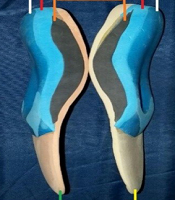
Participants used similar shoes (ASICS Gel-Rocket 7, Men's Volleyball Shoes, model B405N, Japan) to prevent the effect of footwear. In the current study, all participants were right-leg dominant. Nine reflective markers (15 mm in diameter) were placed on the anatomical points of the dominant leg. The anatomical points included the anterior superior iliac spine (ASIS), posterior superior iliac spine (PSIS), medial and lateral condyles of the femur, medial and lateral condyles of the tibia, medial and lateral malleoli, and the second metatarsal head (on the shoe). Markers were placed on the anatomical points before measurements. Three-dimensional (3D) motion data was captured at a frequency of 200 Hz using six digital cameras and Cortex software (Motion Analysis Corp., Santa Rosa, CA, USA) version 2.5.0.1160. Two types of foot orthoses (semi-hard and hard) with four layers were used in the study. The orthoses were similar except for the first and upper layers. The second to fourth layers in both foot orthoses were made of (polypropylene, bone POLYFOAM, and hard POLYFOAM), respectively. The first layer in semi-hard foot orthoses was made from semi-hard foam, while the first layer of hard foot orthoses was made from hard foam. The thicknesses of the layers of foot orthoses were: semi-hard foam and hard foam 5 mm, polypropylene was 1.5 - 2 mm, and hard polyfoam was 7 mm. Also, in general, the thickness of foot orthoses under the heel was 6 to 10 mm, and in the center of the inner longitudinal arc, it reached 25 to 30 mm. It should be noted that the molecular composition of the material used to make the foams was the same. But the factor that made hard foam, for example, harder in terms of hardness was that the amount of material poured into the mold was higher during the manufacturing of hard foam (Figure 1).

The right image of the semi-hard foot orthoses and the left-hand side of the hard foot orthoses with a four-layer profile used in this study.
3.3. Procedures
Subjects were required to do a 5-minute warm-up and given time to practice drop-landing maneuvers before recording data. Participants were requested to stand on a 0.3 m high step on two legs, with both hands on iliac crests. The drop-landing operation was performed with the dominant leg at 20 cm from the front edge of the platform. Subjects were trained to easily roll off the platform and perform the most natural possible landing. Subjects were trained to maintain their balance for at least 2 seconds after landing. Drop-landing was performed in three positions in a randomized manner: including without foot orthoses, with semi-hard foot orthoses, and with hard foot orthoses. A successful landing was defined as a landing without jumping up or forward or landing on both legs. Six successful trials were recorded for each orthosis.
3.4. Measures
The maximum knee angle was measured on the sagittal and coronal planes. Kinematics data were automatically filtered by a fourth-order zero-lag Butterworth 12-HZ low-pass filter (23). A kinematics model was proposed based on the lower limb to describe knee joint angles during landing. The hip joint center was estimated based on the methods described by Vaughan et al. (24). In the standing trial, joint centers for the knee and ankle were determined (25). The femur’s center was defined as the midpoint between the medial and lateral condyles of the femur. The center of the tibia was defined as the midpoint between the medial and lateral condyles of the tibia. The center of the talocrural joint was defined as the midpoint between medial and lateral malleoli. Knee flexion and valgus angles were calculated based on the GROOD and SUNTAY method (26). Maximum knee flexion and maximum knee valgus angles within landing were calculated for each trial during the landing phase. The fool landing phase was defined from the primary ground contact of the dominant foot until maintaining balance without motion and with no ground contact in the opposite foot for at least 2 seconds.
3.5. Statistical Analysis
The variables of interest were maximum knee valgus angle and maximum knee flexion angle during the whole phase of landing. The mean of the six measurements in each trial condition (without foot orthoses, semi-hard foot orthoses, and hard foot orthoses) was calculated. The mean values were used for the analysis. The Shapiro-Wilk test (P > 0.05) was used to check the normality of continuous variables. Continuous variables were presented using mean and standard deviation (SD). One-way repeated measures analysis of variance (ANOVA) was used to compare the three trials. Pairwise comparisons were made using Bonferroni correction. The η2 was calculated as effect size. The η2 value greater than 0.25 is considered as large effect size, while moderate and small effect size are defined as η2 vale between, 0.09 and 0.01 and lesser than 0.01, respectively (27). Data was analyzed using the statistical package for social sciences (SPSS) software (IBM Inc, USA) version 22.
4. Results
4.1. Knee Valgus Angle
There was a significant difference in knee joint valgus angle between the three trials (P < 0.001). Post-hoc tests showed that knee valgus angle was significantly smaller in the mid-hard foot orthoses compared to hard foot orthoses (P < 0.001) or without orthosis trials (P < 0.001). Furthermore, the post hoc test revealed a significant difference in knee valgus angle between without foot orthoses and hard foot orthoses conditions (P < 0.001) (Table 1).
Mean (SD) of Knee Valgus and Flexion Angles
4.2. Knee Flexion Angle
There was a significant difference in knee flexion angle between the three trials (P < 0.001). Post-hoc tests revealed greater knee flexion angle in the mid-hard foot orthoses trial, compared to hard foot orthoses (P = 0.000) or without orthoses trials (P = 0.000). Furthermore, the post-hoc test revealed a significant difference in knee flexion angle between without foot orthoses and hard foot orthoses trials (P = 0.002) (Table 1).
5. Discussion
To the best of our knowledge, this study was the first research that assessed the effect of two different foot orthoses (mid-hard foot orthoses and hard foot orthoses) on knee valgus angle during single-leg drop-landing. As we hypothesized, the mid-hard foot orthoses resulted in a significant reduction in maximum knee valgus angle and a significant increase in maximum knee flexion angle during single-leg landing. These findings indicate that foot orthoses alter the kinematics of knee during single-leg landing.
5.1. Effects of Foot Orthoses on Knee Valgus Angle
The results of this study demonstrated that mid-hard foot orthoses reduced knee valgus angle during single-leg drop landing compared to hard foot orthoses or without foot orthoses trials. No study has yet compared lower limb kinematics during landing with different foot orthoses. Several studies have reported kinematic changes in lower extremity joints during walking and running due to foot orthoses (16, 19, 28-30).
Subjects wearing foot orthoses were shown to exhibit kinematic changes in the frontal (knee), and transverse (ankle and hip) planes. Foot orthoses were shown to reduce knee joint valgus (knee abduction) in a group of female basketball players (31). Previous studies showed that little changes in knee valgus angle could extremely alter knee valgus load. McLean et al. showed that a two-degree change in knee valgus angle resulted in 40 N.M enhancement in knee valgus moment (32). Furthermore, Chaudhari and Andriacchi reported that knee abduction (frontal plane position) as little as 2° has the potential to cause ACL injury (33). Our findings suggest that landing with mid-hard foot orthoses could decrease the valgus load on the knee through decreasing the knee valgus angle.
5.2. Effects of Foot Orthoses on Knee Flexion Angle
Our research also showed the maximum knee flexion angle was achieved when landing with a semi-hard foot orthosis. Previous studies showed that the maximum damage to ACL occurs at an angle close to full extension (8). Increase in knee flexion during sports activities reduces the force on the ACL. The anterior shear force is the main predicting factor of the load on ACL (12). When the knee flexion increases, the angle between the patella and tibial tendon increases, and the quadriceps contraction force produces less anterior shear force in the proximal tibia (34). Therefore, the increase in knee flexion is accompanied by a decrease in force on the ACL. On the other hand, the increase in knee flexion is accompanied by a reduction in the posterior ground reaction force. This finding is due to the direct relation between anterior shear force and posterior ground reaction force (35). Therefore, it can be concluded that the increase in knee flexion will reduce the force on the ACL. Southard et al. used a biomechanical model and compared biomechanical factors in both normal landing and landing with a bending knee and showed that tension on ACL is reduced by 10% in landing mode (36). Concerning findings of previous studies, the findings suggest that landing with a semi-hard foot orthosis can reduce tension on the ACL.
5.3. Conclusions
Enhanced knee valgus angle increased the risk of ACL damage during landing among athletes. Our study showed that mid-hard foot orthoses decreased KVA. The results of this study were based on a small number of subjects. There is need for more studies on the effectiveness of foot orthoses on kinematics in coronal and sagittal planes. A foot orthosis that has the potential to optimally change lower limb motion or neuromuscular recruitment models will be promising in decreasing the incidence of non-contact ACL damage in a male sportsman.
