1. Background
In terms of improving athletic performance, high-intensity interval training (HIIT) leads to reduced weekly running distances and an increase in mean running intensity without impairing performance (1). The primary objective of HIIT is to accumulate activity at an intensity that is too high for the participant to maintain for an extended period of time (i.e., 80 - 95% of peak oxygen consumption (VO2peak) or > 90% of maximum heart rate (HRmax). Therefore, the recovery time should be sufficient to allow the subsequent interval to be completed at the desired intensity (2). It is essential to plan training adequately and to implement a training load monitoring and adjustment system that incorporates biofeedback for the training to take place (3) effectively. Sports practitioners are continually searching for ways to design training programs that are most objectively, quickly, and cost-effectively suited for each individual participant. Based on individual adaptation, these programs aim to maximize long-term performance gains while avoiding excessive fatigue, overtraining, and injuries during the recovery period after training sessions. Recovery and super-compensation of body functions serve as indicators of long-term gains in performance (4-7).
Muscle fatigue is defined as the reduction of maximal voluntary contraction (MVC) or the inability to maintain the desired force during exercise. In HIIT exercise, essential causes of muscle fatigue are increased production of lactate, proton ions, and depletion of creatine phosphate (8). Accurate control of the concentration of these fatigue factors, which are indicators of exercise load, can be effective in monitoring fatigue in the short-term and muscle adaptation in the long term (9). An effective way of achieving this goal is to choose a more accurate indicator of training load.
To control fatigue, a number of variables, including training intensity and recovery, must be considered in individualized HIT programs. Accordingly, an HR-based or speed-based approach is based on the final speed reached in a 30 - 15 intermittent fitness test (30 - 15IFT, VIFT) or on the maximal heart rate (HRmax) (10).
This is more difficult to accomplish in practice, especially in a team setting where players have been prescribed a similar training regime but have individualized responses to stimuli (11). There are a number of potential markers that can be used to determine the training load and its effect on the athlete. Several external tools for quantifying and monitoring load have been investigated, such as speed and time-motion analysis, as well as internal load metrics, such as heart rate, heart rate recovery, and blood lactate. An athlete's fatigue level can be determined by dissociating external and internal load units (12).
Training adaptations and competition performance both depend on the correct titration of fatigue (13). It is also essential to develop a program that is individualized to ensure that the athlete's internal load reflects the one planned by the coach (14). Monitoring HR may serve as one of the best means of elucidating fatigue in athletes (12). However, controlling the training load using speed/time indices does not seem to take into account the individual physiological and psychological differences of the athletes.
2. Objectives
To help clarify the individuals' physiological differences for optimal intensity prescription, the objective of this study was to compare HTHR and HTST on some fatigue-associated factors in active men. Given that the heart rate is constantly affected by various physiological factors, we assume that during HTHR, recovery time, training intensity, and/or output change during training.
3. Methods
3.1. Participants
Twenty young male athletes (age 22.75 ± 2.5 years, weight 76.60 ± 8.85 kg, percent of body fat 17.65 ± 4.44) participated in all components of the study. All participants were nonsmokers, had no musculoskeletal body injury, and had not consumed supplements in the previous 2 years.
VO2max by inserting the calculated maximum speed of the individual in the estimation formula, the aerobic capacity of the subjects was estimated (15) (Table 1).
| Variables | Mean ± SD |
|---|---|
| Age (y) | 22.75 ± 2.51 |
| Height (cm) | 179.73 ± 6.52 |
| Weight (kg) | 76.60 ± 8.85 |
| Body fatty (%) | 17.65 ± 4.44 |
| Maximum heart rate | 191 ± 1.92 |
| VIFT | 16.85 ± 1.31 |
| VO2maxa | 46.37 |
a By inserting the calculated maximum speed of the individual in the estimation formula, the aerobic capacity of the subjects was estimated.
VO2max (mL.kg-1.min-1) = 28.3 - (2.15 * G) - (0.741 * A) - (0.0357 * W) + (0.0586 * A * VIFT) + (1.03 * VIFT): VIFT is the final running speed; G refers to gender (male = 1; female = 2); A for age (in years); W for weight (in kilograms).
3.2. Experimental Design
A randomized crossover study with a 9 a.m. - 12 p.m. start time was used for the experiment. During the week before the experiment, the subjects entered the research protocol after completing the consent form and were fully familiar with the process of implementing the research. Subjects were asked to have breakfast at least 3 hours before body composition assessment. At first, the subjects were evaluated for body composition (Bocax1 body composition assessment device, Korea), height (SECCA, Japan), and weight (SECCA, Japan) at a specific time of day. Then, to elicit heart rates (HR), VO2max, anaerobic capacity, and inter-effort recovery capacity, participants performed the 30-15IFT (16). In the following, participants were randomly divided into two groups, participants performed HTHR or HTST in the first session. Before and immediately after exercise, physical and physiological assessments were performed, and blood samples were collected. After the first session, as a one-week washout period, participants were prevented from strenuous exercise and using supplementation. And on the second session, participants who performed HTHR were asked to perform HTST and vice-versa. For exclusion criteria in this study, drug or supplementation intake in the interval between two tests, injury, and participation in a strenuous exercise before the second test was considered.
3.3. Protocol
In order to estimate aerobic capacity (VO2max) and running velocity (VIFT) for the last completed stage (15), a suitable alternative to vVO2max (16), the subjects performed the 30 - 15 intermittent fitness test (30 - 15IFT) (Metronome device, China). Also, the maximum heart rate of individuals was estimated by the HRmax estimation formula provided by Tanaka et al. for active men (HRmax = 207 - 0.7 × age) (17). In the next step, two HIIT acute running exercises were performed on the treadmill (H/P/cosmos, Germany) with control based on heart rate and speed/time.
3.3.1. HIIT Exercise Based on Speed/Time (HTST)
In this method, the ratio of work to rest duration was considered 1 to 3 (work 1: 3 rest). In such a way that during activity, the subjects ran with an intensity of 90% of maximum speed for 30 seconds, then they ran with an intensity of 55% of maximum speed during rest for 90 seconds alternatively. During exercise, the individual's monitoring heart rate was recorded every 30 seconds (18). This exercise was performed for 40 minutes.
3.3.2. HIIT Exercise Based on Heart Rate (HTHR)
In this exercise, the heart rate of the subjects was visible on display at any time due to the wearing of a Polar heart rate monitor chest strap. The exercise started at 1 km/h, and every 20 seconds, 1 km/h was added to the treadmill speed to reach 90% of the maximum heart rate (HRmax). Immediately, the instantaneous speed and time elapsed from the start of the exercise were recorded. Then every 20 seconds, the treadmill speed was reduced by 1 km/h until the heart rate reached 55% of the maximum heart rate (HRmax). At this moment, the instantaneous speed and the time elapsed from the beginning of the exercise were recorded (19). This cycle lasted for 40 minutes. Heart rate was also recorded every 30 seconds throughout the workout.
3.4. VIFT Test
Briefly stated, the 30 - 15IFT involved running shuttles for 30 s (40 m) interspersed with time for passive recovery for 15 s. The starting velocity was 8 km/h for the first 30 s, then increased by 0.5 km/h each 30 seconds afterward. An audio signal controlled the running pace. As many 30 s "stages" as possible were required of the subjects during the test, with the test ending when they were unable to maintain the required pace (that is, when players were unable to reach a 3 m zone near each marked line each time the audio signaled three times). As part of the test, performance was expressed in terms of estimated VO2max (mL × kg × 1 × min × 1), speed (km × h × 1), and time (min). During the test, the subjects were advised orally to exert maximum effort (20).
3.5. Blood Samples and Analyses
Venous blood samples were taken from the brachial vein before and at the end of the exercise. Immediately, drawn blood in a syringe was transferred exactly 7 mL and 3 mL of blood into the clot activator tube and the evacuated tube, respectively. Then the tube was shaken vigorously to mix. The whole blood specimen was sent in the original collection container at refrigerated temperatures and was sent immediately to the laboratory. All of the kits had been bought from Pars Azmoon company (21).
3.6. CK
NAC. Kinetic UV. The liquid in serum or plasma was used to measure the creatine kinase. The diameter of the cuvette was 1 cm. The wavelength at 25, 30, and 37°C was set at 340 nm. The sensitivity of this method was 10 U/L.
3.7. Lactate
Enzymatic UV method (SFBC) for the determination of lactate dehydrogenase (LDH) in serum or plasma was used to measure the lactate concentration. The diameter of the cuvette was 1 cm. The wavelength at 37 C was set at 340 nm. The sensitivity of this method was 1 mg/dL.
3.8. Pyruvate
A quantitative enzymatic UV-test in serum or plasma was operated to measure the creatine kinase. The diameter of the cuvette was 1 cm light path. The wavelength at 30 and 37 C was set at 340 nm. The lower detection limit (sensitivity) was 0.1 mg/dL.
3.9. Glycerol
The level of glycerol was measured by enzymatic photometric assay. The diameter of the cuvette was 1 cm. The wavelength at 20 - 25 C/37 C was set at 546 nm.
3.10. Glucose
A glucose oxidative GOD-POD in serum or plasma was operated to measure the glucose levels. The diameter of the cuvette was 1 cm light path. The wavelength at 37 C/15 - 25 C was set at 340 nm. The sensitivity of this method was 1 mg/dL= 0.0039 (A).
3.11. Statistical Analysis
The normality of the distribution of the variables was tested using the Shapiro-Wilk W-test. Pre-test and post-test changes of each variable were analyzed using a dependent t-test. The univariate test was used to investigate the two types of HIIT. Statistical analysis was performed using IBM SPSS statistics (version 24, IBM). P-values < 0.05 were considered significant.
4. Results
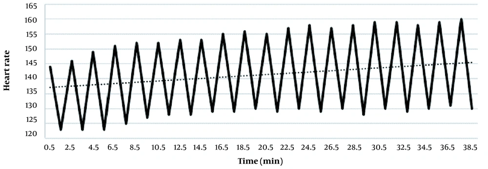
There were significant differences between maximal, minimum, and mean heart rates at the beginning of the HTST and the end of the training (Figure 1)
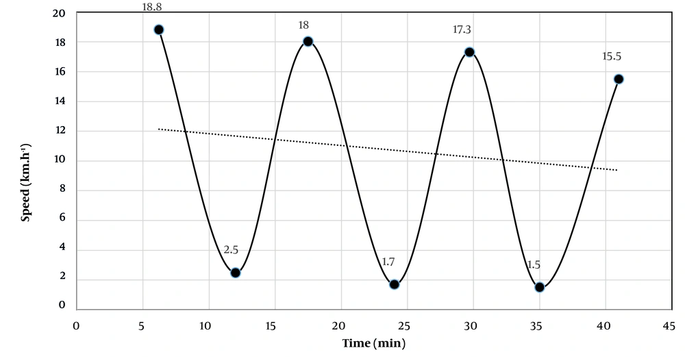
Also, there were changes between maximal, minimum, and mean speed of the beginning of HTHR and the end of the training (Figure 2)
4.1. Blood Analysis
Table 2 shows that the rate of change [posttest-pretest] levels of lactate, creatine kinase, and lactate to pyruvate ratio in HTHR exercise increased significantly compared to HTST exercise (P < 0.05). Also, in the HTHR, the pH and pyruvate variables decreased significantly more (P < 0.05).
| Variables | HTHR | HTST | P | ||||
|---|---|---|---|---|---|---|---|
| Pre | Post | Δ | Pre | Post | Δ | ||
| La- (mg/dL) | 5.80 ± 0.102 | 56.05 ± 1.287 | 50.25 | 5.95 ± 0.15 | 50.90 ± 1.40 | 44.95 | < 0.01 |
| CK (mg/dL) | 126.9 ± 3.71 | 231.3 ± 4.29 | 104.4 | 138.1 ± 3.91 | 221.4 ± 5.40 | 83.3 | < 0.01 |
| Pyr (mg/dL) | 0.670 ± 0.007 | 0.547 ± 0.009 | - 0.123 | 0.647 ± 0.011 | 0.544 ± 0.016 | - 0.103 | < 0.01 |
| La-/Pyr | 8.69 ± 0.84 | 102.9 ± 12.93 | 94.21 | 9.24 ± 1.35 | 93.8 ± 13.06 | 84.56 | < 0.05 |
| pH | 7.41 ± 0.004 | 7.05 ± 0.01 | - 0.36 | 7.40 ± 0.003 | 7.11 ± 0.02 | - 0.29 | < 0.01 |
| Glyc (mg/dL) | 160.6 ± 4.19 | 137.8 ± 4.30 | - 22.8 | 163.7 ± 5.05 | 164.7 ± 5.02 | 1.7 | < 0.01 |
| Glu (mg/dL) | 82.70 ± 1.69 | 62.05 ± 0.87 | 20.05 | 87.95 ± 2.08 | 84.85 ± 1.28 | - 3.1 | < 0.01 |
Abbreviations: La-, lactate; CK, creatine kinase; Pyr, pyruvate; La-/Pyr, lactate to pyruvate ratio; Glyc, glycerol; Glu, glucose.
In the HTHR, the blood glycerol level decreased significantly (P < 0.05), but in the HTST, the glycerol level increased significantly (P < 0.05). Since the Vmax (intensity) of HTHR was higher, these results had been predicted. However, it should be noted the standard deviation of the post-blood variables in two types of training; thus, it is lower in HTHR than it in HTST. This means that the blood factors of HTHR were more concentrated around the mean.
Table 3 shows that the covered distance in HTST is significantly longer than in HTHR. Therefore, considering that the total training time was equal in both types of training, the average speed of the subjects in HTST is higher. In the same way, the average HR in HTST was higher. Although the mean of HR and speed exceeded in HTST, blood factor variations were lower. These data show that the relative intensity of each exercise can't be calculated by the average of indexes and covered distance. In contrast, between maximum HR and speed with the intensity of the exercises exists a powerful relation. It’s while the intensity of HTHR was higher, mean blood pressure was lower (Table 4). This finding is in contrast to previous studies' results.
| Variablea | HTHR (n = 20) | HTST (n = 20) | Δ | P |
|---|---|---|---|---|
| Distance (m) | 6200 ± 759 | 7119 ± 540 | 919 | < 0.01 |
| Average speed (km.h-1) | 9.30 ± 1.13 | 10.68 ± 0.81 | 1.38 | < 0.01 |
| Maximum speed (km.h-1) | 18.85 ± 2.51 | 16.85 | 2 | < 0.01 |
| Minimum speed (km.h-1) | 1.50 ± 1.2 | 9.2 ± 10 | - 7.7 | < 0.01 |
| Average HR | 137.45 ± 2.74 | 140 ± 12 | 3 | < 0.01 |
| Maximum HR | 193 | 160 ± 14 | 33 | < 0.01 |
| Minimum HR | 105 | 122 ± 12 | - 17 | < 0.01 |
| Variable | HTHR | HTST | P | ||||
|---|---|---|---|---|---|---|---|
| Pre | Post | Δ | Pre | Post | Δ | ||
| Values | 92.55 ± 10.17 | 93 ± 7.71 | 0.45 | 91.85 ± 7.7 | 96.2 ± 8.34 | 4.35 | < 0.01 |
5. Discussion
It has become increasingly important to monitor athlete loads in order to assess whether athletes are adapting positively or negatively to the collective stress of training and competition because of the importance of managing athlete fatigue (22). However, to the authors' knowledge, the comparison of the recovery, intensity, internal pressure, training output, and blood factors in the HTHR and HTST and associating them to fatigue has never been examined on active men. Heart rate, speed, and time are important and practical indicators for controlling training load in athletes. The data from this study show that controlling the exercise load by speed/time indicators leads to an increase in heart rate response during exercise. We can see this response both in the low-intensity phases and in the high-intensity phases. It has been shown in some previous studies that change in internal load with respect to a standard external load may be used to infer an athlete's fitness or fatigue over time or in comparison with that of their peers (23, 24). The activities performed by athletes represent an external load (speed, distance, and time), yet the physiological adaptations come about because of internal load (heart rate as one of the indicators of internal load), and this is primarily in the form of biochemical stresses (25). Heart rate rises, and stroke volume falls over time when exercise lasts longer than 15 - 20 minutes, which is known as cardiovascular drift (26-28). Cardiovascular drift may be modulated by a number of factors, including exercise intensity, hyperthermia, dehydration, and ambient temperature (26). During an exercise session lasting an hour, Ekelund identified the physiological changes that took place in 18 individuals. Gradually, HR increased over the hour, but the largest increases were observed within the first 30 minutes. The increase in HR over the first hour was 15%. One hour of cycling constant work rate was required by Mognoni et al. (29). As the exercise duration increased from 10 minutes to 60 minutes, the heart rate increased from 135 beats/min to 150 beats/min (11%) (30).
Figure 2 shows that in HTHR, the subjects' speed decreased gradually. This means that the rate of increase in heart rate has increased in a disjointed manner. A study that has already shown this has not been found, but it also seems to be related to cardiovascular drift. Furthermore, glycogen depletion, increased catecholamine hormones, and fatigue factors may be the causes. As we have assumed, in this exercise, the output of the exercise will probably decrease with increasing internal pressure (load). This phenomenon is important in team sports; because before and during training, there are internal load differences between individuals for various reasons. Therefore, coaches are more likely to ignore these individual differences when training based on speed, time, and VO2max. These individual differences can be due to variable and unpredictable factors, including nutrition, psycho-physiological condition, weather conditions, rest, and previous exercises.
Comparing the variables of distance, speed (average, maximum and minimum) and blood factors can provide us with useful information. A comparison of all these variables shows that individualization is better done in HTHR; in this exercise, the standard deviation and also the difference between the maximum and minimum average speed are more than in HTST. On the other hand, the standard deviation of fatigue-related blood factors (lactate, pH, creatine kinase, lactate/pyruvate, glycerol, and glucose) is less in HTHR (Table 2). This can be one of the reasons supporting our hypothesis that with HTHR, the principle of individual differences is considered more. Because although the subjects exercised with more differences in speeds, the accumulation of fatigue factors in them was closer to each other (concentrated around the means). Previous studies have demonstrated that lactate production is associated with muscular fatigue, and is a major limitation in athletic performance. This fatigue is partially due to the production of H+ ions which depresses muscle function (31). Increased production of proton ions, lactate, creatine kinase, and lactate/pyruvate ratio during exercise is directly related to its intensity (32). Since a higher maximum speed (intensity) was observed in the exercise based on heart rate, the rate of increase of these variables was higher in HTHR than in HTST. In this regard, increasing the level of glycerol in HTST and decreasing it in HTHR indicates a higher intensity and fatigue in exercise based on heart rate; because changes in glycerol with increasing intensity of exercise are first increasing and then decreasing (32).
Our results show that although average speed, average HR, and covered distance in HTST were more, however, its intensity was performed at a lower level according to the blood agents’ variations. In other words, it seems the mentioned variables do not have use for the determination of intensity, while potent relation exists between blood factor variations with maximum speed and maximum HR in the two types of exercise.
Although HTHR was performed at a higher intensity, blood pressure was significantly lower at the end of training. While many previous studies demonstrated that MAP increases with an increasing workload (33, 34).
In general, by gathering all mentioned details together, it can be concluded that training based on HR, in contrast to speed/time, decreases internal load differences and increases external load differences between individuals.
5.1. Conclusions
The work-to-recovery ratio seems to be more precisely controlled in HTHR. It also adjusts the external load in subjects by keeping the internal load constant. Probably because of this, there are fewer differences in blood factors (standard deviation) among the subjects. However, in HTST, the internal pressure increases as the external pressure is constant. Also, the deviation from the average blood factors of the subjects in this exercise is more. In general, in order to accurately control the intensity and recovery of acute training, the use of the heart rate index is more appropriate than the speed/time index.


