1. Background
Adequate flexibility is important for injury prevention in athletes. Flexibility is often evaluated according to the range of motion (ROM), which is measured either actively or passively. Active ROM is defined as the movement of an individual’s voluntary muscles, whereas passive ROM is defined as therapist- or device-assisted movement, possibly at a joint or group of joints. Both measures greatly depend on the stretch tolerance of participants (1). Previous studies (2, 3) have reported that limited ROM is a risk factor for injury occurrence.
Several studies have shown that foam rolling intervention could increase the hip, knee, and ankle ROM without impairing muscular strength (4-8), whereas others reported no changes in ROM after the intervention (9, 10). Applying the foam rolling intervention to the quadriceps and hamstrings has reportedly resulted in up to 5 – 12% and 9% increased ROM for knee and hip flexion, respectively, as compared with the values observed before the intervention (6, 11). Therefore, foam rolling is considered an effective method for athletes to perform their own conditioning.
Pole vaulting is a track and field event. A large hip flexion and extension angle during pole vaulting is considered an effective method for injury prevention. Rebella (12) reported that the lower back was the most common location of injury among collegiate pole vaulters. A previous study reported that the flexed type of low back pain was the most prevalent, and its occurrence was associated with low hip flexion and extension ROM among collegiate pole vaulters and decathletes (2). In addition, pole vaulters with chronic low back pain were shown to have lower active straight leg raise (SLR) angles than healthy pole vaulters, suggesting poor trunk stability or incompetence of the rest of the kinetic chain required when raising the lower limbs (3). Thus, it is likely that athletes must improve their hip ROM in some way. Nonetheless, whether the improvement in hip flexion and extension ROM changes their hip joint angle during pole vaulting remains unclear.
2. Objectives
The present study aimed to clarify the acute effects of intervention for hip flexion and extension ROM in pole vaulters on the maximum hip joint angle during pole vaulting. We performed an intervention to improve the hip ROM in male pole vaulters and compared the pre and post-intervention hip flexion angle during pole vaulting. We hypothesized that increased hip ROM would also increase the maximum hip joint angle during pole vaulting.
3. Methods
3.1. Research Design
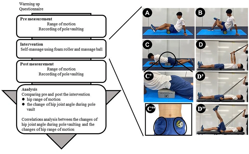
We performed the same intervention to improve the hip flexion ROM. All measurements and interventions shown in Figure 1 were performed within a single day.
General procedure followed during the present study and interventions to improve hip range of motion (ROM). A, Foam rolling for the hamstrings (5 minutes on each side). B, Foam rolling for the buttocks (5 minutes on each side). C, Self-massage of the muscle groups in the anterior pelvis (C’’) using a massage ball and baller box (10 minutes on both sides). D, Active straight leg raise exercise (20 times on each side): Athletes extend their shoulder (D’) and raise their straight leg (D’’).
3.2. Population
Male pole vaulters aged ≥ 18 years as of March 2019 who participated in a game organized or co-sponsored by the Inter-University Athletic Unions of Tokai or Central Japan Industrial Track and Field Association in 2019 were recruited. Among 30 recruited athletes, those who did not provide written consent (n = 13) and experienced pain during measurements (n = 0) were excluded. The evaluation was performed independent of whether the athletes had low back pain. The present study was conducted in accordance with the ethical guidelines of the 1975 Declaration of Helsinki, as reflected in a prior approval obtained from the Chukyo University Research Ethics Committee (approval no. 2019 - 004). Informed consent was obtained from each pole vaulter included in this study.
3.3. Questionnaire
A self-report questionnaire was used to collect data on participants’ demographics, such as age, height, body weight, personal best record, and competition history in pole vaulting. The take-off leg was also recorded in the same questionnaire and was defined as the leg used during pole vaulting. The other leg was defined as the lead leg.
3.4. Medical Analysis
A procedure employed in previous studies (2, 3) was used to measure both active and passive hip flexion and hip extension ROM in participants lying on a bed. All measurements were recorded using a camera (EX-F1, CASIO, Tokyo, Japan) and analyzed using image analysis software (NIH ImageJ version 14.4). The hip flexion-extension angle was calculated as the angle between the line connecting the greater trochanter and the lateral epicondyle of the femur and a line parallel to the trunk. Passive ROM measurement was performed by an athletic trainer.
3.5. Intervention Program
We used the intervention program suggested by Markovic (11) with additional exercises to improve hip flexion and extension ROM. Self-massages were used to improve the hip flexion and extension ROM (Figure 1A – C). Self-massages of the hamstrings (Figure 1A) and buttocks (Figure 1B) using a foam roller were combined, and the participants could massage themselves for 5 minutes per leg. Self-massages of the muscle groups in the anterior pelvis using a massage ball and ball box (Figure 1C) were also performed for 5 minutes per leg. Active SLR exercise was performed to improve trunk stability and the kinetic chain (Figure 1D). Each participant carried out a set of 10 repetitions during the active SLR exercise. We completed all intervention programs in approximately 25 min on an experimental day, with the examiner monitoring the interventions for all participants.
3.6. Biomechanical Analysis
A procedure employed in a previous study (13) was used for biomechanical analysis. The video for motion analysis was recorded from four directions with the planting box at the center using four high-speed cameras (GC-LJ20B, JVC, Kanagawa, Japan), recording at a rate of 240 fields/s. All the videos were synchronized by recording LED lights on them. The calibration area was set using the left edge as a reference, with a depth of 5 m and 2.5 m on the runway and mat sizes, respectively; a width of 1.25 m and 2.5 m on the left and right sides of the runway, respectively; and a height of 5 m. Calibration poles of 5 m in height (0.5 m between marks) were set up at 10 points in the range and included in the video. For the experimental trials, the participants vaulted over bungee bars that were set at the height of 90% of the athletes’ personal best record until the pole vaulters were able to jump over it. This trial was used for the analytical trials. The poles and the number of steps for each trial were selected by the pole vaulters themselves.
Video analysis was conducted from the moment of touchdown of the last step of the run-up to the moment of pole straightening (14). Digitization of body measurements was performed manually at a rate of 240 fields/s using a motion analysis system (Frame-DIAS V, DKH Inc., Tokyo, Japan) to assess the knee and hip joints in both lower extremities and the 12th rib in both upper extremities. We affixed markers to the knee and hip joints of both lower extremities and the 12th rib of both upper extremities of the athletes. The global coordination system was constructed using Gy in the horizontal direction and Gz in the vertical direction of the run-up, with Gx as the cross product of Gy and Gz. The maximum standard errors of the control points in each axis were 0.023 m, 0.020 m, and 0.011 m along the x-, y-, and z-axes, respectively. Data smoothing was performed by determining the optimal cut-off frequency (14.6 – 41.7 Hz) using a low-pass Butterworth digital filter (15). The joint angle during pole vaulting was calculated from digitized data using a three-dimensional direct linear transform algorithm. In this study, only joint angles within the Y-Z plane were calculated to determine hip flexion and extension on each side because the hip angles in the sagittal plane were measured in ROM measurement. The detailed method for calculating the joint angles is described below.
3.6.1. Hip Joint Angle (Both Legs)
For the migration coordinate system of the lower torso segment, the unit vector from the left great trochanter to the right great trochanter was defined as xlt, and the unit vector from the center of the great trochanter to the center of the lower end of the ribs was defined as zlt. Further, ylt was determined by extrapolating xlt and zlt, and xlt’ was determined by extrapolating ylt and zlt. The hip angle was defined as the angle formed by projecting vector zth from the abductor to the knee joint onto the plane defined by yltzlt, which was defined as -zlt. From the upright position, flexion was considered positive, whereas extension was considered negative.
3.7. Statistical Analysis
All data analyses were performed using SPSS version 23 (IBM Corp., Armonk, NY, USA). The normality of all data was analyzed using the Shapiro–Wilk test. A paired t-test was used to compare the pre and post-intervention hip ROM. Results were reported as mean ± standard deviation and 95% confidence interval, and Cohen’s d was set as the effect size (16). All tests were 2-sided, and P < 0.05 was considered statistically significant. The effect sizes were divided into five categories as follows: < 0, adverse effect; 0 – 0.20, no effect; 0.20 – 0.50, small effect; 0.50 – 0.80, intermediate effect; and ≥ 0.80, large effect (17). Pearson’s correlation analysis was used to test the correlation between the change in ROM due to the intervention and the change in joint angle during pole vaulting. Correlations were analyzed to clarify whether hip joint angles during pole vaulting also changed according to the changes in hip ROM caused by the intervention.
4. Results
This study included a total of 17 male pole vaulters (mean ± standard deviation: Height, 1.73 ± 0.1 m; body weight, 66.8 ± 6.7 kg; age, 22.6 ± 3.5 years; personal best record in pole vaulting, 5.0 ± 0.3 m; period of pole vaulting, 10.0 ± 2.6 years).
The pre and post-intervention hip ROM is shown in Table 1. The active hip flexion ROM on the lead leg was significantly decreased post-intervention (109.08 ± 5.74 degree) compared to pre-intervention (111.20 ± 6.57 degree). No significant improvement in hip ROM was observed post-intervention.
| Range of Motion (deg) | Pre-intervention (N = 17) | Post-intervention (N = 17) | P-Value | Cohen’s D | ||
|---|---|---|---|---|---|---|
| Mean ± SD | 95% CI | Mean ± SD | 95% CI | |||
| Take-off leg | ||||||
| Active | ||||||
| Hip flexion | 108.29 ± 7.24 | 104.56 - 112.01 | 107.84 ± 6.60 | 104.45 - 111.23 | 0.490 | 0.24 |
| Passive | ||||||
| Hip flexion | 117.07 ± 7.32 | 113.30 - 120.83 | 117.26 ± 8.45 | 112.92 - 121.60 | 0.848 | 0.07 |
| Hip extension | 16.05 ± 5.69 | 13.12 - 18.97 | 15.20 ± 5.31 | 12.47 - 17.93 | 0.247 | 0.41 |
| Lead leg | ||||||
| Active | ||||||
| Hip flexion | 111.20 ± 6.57 | 107.82 - 114.58 | 109.08 ± 5.74 | 106.13 - 112.04 | 0.032a | 0.81 |
| Passive | ||||||
| Hip flexion | 118.32 ± 6.95 | 114.74 - 121.89 | 118.25 ± 7.85 | 114.21 - 122.28 | 0.938 | 0.03 |
| Hip extension | 16.09 ± 4.89 | 13.58 - 18.60 | 15.22 ± 4.84 | 12.73 - 17.71 | 0.407 | 0.29 |
Abbreviation: CI, confidence interval.
a Significant difference at P < 0.05
The maximum hip joint angle during pole vaulting pre- and post-intervention is presented in Table 2. No significant improvement in the maximum hip joint angle was observed post-intervention.
| Range of Motion (deg) | Pre-intervention (N = 17) | Post-intervention (N = 17) | P-Value | Cohen’s D | ||
|---|---|---|---|---|---|---|
| Mean ± SD | 95% CI | Mean ± SD | 95% CI | |||
| Take-off leg | ||||||
| Hip flexion | 113.32 ± 15.45 | 105.38 - 121.26 | 112.66 ± 10.98 | 107.02 - 118.31 | 0.809 | 0.08 |
| Hip extension | 31.01 ± 8.48 | 26.65 - 35.37 | 31.73 ± 7.21 | 28.02 - 35.44 | 0.632 | 0.17 |
| Lead leg | ||||||
| Hip flexion | 124.03 ± 12.11 | 117.80 - 130.26 | 122.72 ± 10.13 | 117.51 - 127.93 | 0.473 | 0.25 |
| Hip extension | 27.13 ± 5.62 | 24.24 - 30.02 | 27.37 ± 5.91 | 24.33 - 30.41 | 0.885 | 0.05 |
Abbreviation: CI, confidence interval.
a Significant difference at P < 0.05.
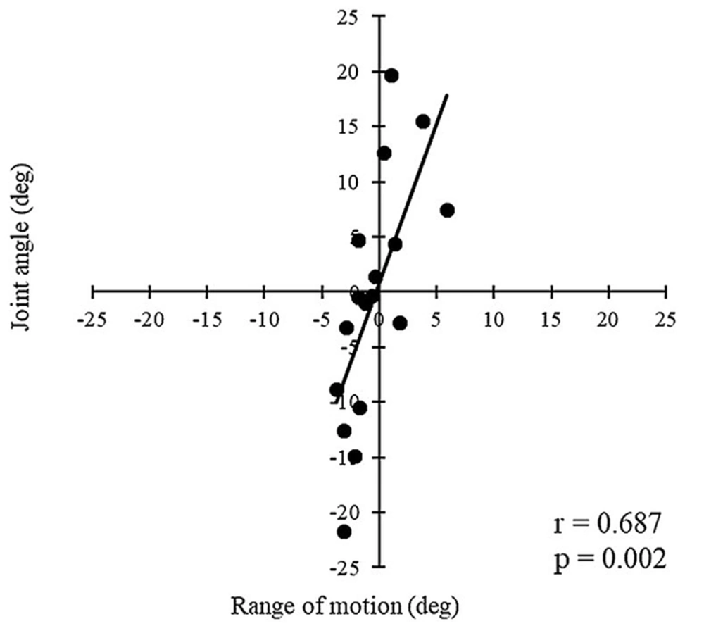
The change in active hip flexion ROM was found to be significantly correlated with the change in maximum hip flexion angle pre-and post-intervention (P = 0.002, r = 0.687, Figure 2). No significant correlations between the changes in other hip ROM and the changes in maximum joint angle were observed (Table 3).
| Range of Motion (deg) | Corresponding Maximum Joint Angle During Pole Vaulting | |||
|---|---|---|---|---|
| R | 95% CI | P-Value | ||
| Take-off leg | ||||
| Active | ||||
| Hip flexion | 0.687 | 0.308 | 0.878 | 0.002a |
| Passive | ||||
| Hip flexion | 0.205 | - 0.306 | 0.624 | 0.430 |
| Hip extension | - 0.366 | - 0.720 | 0.139 | 0.148 |
| Lead leg | ||||
| Active | ||||
| Hip flexion | - 0.008 | - 0.487 | 0.474 | 0.975 |
| Passive | - 0.119 | - 0.567 | 0.383 | 0.649 |
| Hip flexion | ||||
| Hip extension | - 0.185 | - 0.611 | 0.325 | 0.902 |
Abbreviation: CI, confidence interval.
a significant correlation at P < 0.05.
5. Discussion
The present study compared the hip ROM and the maximum hip joint angle during pole vaulting pre and post-intervention. No significant improvement in hip ROM and maximum joint angle was observed post-intervention. Nonetheless, the change in post-intervention active hip flexion ROM was significantly correlated with the change in post-intervention maximum hip flexion angle during pole vaulting. This result supports our hypothesis.
No significant correlation was observed in changes of both legs’ hip flexion between the passive ROM and maximum joint angle. In contrast, the change in active hip flexion ROM was significantly correlated with the change in the maximum hip flexion angle of the take-off leg after the intervention. A previous study reported that pole vaulters with chronic low back pain had lower active SLR angles than healthy pole vaulters, suggesting poor trunk stability or incompetence of the kinetic chain required when raising the lower limbs (3). In addition, another previous study (18) reported that the maximal hip extension torque was significantly correlated with the intra-abdominal pressure, and the relationship was still significant even when the anatomical cross-sectional area of the hamstring and the thickness of the gluteus maximus were adjusted statistically. Therefore, trunk stability and the kinetic chain may be a factor that can be used to improve the active ROM. The psoas major muscle connects the intervertebral discs of the rib processes or the lumbar vertebrae to the femur. When the psoas major muscle works as a hip flexor, it is possible that the lower limb cannot be raised because of poor trunk stability or incompetence of the kinetic chain. Hodges and Richardson (19) reported that the central nervous system initiates the contraction of the abdominal muscles and lumbar multifidus muscles in a feedforward manner ahead of the prime mover of the lower limb. The active SLR exercise in the intervention of this study was performed to improve the kinetic chain and core activation. Therefore, we considered that the intervention in this study improved the kinetic chain and core activation of participants and increased the maximum hip flexion angle during pole vaulting. In addition, to change the vaulting motion to prevent injuries or improve, active ROM (instead of passive ROM) may need to be changed, and coaches and athletic trainers should keep on assessing and improving active ROM. However, it is difficult to consider that the active ROM is greater than the passive ROM, and it is deemed necessary to acquire a large passive ROM as a basis.
The hip ROM did not significantly improve after the intervention. Many studies have shown that intervention using foam rolling massage may increase the hip, knee, and ankle ROM without impairing muscular strength (4-8). In addition, a previous study (20) reported that the vibration foam rolling group showed substantially greater improvements in passive hip extension ROM compared with the non-vibration foam rolling group. All intervention programs in this study lasted one session of approximately 25 min using a non-vibration foam roller and massage ball, which may have been insufficient to improve the hip ROM. The effectiveness of the intervention may also be influenced by the fact that different athletes’ hip ROMs are limited by different factors and depend on each athlete’s stretch tolerance. In addition, the active hip flexion ROM on the lead leg was significantly decreased post-intervention compared to pre-intervention. The athletes performed a high-stress exercise (i.e., pole vaulting) between ROM measurements pre and post-intervention in this study. Baroni et al. (21) reported a decreased ROM in the elbow joint immediately after arm curl training that consisted of 4 sets of 10 concentric-eccentric repetitions on unilateral elbow flexion using a dumbbell resistance equal to 80% of 1 repetition maximum. Therefore, the deterioration caused by the stress of pole vaulting may be greater than the improvement in ROM caused by the intervention in this study.
This study has several limitations. First, the sample size for this study was limited, with only 17 participants. Second, to prevent male and female sexual characteristics from affecting the performance of pole vaulting and, consequently, the results, only male pole vaulters were recruited in this study. Hence, the findings of this study might not be entirely applicable to female pole vaulters. Lastly, no control group was formed because of the limited number of participants included in the present study, and the intervention might have prevented a reduction in ROM, which could not be ascertained from the results of this study.
5.1. Conclusions
The change in active hip flexion ROM was found to be significantly correlated with the change in maximum hip flexion angle during pole vaulting pre-and post-intervention. Athletes may need to improve their active ROM to prevent injuries and improve their performance. Coaches and athletic trainers may also need to adopt active ROM as an indicator to control athlete conditioning.

