1. Background
Medial tibial stress syndrome (MTSS) is a very common injury to lower leg in both athletic and military populations (1); with an incidence rate between 4% and 35% reported in the past four decades (2-4). MTSS is a common exercise induced injury that causes a tender and painful area in the distal two-third of the posterior medial edge of tibia, the pain is relieved with rest but it reappears with exercise (5, 6). So far, the characteristic signs and symptoms of MTSS are fully defined, but the pathophysiology of this disorder is not exactly known. Most of previous studies define MTSS as an inflammation in posterior medial edge of tibia due to repeated tension to lower extremity and bone overload (7). Other theories suggest periostalgia (8), periostaitis (9, 10), bone stress reaction (11-14), and low bone mineral density (15, 16). There are also different theories to describe the pathomechanism of this syndrome; Sommer and Vallentyne proposed pronation of foot at subtalar joint due to a varus malformation induced by posterior tibialis tightness or peroneus longus weakness as a cause for MTSS (17). Viitasalo and Kvist found out that over-inversion/eversion of subtalar joint and also significant increase in calcaneal angles is present in people with MTSS (18). Raissi et al. (19) and Bennett et al. (3) in separate studies revealed that navicular drop test is related to MTSS and this syndrome is more prevalent in females. White and Yates showed that those with MTSS have less dorsiflexion due to tightness of soleus and gastrocnemius muscles (4); Beck and colleagues used the same theory to explain tibial bending by sharpy’s fibers that leads to MTSS (5). Moen et al. reported that higher internal rotation range, more plantar flexion and positive navicular drop test are related to prevalence of MTSS while higher BMI just prolongs recovery time (20). Bouche and Johnson studied pathomechanics of MTSS and reported that tension introduced to deep crural fascia due to contraction of posterior compartment muscles is the reason of MTSS (21). Bartosik et al. revealed that limb length disparency might be responsible for MTSS. The shorter side could have foot equinus, hip joint drop, over-extension of knee joint, while the longer side might have over-flexion of hip and knee joint, and over-pronation at subtalar joint (22). The same study showed that those with MTSS have lower ankle dorsiflexion. Messier and Pittala showed that in those with MTSS foot pronation and the speed of this action both are increased (23). MTSS is one of the most common injuries between military recruits (4). Different studies have shown different associations and risk factors such as over-pronation of midfoot (4), high BMI (24), female gender, lower calf girth, increased internal and external rotation (25), and higher range of plantar flexion (26). These risk factors are not the same in all the studies and there is contradiction. Imaging studies of MTSS with MRI have shown a high signal line in posterior edge of tibia in MTSS patients (27). Thacker and colleagues showed that strengthening soleus, controlling over-pronation, using appropriate insole, and low tension exercise can help preventing MTSS (28). It is reported that patients with MTSS have lower bone density compared to normal subjects (15), it is also shown that after treatment of MTSS bone density was normal again (16); so it could be the cause or result of MTSS.
2. Objectives
Since factors inducing MTSS are not fully known based on the previous studies and also the pathomechanical theories that are already presented, it was needed that we study anthropometric and anatomic factors and their relations with MTSS in military recruits, a population in which MTSS is reported to be high.
3. Patients and Methods
In this retrospective cross-sectional study 200 male military personnel were randomly selected from an expert training brigade. Nineteen subjects with previous history of lower extremity fracture or surgery were excluded from the study which left us 181 subjects to perform the study on. All the subjects were informed about the study and they signed the written consent form. The study was approved by the Ethical Committee of Baqiatallah University of Medical Sciences, Tehran, Iran. Diagnostic criteria for MTSS were based on the description by Yates and White that includes a pain history which is induced by exercise and lasts for a couple of hours to several days after exercise. This pain is located on the distal two-thirds of posteromedial border of tibia. The patient must not have any history of paresthesia or neurovascular symptoms. The pain must cover an area of at least 5cm and palpation of this area could cause a diffuse vague pain. The measures were taken two times with a one-weak interval; the mean of these two measurements was recorded for each parameter. Demographic data of each subject (age, weight, and height) were collected by four skilled examiners (with an average of 4 years of experience). Weight was measured by a digital Seca scale (Germany, 100 grams accuracy) with light clothing and bare feet. Height was measured with Seca stadiometer (Germany, to the nearest 0.1 cm) with bare feet. Then the patient was examined by an experienced sports medicine specialist in order to gather the following data based on the studies by Moen et al. and Raissi et al. All angles were measured by one goniometer and all static measurements of lower extremity were taken using a tape meter to the nearest 0.5 cm. Leg length discrepancy: using a tape meter (nearest 0.5 cm), lower extremity length was measured from the anterior superior iliac spine to the most prominent part of medial malleolus (7, 19). Hip range of motion: the subject was in sitting position with knee and hip joints flexed at 90°; hip joint was rotated internally and externally to a firm end feel. The angles were measured in degrees relative to the initial position (19). Intercondylar interval: the interval was measured with a gauge (accuracy of 0.1 mm) while the subject was standing upright and the feet were paired and in one direction (19). Quadriceps (Q) angle: the subject was standing with its knee and hip joints extended and quadriceps muscle at rest. The center of goniometer was at patella with lower hand at tibial tubercle (which was found with palpation), and the other hand was toward anterior superior iliac spine (ASIS). The angle was measured in degrees (19).
Calf girth: the subject was standing relaxed and upright. Using a tape meter the maximum circumference of the relaxed calf was measured and recorded (20).
Ankle girth: in the same position as for the calf girth measurement, proximal to the internal malleolus, the minimum circumference was measured and recorded (29).
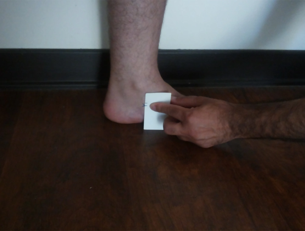
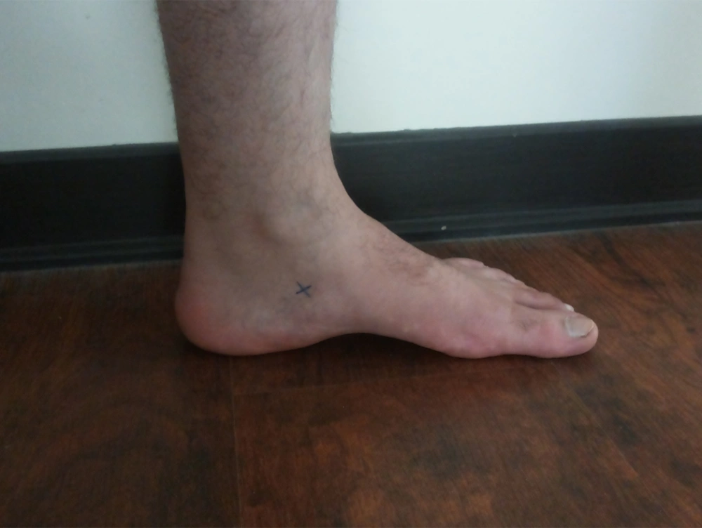
Navicular drop test: while the subject was in a sitting position, with knees flexed at 90° and ankle joint in a neutral position, the tuberosity of navicular bone was marked with a non-toxic marker. Then the individual was asked to stand without changing the position of the foot. The difference between heights of the tuberosity of navicular bone in these two positions was referred as navicular drop and was measured in mm (20).
Iliospinale height, lateral tibial height, and trochanteric-lateral tibial height were measured based on Kinanthropometric Assessments; Guidelines for Athlete Assessment in New Zealand Sport (29).
Body composition: the proportion of fat tissue mass to lean body mass was measured based on bioelectrical impedance analysis method using Tanita BF 350 (Japan).
Intratest reliability: in order to determine the intratest reliability a pilot study was performed (kappa = 0.75). Based on the measurement methods described above, we measured 30 lower extremities of 20 subjects 3 times during one week which were randomly selected; these measurements were blinded to the subjects.
Statistics: Using SPSS software for Windows, version 19 (SPSS, Chicago, IL, USA), we performed the statistical analyses. Data was expressed as mean ± SD and normal distribution was determined by Kolmogrov-Smirnov method. Then using independent sample test and Kolmogorov-Smirnov test data was analyzed. Statistical significance was accepted if P value ≤ 0.05.
4. Results
Of 181 army recruits who were included in this study with a mean age of 30.7 ± 4.68 years, 30 participants (16.6%) were in MTSS group (age: 29.52 ± 3.88 years, height: 173.76 ± 6.57 cm, and weight: 80.20 ± 11.63 kg) and the other 151 participants (83.4%) were in control group (age: 30.26 ± 4.83 years, height: 174.40 ± 7.84 cm, and weight: 83.10 ± 17.48 kg). Factors like lower extremity length, intercondylar interval, Q angle, tibial lateral height, calf girth, ankle girth and fat mass of the subject were measured and analyzed but there were no significant differences between the two groups. There was significant difference in other factors like navicular drop test, internal and external rotation, iliospinale height, and trochanteric tibial lateral length. Mean ± SD of all of the studied factors and also the P values are shown in Table 1.
| Variable | Normal Group | MTSS Group | P Value |
|---|---|---|---|
| Age, y | 30.26 ± 4.83 | 29.52 ± 3.88 | 0.154 |
| Height, cm | 174.40 ± 7.84 | 173.76 ± 6.58 | 0.298 |
| Weight, kg | 83.10 ± 17.48 | 80.20 ± 11.63 | 0.337 |
| Right leg length, cm | 87.53 ± 3.54 | 88.84 ± 4.29 | 0.448 |
| Left leg length, cm | 87.65 ± 3.59 | 88.91 ± 4.36 | 0.463 |
| Intercondylar interval, mm | 1.08 ± 1.46 | 1.29 ± 1.56 | 0.475 |
| Q angle, degree | 14.56 ± 5.89 | 14.51 ± 5.61 | 0.809 |
| Hip external rotation, degree | 45.23 ± 3.34 | 41.42 ± 3.82 | 0.000 |
| Hip internal rotation, degree | 38.80 ± 4.61 | 37.07 ± 2.80 | 0.004 |
| Navicular drop test, mm | 6.24 ± 2.79 | 4.22 ± 3.22 | 0.015 |
| Iliospinale height, cm | 51.07 ± 3.02 | 53.14 ± 3.07 | 0.017 |
| Trochanteric tibiale lateral height, cm | 43.22 ± 2.53 | 44.69 ± 1.68 | 0.022 |
| Tibiale lateral height, cm | 45.26 ± 2.61 | 45.80 ± 2.10 | 0.360 |
| Calf girth, cm | 38.01 ± 3.25 | 38.35 ± 3.23 | 0.691 |
| Ankle girth, cm | 22.87 ± 1.53 | 22.83 ± 1.37 | 0.575 |
| Body fat mass, percent | 20.09 ± 5.27 | 19.97 ± 4.83 | 0.677 |
5. Discussion
Based on this study the prevalence of MTSS in these 181 military recruits was 16.6% (30 participants). Navicular drop test, internal and external rotation, iliospinale height, and trochanteric tibial lateral length were the only five factors that were significantly different btween the two groups. The results of this study were in concordance with those of Raissi et al. (19), Bennett et al. (3), Moen et al. (7) and Yates and White (4). However, the findings of this study about navicular drop test is different from those of Plisky et al. (24), who found no association between navicular drop and MTSS in competitive adult runners. The difference between some findings of this study and the previous ones (and also between the previous ones) might be due to variation in target groups (expert military recruits, runners, female, or male participants) and measurement techniques. Navicular drop test is an indicator of pronation of foot. In our study mean of NDT was significantly lower in MTSS group (NDT: 4.22 ± 3.22 mm) than in control group (NDT: 6.24 ± 2.79 mm); though both were in normal ranges (P value: 0.015). So far, NDT has been investigated in seven studies, five of them found association between positive NDT and MTSS (3, 4, 19, 20, 30), while the other two have found no association (24, 25). We found a significant decrease in range of hip internal and external rotation in association with MTSS in this study (P value: 0.004 and 0.000 respectively). This finding was in agreement with that of Moen et al. (7) and in contrast with Burne et al. (31). No obvious mechanism is established to define the relation between MTSS and hip range of motion. Burne et al. stated a mechanism in which those with increased hip internal range of motion have a specific pattern of running which causes extra load on their tibia that might lead to MTSS. The same pathomechanism might be induced by increased hip external range of motion (31).
In this study there was no association between weight, calf girth, and ankle girth with MTSS (P value: 0.337, 0.691, and 0.575, respectively), despite the results of Hubbard’s study (25). Plisky et al. found that BMI > 20.2 was associated with MTSS (24). Another study found that in those with MTSS lean calf girth was significantly lower in their right leg (P value: 0.044) (20). Moen et al. found a relation between higher BMI and longer time to full recovery (P value: 0.005) (32). We found no significant difference between BMI in the two groups (P value > 0.05); the reason might be that all the personnel involved in this study were in shape and had no significant difference in their BMIs.
In a cohort study on 77 cross country runners in the united states, runners with navicular drop score of more than 10 mm were almost 7 times more likely to incur medial exercise related leg pain (ERLP) than those with with a less than 10mm navicular drop score (OR:6.6, 95% CI = 1.2-38.0) (33). Navicular drop test (NDT) which is a valid measure of foot pronation (26, 34), is defined as the difference between height of navicular tuberosity at neutral position of subtalar joint and its height in relaxed stance. It was measures based on description by Brody et al. (35). Relatively low prevalence rate of MTSS in our target group was not probably due to type of military shoes or exercise area surface (none of them were standardized); it could be due to low intensity trainings and the long intervals between training sessions. One more logical hypothesis was mentioned by Bouche and Johnson as tibial fasciitis, which proposes that this syndrome is due to the tension on the site of deep crural fascia insertion to tibia; that is mainly from posterior compartment muscles especially the soleus muscle (21). One evidence in favor of this theory is that the most effective surgical procedures involve release of the deep posterior compartment, including the soleus sling and removal of a strip of posteromedial tibia periosteum (36). This tension on insertion site of deep crural fascia to tibia caused by posterior compartment muscles could be due to the different growth rate of muscles and deep crural fascia and flexibility of this fascia. Since muscle growth rate is probably more than fascial growth rate and collagen synthesis by fibroblasts, a tension is introduced to deep crural fascia and guided to its insertion site on tibia from posterior compartment muscles; especially in those who have recently started exercise and had no time for adaptation of their deep crural fascia to the relatively rapid growth rate of calf muscles. In this study we found that MTSS is associated with some of the investigated factors but in order to fully understand the pathomechanism of MTSS further research with new methods is needed to investigate the possible risk factors of MTSS among military recruits with focus on the tension induced by hypertrophy of the posterior compartment muscles to insertion site of deep crural fascia.

