1. Background
Fatigue may be defined as a decrement in performance during continuous or intermittent exercise (1). A soccer player experiences fatigue or reduced performance at three different game conditions: after short-term intense periods of activity, at the start of the second half and towards the end of the 90 min game (2). The effect of fatigue on soccer technical skills, especially kicking, has been poorly investigated with only three studies having been reported in the literature. Lees and Davies (3) used a 6-minutes step protocol to induce fatigue and reported higher maximum foot speeds and thigh angular velocities, but lower ball speed and foot/ball ratio of the kicking leg after fatigue. These results were attributed to lack of co-ordination before and during ball impact. Apriantono et al. (4) reported that an isokinetic fatigue protocol resulted in a less powerful soccer kick by reducing speed of the ball, the toe and the kicking leg. These findings were attributed to muscle strength capacity reduction and an alteration of inter-segmental co-ordination following fatigue. Kellis et al. (5) also found that a 90-min fatiguing protocol, simulating realistic game conditions reduced fast kick performance. Slower kick performance was attributed to a reduction in knee extension angular velocity and a linear velocity of the ankle. However, all the aforementioned studies examined fatigue responses of soccer male players. Despite the vast growth in female participation in soccer, few studies have examined female kicking skill (6, 7). To our knowledge there are no studies on the effect of fatigue on female kicking performance. If fatigue responses are gender dependent, then differences in soccer kick performance will be expected. In this case, a different approach to the design of training programs for male and female players would be necessary.
While it is known that intense muscular effort reduces muscle strength (8) and has been shown to have a detrimental effect on skilled soccer performance, the effect of short-term intensive exercise is less well established. To our knowledge, only Lees and Davies (3) have investigated changes in kick mechanics after a short-term fatiguing protocol, which was not specific to soccer, and reported that some key aspects of performance improved rather than deteriorated after fatigue. Therefore, more information on the effect of intense running fatigue on kicking is necessary.
Another limitation of previous studies is that the time needed for a player to recover powerful kick performance after fatigue was not examined. In particular, previous studies analyzed only one trial (3), the average ball speed after several kicking trials (4) or the trial that produced the highest ball speed post-fatigue (5). Therefore, even if fatigue causes an immediate effect on soccer kick performance, it is not clear how fast players can recover performance and technique. From a practical point of view, examining the mechanisms underpinning short-term fatigue using actions which are commonly performed during a soccer game would provide useful information to coaches for the design of appropriate training programs. In addition, if soccer kick performance recovers fast after fatigue, then the effects of fatigue on kicking have been previously overestimated.
The recovery of performance after intense fatigue game events, such as intense running events, has never been estimated, thus leaving several questions regarding the effect of fatigue on game performance unanswered. If the occurrence of fatigue has a practical meaning and impairs players’ kicking performance, then the examination of performance recovery after fatigue would provide additional information to coaches and players as to which kicking parameters are most affected by fatigue and what type of training should be followed to allow more effective performance during the game. Such information is even more useful when examining female players due to the highly increasing number of active female soccer players.
The aim, therefore, of this study was to investigate powerful kick recovery after fatigue produced by intense periods of running. A secondary objective was to examine whether fatigue responses are gender specific.
2. Objectives
Our main hypotheses were firstly, that soccer kick performance would be reduced during each of the three consecutive kicks performed immediately after fatigue and secondly, gender differences in fatigue responses would be observed.
3. Patients and Methods
3.1. Participants
Ten male (age: 26.3 ± 4.9 years, height: 178.1 ± 5.1 cm, mass: 81.3 ± 8.1 kg) and ten female (age: 24.4 ± 4.2 years, height: 169.7 ± 5.7 cm, mass: 61.8 ± 5.1 kg) amateur soccer players participated in the study. Each participant had a minimum of 8 years of experience, trained three times and played one game per week. All players were right footed and had no history of neurological diseases or musculoskeletal abnormalities, and none was taking any medication during the course of the study. Participants were fully informed of the procedures of this study and provided written consent which was based on the Declaration of Helsinki. The University Ethics Committee approved the protocol.
3.2. Procedures
The protocol used in the present study has been previously applied by Abdul-Aziz et al. (9). Specifically, players had to run on a treadmill till exhaustion. They start running at a speed of 10.0 km h-1 for 2 minutes, followed by an increase to 12.0 km h-1 for another 2 min. Thereafter, inclination was systematically increased by 2% every minute until a maximum of 12% was achieved. If player’s exhaustion was not achieved by this time, the speed was increased by 1.0 km h-1 every minute thereafter until the player attained volitional exhaustion.
All testing procedures were conducted in a standardized laboratory environment (temperature 22-25ºC and humidity 55-65%) during two visits to the laboratory. On the first visit participants familiarized with the protocol, while on the second visit the main protocol was performed. All tests were performed during the same time of the day (6-9 p.m.) to avoid any chronobiological effect. The tests were conducted during the pre-season period. Participants were instructed to refrain from any vigorous exercise 48 hours before the tests. One day before the main protocol participants were asked not to undertake any training. All players followed their regular nutritional intake and maintained regular sleeping habits before the tests. The kicking trials were performed on an artificial turf, similar to the turf used during games, while all participants wore their own soccer shoes.
A 10-minute warm-up consisting of jogging, stretching exercises and several familiarization trials was performed. The participants performed instep kicks prior to and after the implementation of the fatigue protocol. Each participant performed 3 consecutive kicking trials of a stationary ball with the instep portion of the foot after a one step angled approach (45º), against a goalpost located 7m in front of the ball. A standard size and inflated ball was used. Participants were instructed to kick the ball as fast and hard as possible aiming at the centre of the goalpost.
A 30 sec rest interval between consecutive kicks was provided to allow examination of the acute effects of fatigue on successful kick performance. A greater rest interval during post fatigue trials would allow quick recovery of performance. Totally, the six kicking trials (3 pre- and 3 post-fatigue) were analyzed.
3.3. Kinematics
Kinematic data were collected with a passive, 6-camera, 3-D Vicon motion analysis system (Oxford Metrics Ltd., Oxford, UK). The cameras were calibrated to a volume of 2.0 m3 and calibration errors were all below 3 mm. Kinematic data were sampled at 120 Hz.
Retro-reflective spherical markers were placed on selected anatomical landmarks of both limbs in order to identify segments and joints of the lower extremities: the head of fifth metatarsal, the heel, the mid-shank, the lateral malleolus, the femoral epicondyle, the mid-thigh, the greater trochanter and the anterior superior iliac spine. Four additional markers were placed on the surface of the ball. Prior to each kicking trial, a standing trial was recorded to establish initial joint angle conditions. Marker position was automatically tracked using the Polygon software. Three-dimensional marker position coordinates were computed using the direct linear transformation (DLT) method. The three-dimensional coordinates were expressed as a right handed orthogonal reference frame, in which Z axis was vertical and pointed upward, Y axis was horizontal and X axis was perpendicular to Z and Y axis. The resulting displacement - time data of each marker were filtered using a low-pass, two-order, Butterworth, dual-pass filter. Optimal cut-off frequencies were chosen by comparing the residuals of the difference between filtered and unfiltered signals at several cut-off frequencies. The cut-off frequency for filtering the kinematic data was set at 16 Hz.
Angles between the segments were examined by viewing the Euler angles decomposed from the rotation matrix which describes the orientation of one segment to another using an X-Y-Z rotation sequence (10). In the present study, the decomposition of the Euler angles for each segment was as follows: the ankle was separated into plantar flexion/dorsi flexion and inversion/eversion angles; the knee was separated into flexion/extension and internal/external rotation angles; and the hip was separated into internal/external rotation and abduction/adduction angles.
The absolute magnitude of ball velocity (Vball) was calculated from the values of its vertical and horizontal components (11). The horizontal component of the ball velocity was calculated as the first derivative of linear regression lines fitted to their non-filtered displacements. The vertical component was calculated as the first derivative of a quadratic regression line with its second derivative set equal to -9.81 m∙s-2 fitted to its non-filtered displacement in the available frames. The velocity of the centre of mass of the foot (Vfoot) was measured from the toe and heel markers coordinate data (12). Kinematic data were recorded prior to start of the kick and ended when the kick was concluded.
3.4. Data Analysis
To simplify the results, kicking motion was divided in two phases: from the toe-off of the kicking leg to ground contact of the support leg (pre-support phase) and from ground contact to initial ball impact (support phase). Each phase was set as 100%. Subsequently, the segmental linear velocities and joint displacements were averaged for every 10% of each phase. In addition to the time-series calculation, maximum ball speed; maximum and at impact linear velocity and angular displacement of the hip, the knee, and the ankle were also examined. These parameters were selected based on previous studies (7, 13), as they are frequently considered to be important biomechanical indicators of soccer kick performance.
3.5. Statistical Analyses
A three-way analysis of variance (ANOVA) with repeated measures (Gender x Fatigue x Trial) was used to examine differences in ball speed and maximum and at impact linear velocity and angular displacement of the hip, the knee, and the ankle joint.
A three-way ANOVA with repeated measures with three within participant variables (Fatigue x Trial x Phase) was used to examine the differences between the two measurements sessions (pre and post) in angular joint displacements and segmental velocities over 10 data points of the pre-support phase. Separate designs were applied for support phase data. Significant interactions were followed up with simple effects tests and, if significant, post-hoc Tukey tests were applied to examine significant differences between pairs of means. Statistical significance was set at P < 0.05.
4. Results
The ANOVA showed a significant interaction (Gender x Fatigue x Trial) effect for the ball speed. Post-hoc analysis indicated that ball speed was significantly lower (P < 0.05) during the first and the second kicking trial post-fatigue compared to pre–fatigue kicking trials for both male and female players (Pre fatigue 1st trial: 21.50 ± 2.01 m/sec; 2nd trial: 21.14 ± 1.74 m/sec; 3rd trial: 21.39 ± 1.60 m/sec and post fatigue 1st trial: 18.99 ± 1.81 m/sec; 2nd trial: 19.86 ± 2.06 m/sec; 3rd trial: 19.96 ± 2.66 m/sec for males and pre fatigue 1st trial: 18.41 ± 1.55 m/sec; 2nd trial: 18.03 ± 0.73 m/sec; 3rd trial: 17.69 ± 0.56 m/sec and post fatigue 1st trial: 15.77 ± 1.82 m/sec; 2nd trial: 16.44 ± 2.20 m/sec; 3rd trial: 17.46 ± 2.12 m/sec for females, respectively).
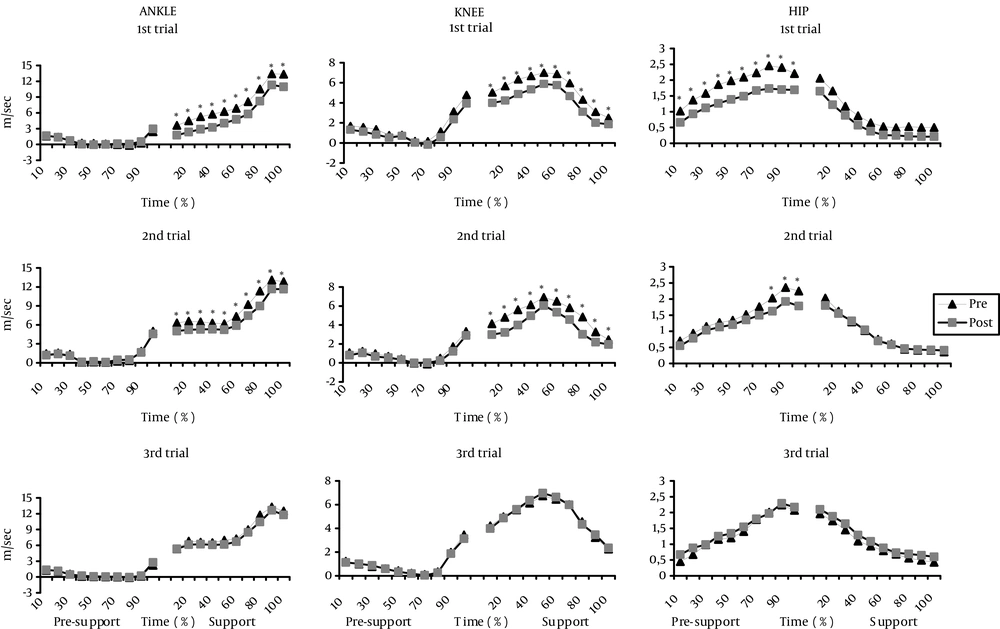
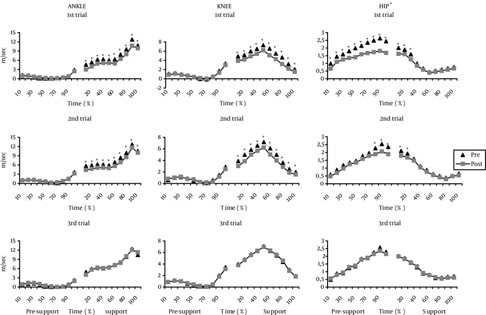
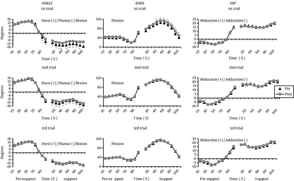
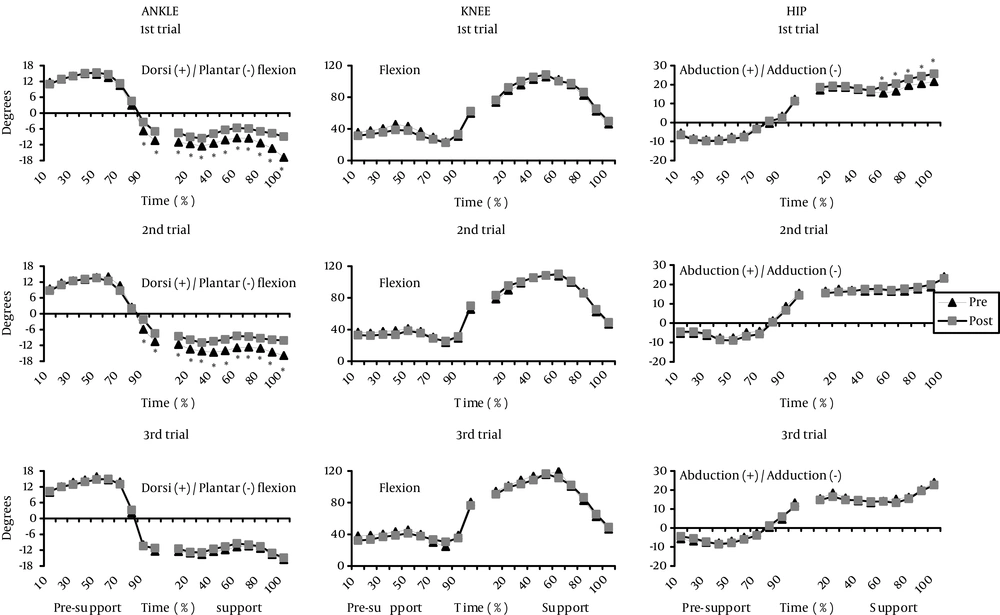
Similarly, a significant interaction (Gender x Fatigue x Trial) effect for the maximum and at impact joint linear velocity and joint displacement (P > 0.05) was found. Post-hoc analysis indicated that maximum ankle, knee and hip linear velocity (Figures 1 and 2) and ankle and knee velocity at impact (Figures 1 and 2) were significantly lower during the first and the second kicking trial post-fatigue compared to pre–fatigue kicking trials for both male and female players (P < 0.05). Similarly, a significantly lower ankle plantar flexion angle at ball impact (Figures 3 and 4) was observed for males and females (P < 0.05). Males demonstrated a lower knee flexion angle (Figure 3), while females showed a higher hip abduction angle during the first post-fatigue trial (Figure 4) compared with pre-fatigue trials (P < 0.05). A non-significant effect was found for the ankle eversion/inversion angles, the knee and the hip internal/external rotation angles at ball impact, the ball/foot speed ratio and the duration of the kick (P > 0.05).
The sagittal ankle, knee and hip linear velocities are presented in Figure 1 and 2, for males and females, respectively. The ANOVA results showed a significant interaction (Fatigue x Trial x Phase) effect for the joints' linear velocity. Post-hoc analysis indicated a significant decline of ankle and knee velocity during the support phase (from 10% to 100%) of the first and the second kick trials post-fatigue in male (Figure 1) and female (Figure 2) players (P < 0.05). Similarly, post-hoc analysis indicated a decline in hip speed of the pre-support phase during the 1st and 2nd (from 80% to 100%) kick trials after fatigue (Figure 1; P < 0.05). Females showed significant decrements in hip speed during most phases of the 1st and 2nd post-kicking trials (whole pre-support phase and initial support phase) (Figure 2; P < 0.05).
The joints' angular displacements are presented in Figures 3 and 4, for males and females, respectively. The ANOVA results showed a significant interaction effect for the ankle angular displacement. Post-hoc analysis indicated that males showed a less plantar flexion angle during 1st (end of pre-support and whole support phase) and 2nd (whole support phase) post-fatigue kicking trials (Figure 3; P < 0.05). Females also showed a less plantaflexed ankle during specific stages of the pre-support and the support phase of the first two trials post-fatigue (Figure 4 ; P < 0.05).
The ANOVA results also showed significant interaction effects for the knee and the hip angles. Post-hoc analysis indicated that males showed less knee flexion angle (Figure 3 ; P < 0.05) during the 1st post fatigue trial (from 40% to 100%), while females showed higher hip abduction angles, from 60% to 100% of support phase of the 1st post fatigue kick (Figure 4 ; P < 0.05).
No significant fatigue differences were found for the ankle inversion/eversion angles, the knee and the hip internal/external rotation angles for both genders during the pre- and post-fatigue kicking trials (Figures 3 and 4 ; P > 0.05).
Males' sagittal linear velocity (m/sec) of the ankle (left diagrams), the knee (middle diagrams) and the hip joint (right diagrams) during the three pre- and post-fatigue kicking trials presented as a percentage (%) of the total time of the pre-support and the support phase (* significant differences between pre- and post-fatigue trials).
Females' sagittal linear velocity (m/sec) of the ankle (left diagrams), the knee (middle diagrams) and the hip joint (right diagrams) during the three pre- and post-fatigue kicking trials presented as a percentage (%) of the total time of the pre-support and the support phase (* significant differences between pre- and post-fatigue trials).
Males' angular displacement (degrees) of the ankle (left diagrams), the knee (middle diagrams) and the hip joint (right diagrams) during the three pre- and post-fatigue kicking trials presented as a percentage (%) of the total time of the pre-support and the support phase (* significant differences between pre- and post-fatigue trials).
Females' angular displacement (degrees) of the ankle (left diagrams), the knee (middle diagrams) and the hip joint (right diagrams) during the three pre- and post-fatigue kicking trials presented as a percentage (%) of the total time of the pre-support and the support phase (* significant differences between pre- and post-fatigue trials).
5. Discussion
The main findings of the present study were, first, that high intensity fatigue had an immediate effect on kicking performance, which recovered to pre-fatigue levels approximately within 1-minute after the end of the fatigue protocol. Second, we noticed minor gender differences in fatigue responses. Consequently, the first research hypothesis of the study is accepted, while the second research hypothesis is rejected.
The exercise protocol applied in the present study has been previously validated (9) and used to simulate high intensity running conditions. Mohr et al. (2) reported that players experience fatigue during intense periods of a soccer game. During the performance of the present protocol players gradually increase their effort, similar to several actions taking place during a real game, at the end of which players are exhausted. This is the main reason for selecting the present protocol as it allows examination of the technique responses after intense periods of running.
The present results are in agreement with the three previous studies which reported a decline in ball speed immediately after various fatigue protocols (3-5). However, only Lees and Davies (3) applied a step exercise protocol that was of similar duration and intensity with the present protocol, thus permitting comparisons. Lees and Davies (3) reported that players performed slower kicks in the post-fatigue condition. It was suggested that timing and co-ordination were impaired as a result of fatigue. The results of the present study did confirm this finding as joint velocities and displacements significantly changed after fatigue (Figures 1 - 4). This indicates that co-ordination was affected and the kicking technique was quite different between pre- and (first and second) post-fatigue kicking trials.
Previous studies analyzed only one trial after fatigue (3) or the average ball speed after several kicking trials post-fatigue (4) or the trial that produced the highest ball speed post-fatigue (5), thus leaving several questions unanswered regarding the acute effects of fatigue on kicking performance and the duration of these effects. In the present study it was clear that players performed less powerful kicks during the first and the second trial post-fatigue, but they were able to perform powerful kicks during the third trial post-fatigue in levels comparable to pre-fatigue kicking conditions. Within the limitations of the present protocol, it could be suggested that fatigue effects on soccer kicking technique and performance probably occur within seconds after fatigue, which should be taken into consideration by both players and coaches.
The results of the present study showed a decline in maximum and at impact joint velocities during the first two, but not during the third post-fatigue trial (Figures 1 and 2). Foot speed at ball impact is crucial for powerful kicking performance (14, 15). Powerful kicking trials require a high foot speed at impact with the ball. The present results indicate that the whole kicking motion became slower immediately post-fatigue, thus explaining the decreased kicking performance during the first two trials post-fatigue.
The quality and the characteristics of foot/ball impact have also been reported as significant determinants of kicking performance (16). A more plantar flexed ankle during instep kicking is associated with increased effective striking mass and consequently with a more powerful kicking performance (17). In contrast, a dorsi flexed ankle would lead to an inappropriate foot/ball collision and lower ball speed. The present results showed a less plantar flexed ankle in males (1st trial) and females (1st and 2nd trial) post-fatigue (Figures 3 and 4). This indicates that fatigue might have altered the quality of ball/foot contact. This is further supported by the recovery of plantar flexion accompanied by increases in ball speed during the third post-fatigue trial.
Kicking performance is also affected by player’s technique (14). The present results showed that both males and females demonstrate significant differences in their technique between pre- and post-fatigue kicking trials (Figures 3 and 4). Males showed a more flexed knee during the pre-support kicking phase after fatigue (Figure 3). Practically this means that during the post-fatigue condition the foot travelled through a shorter trajectory until ball impact, which might reduce final foot velocity. Similarly, males showed a smaller ankle plantar flexion post-fatigue (Figure 3), which might be indicative of a less optimal foot position at ball impact for achieving a powerful kick after fatigue. In addition, females showed a more abducted hip after fatigue which is also a sign of unnecessary movement initiation as a result of fatigue (Figure 4). It seems, therefore, that both technique and co-ordination were affected in the presence of fatigue, which subsequently leads to decreased kicking performance. However, the effects disappeared after a minute of rest, during the third post-fatigue trial.
In the present study, we have found minor gender differences in fatigue responses during kicking. Studies examining gender differences in fatigue responses in non-soccer players have yielded conflicting results as some studies found greater resistance to fatigue for females compared to male counterparts (18), while other studies reported no gender effects (19). Our observation, that fatigue affected the ankle and the knee in males and the ankle and the hip kinematics in females, is not sufficient to provide conclusive evidence that there are gender differences in fatigue responses in kicking technique.
A limitation of the study was that fatigue was assessed by recording the perceived exhaustion of the players. Monitoring the biochemical indices of fatigue or recording the heart rate might have provided a more standardized level of fatigue for each participant. Even in this case, the protocol would have led to the exhaustion of the player, defined by his/her inability to continue running at the same rate. Therefore, any changes in the effects of this high intensity fatigue task on kicking performance seem unlikely. Finally, the participants of the present study were amateur soccer players. The same protocol might have different effect on younger or professional players.
The results of the present study indicated that a generic task, such as continuous running, did affect kicking performance, but players were able to perform powerful kicks after at least a minute of rest. This has a practical meaning for both players who have to perform kicks under fatigue conditions and their coaches. When a player performs a high intensity running during a game, he/she needs to then “gain” time before performing a powerful kick. Based on the results and the skills and abilities of the participants of the present study, a rest interval greater than a minute would permit players to perform a powerful kick. Such information may be used by coaches to design soccer-specific training games and exercises so that players could improve their ability for fast recovery after fatigue. Such information can also assist coaches to select the most suitable players for performing kicks under set play conditions.
High intensity running till exhaustion resulted in reduced foot speed and joint velocities at impact and reduced ball speed when players performed two kicks immediately after the end of a fatigue protocol. However, performance and technique alterations recovered during the third kicking trial after approximately 1 minute of the ending of the fatigue protocol. Furthermore, fatigue responses were similar for both males and females. These results indicate that any fatigue effects on soccer kicking technique and performance probably occur within seconds after fatigue inducement activity ends. Players, therefore, should gain time after intense periods of running before performing a kick, and coaches should plan appropriate training sessions in order to minimize these fatigue effects.


